
Prognostic and diagnostic glycan-based biomarkers of brain damage

a biomarker and brain damage technology, applied in the direction of peptide/protein ingredients, peptide sources, instruments, etc., can solve the problems of complex brain injury, unexplored pathological condition, and inability to diagnose brain damage, so as to achieve the effect of diagnosing, monitoring or prognosing a brain injury

Active Publication Date: 2020-08-11
MEDICORTEX FINLAND OY
View PDF10 Cites 1 Cited by
  • Summary
  • Abstract
  • Description
  • Claims
  • Application Information

AI Technical Summary

Benefits of technology

The present invention provides a method of diagnosing, monitoring, or predicting a brain injury in a subject by measuring the level of at least one glycan-based biomarker in a sample of bodily fluid from the subject. The method can confirm the presence or absence of brain injury and can also predict the outcome of brain injury. A kit or device for use in this method is also provided, which includes at least one lectin, antibody, or combination thereof that selectively binds to a glycan-based biomarker and a control for comparison.

Problems solved by technology

Brain injuries are complex and can have multiple severe clinical outcomes.
Despite the broad range of the population affected, TBI is still under-served and remains an unexplored pathological condition.
However, the use of the GCS as a diagnostic tool is subject to a number of important limitations.
Recent research has provided evidence that the use of sedative drugs precluded accurate GCS assessment during the first 24 h. Further challenges to diagnosis are presented by the evolving nature of some brain lesions, which can lead to further neurological impairment.
Still further challenges include the trauma subject's possible unconsciousness or inability to communicate.
However, CT scanning has low sensitivity to diffuse brain damage, and availability and utility of MRI is limited.
MRI is also very impractical to perform if subjects are physiologically unstable, and can lead to inaccurate diagnoses in military injuries in which metal fragments are common.
Moreover, mild and moderate TBI represent more than 90% of TBI injuries; this injury range represents the greatest challenges to accurate acute diagnosis and outcome prediction.
However, to date, there are no approved biomarkers for the diagnosis or prognosis of TBI.
This is because of several obstacles to the development of reliable blood biomarkers of TBI.
For instance, the blood-brain barrier (BBB) hinders the assessment of biochemical changes in the brain by use of blood biomarkers in mild TBI, although impaired BBB integrity, as seen in severe TBI, can increase the levels of brain-derived proteins in the blood.
Moreover, some potential biomarkers undergo proteolytic degradation in the blood, and their levels might be affected by clearance from blood via the liver or kidney.
As a consequence, reliable blood biomarkers have been extremely difficult to identify.

Method used


Image

  • Prognostic and diagnostic glycan-based biomarkers of brain damage
  • Prognostic and diagnostic glycan-based biomarkers of brain damage
  • Prognostic and diagnostic glycan-based biomarkers of brain damage

Examples


example 1

[0074]A mouse model of experimental closed head injury disclosed by Yatsiv et al. in FASEB J. 2005, 19:1701-1703 is employed for identifying changes, increase or decrease, in the level of glycan-based biomarkers after a head trauma caused by a weight-drop onto an exposed skull.

[0075]Severity of the injury is assessed according to the Neurological Severity Score disclosed by Beni-Adani et al. (J. Pharmacol. Exp. Ther. 2001, 296:57-63) on the basis of ten individual tasks reflecting motor function, alertness, and behaviour. Severity assessment is carried out 1 hour and 7 days after the trauma in order to allocate the mice into comparable study groups in order to find a correlation between the severity of damage and level of detected biomarker.

[0076]After euthanization, body fluids (including urine, blood plasma or serum, and CSF) from normal and brain injured animals are collected and analysed for glycan-based biomarkers using a lectin assay, and the brains are evaluated histologicall...

example 2

[0077]A rat model of experimental closed head injury disclosed by Bilgen et al. (Neurorehabil. Neural Repair, 2005, 19:219-226) with some modifications is employed for identifying changes, increase or decrease, in the level of glycan-based biomarkers after a head trauma.

[0078]In vivo T2 weighted magnetic resonance imaging (MRI) is performed on the animals to depict the pathologies, including lesion size, tissue viability, and brain oedema, of the resulting injuries in the corresponding neuronal tissues at 24 h and day 3.

[0079]After euthanization, histological evaluation of the injury in the cortex and hippocampus is performed. Additionally, body fluids (including urine, blood plasma or serum, and CSF) from normal and brain injured animals are collected and analysed for glycan-based biomarkers using a lectin assay.

example 3

[0080]A controlled cortical impact was carried out to exposed dura of anesthetized rats according to Bilgen et al. (Neurorehabil. Neural Repair, 2005, 19:219-226) with modifications. The animals were terminated at various times elapsed after the operation. Samples of urine, blood plasma, saliva and cerebrospinal fluid (CSF) were collected and processed using regular methods well known to professionals skilled in the art.

[0081]The samples were incubated on a lectin array, and after washing, a fluorescent conjugate was allowed to bind to the captured glycans or glycan-containing complexes. The spots were visualized and the fluorescence intensities were recorded with a laser scanner.

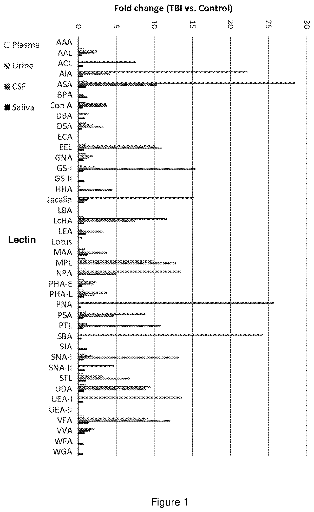
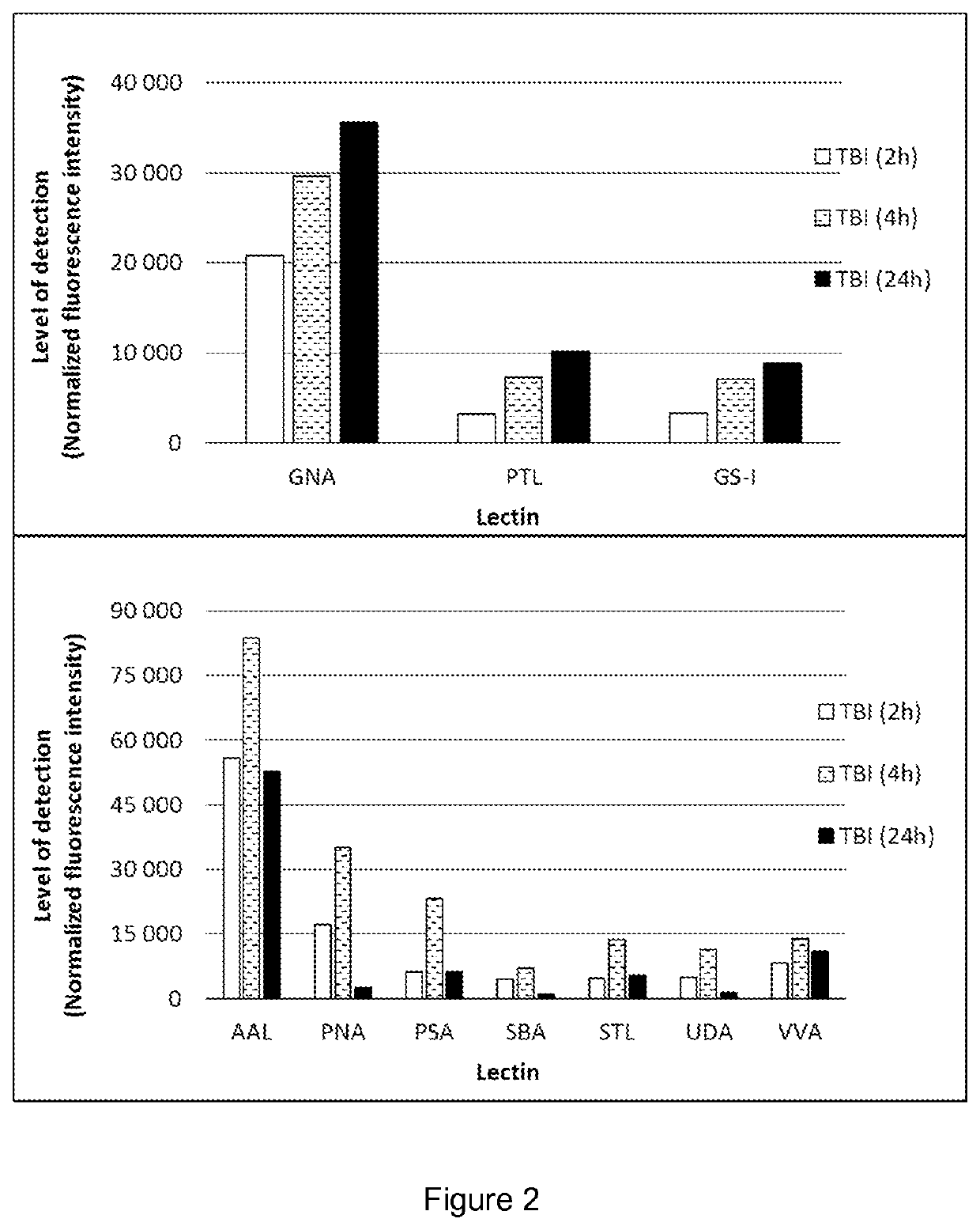
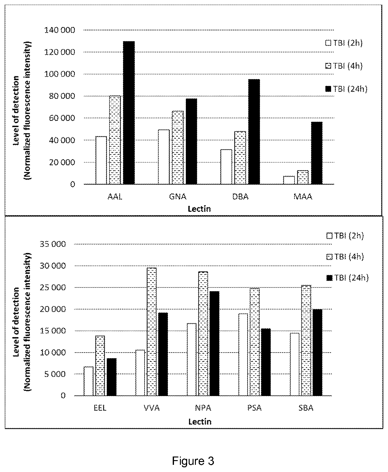
[0082]The results summarized in FIGS. 1 to 3 prove that the body fluids of TBI animals contain glycans or glycan-containing complexes showing significantly elevated binding to particular lectins, compared with the fluids of the sham animals. By selecting appropriate lectins, the TBI detection kit can be adj...


PUM

PropertyMeasurementUnit
CTaaaaaaaaaa
magnetic resonance imagingaaaaaaaaaa
MRIaaaaaaaaaa

Abstract

The present disclosure relates to glycan-based biomarkers for the diagnosis or prognosis of brain damage, such as traumatic brain injury (TBI).

Description

FIELD OF THE INVENTION[0001]The present disclosure relates to glycan-based biomarkers for the diagnosis and prognosis of brain damage, such as traumatic brain injury (TBI), subclinical brain injury (SCI) and acquired brain injury (ABI). The glycan-based biomarker protocol may also be used as an end point in clinical trials. More specifically, the biomarkers of the present invention can be used in diagnostic tests to determine, qualify, and / or assess brain injury status, for example, to diagnose brain injury, in an individual, subject or patient. In particular embodiments, brain injury status can include determining a subject's subclinical brain injury status or SCI status, for example, to diagnose SCI, in an individual, subject or patient (conscious or not).BACKGROUND OF THE INVENTION[0002]Brain injuries are complex and can have multiple severe clinical outcomes. Traumatic brain injury (TBI) is the leading cause of central nervous system impairment in these days. More than 1.7 milli...

Claims


Application Information

Patent Timeline
Patent Type & Authority Patents(United States)
IPC IPC(8): A61K38/00C12Q1/68G01N33/567G01N33/53G01N33/68
CPCG01N33/6896G01N33/5308G01N2800/28G01N2400/12G01N2400/38G01N2333/42C07K14/42G01N33/53G01N33/48
Inventor HAREL, ADRIAN
Owner MEDICORTEX FINLAND OY
Learn More