
Eureka
For R&D, Eureka makes reading and utilizing patents & technical documents easy.
Eureka AIR
Designed for self-driven R&D workflows. Generate viable solutions, solve complex R&D challenges, empower your innovation with AI.

Eureka Materials
Designed for material experts only. Revolutionize your material R&D, from search, analyze, to developing new materials.

TechResearch
Generate reliable direction feasibility study reports for your R&D in just a few steps.

TechSeek
Discover and master advanced knowledge NOW. Basics, ideas, possibilities, all at once.

TechMind
As an expert in R&D Theories, TechMind can generates customized viable solutions instantly.

TechRisk
Analyze your overall solution with one click, know your potential R&D risks in advance.

TechMonitor
Get weekly tech updates, stay abreast of the latest tech innovations and key insights.
Retinal blood vessel oximetry using retinal auofluorescence
- Summary
- Abstract
- Description
- Claims
- Application Information
AI Technical Summary
Benefits of technology
Problems solved by technology
Method used
Image

Examples
example 1
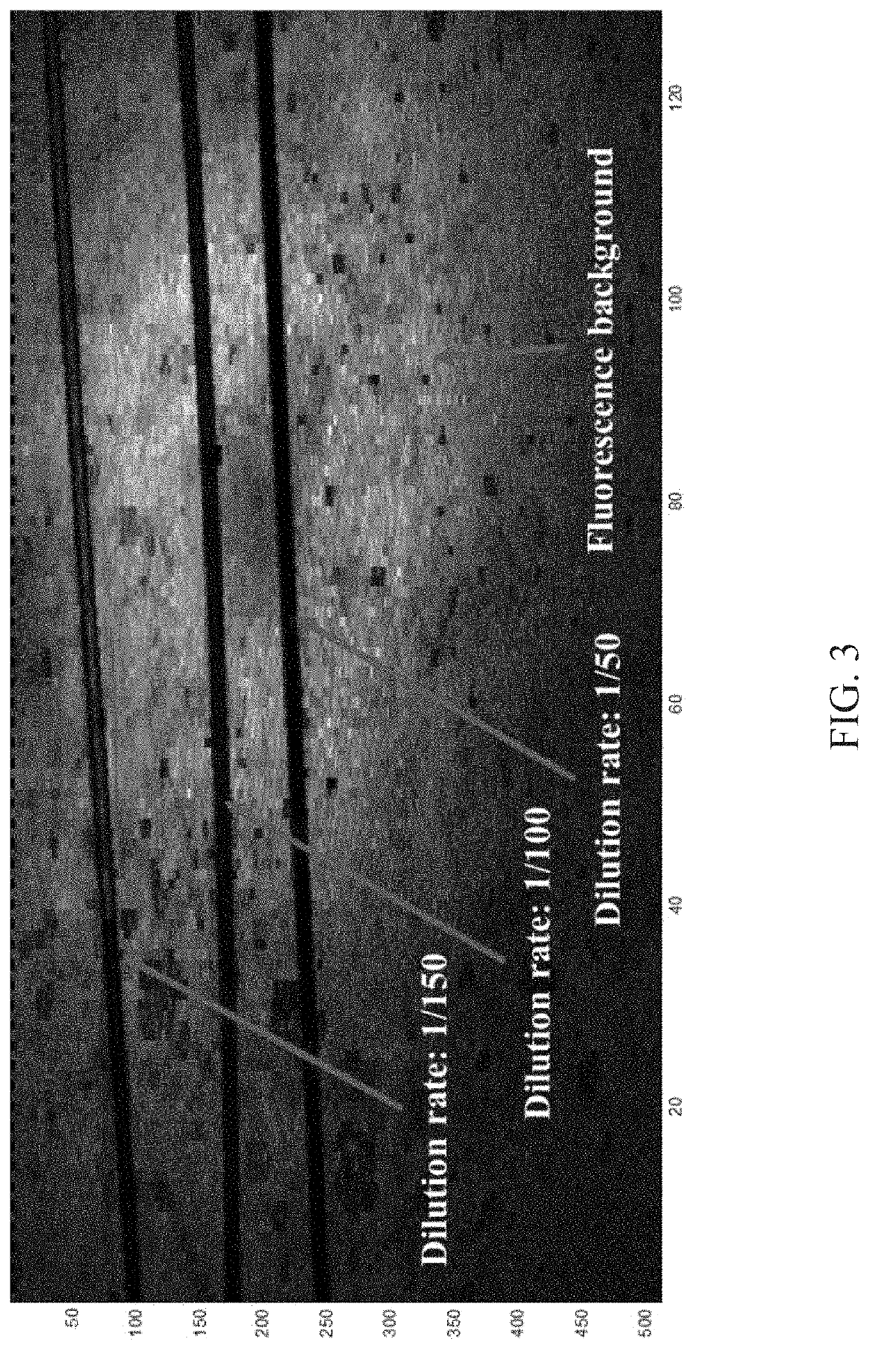
[0040]An experiment was performed using phantom dye. To design the phantom dye, an original Red 40 dye solution was diluted to the relative concentrations of 1 / 50, 1 / 75, 1 / 100, and 1 / 150, respectively. Capillary tubes with inner diameter of 100 micrometer (μm), which was measured with an optical coherence tomography (OCT) scan, were filled with the diluted dye solutions. One piece of paper was immersed into fluorescein solution and dried. The tubes were the placed on the paper to mimic the vessels. FIG. 3 shows en face imaging of the phantom dye.
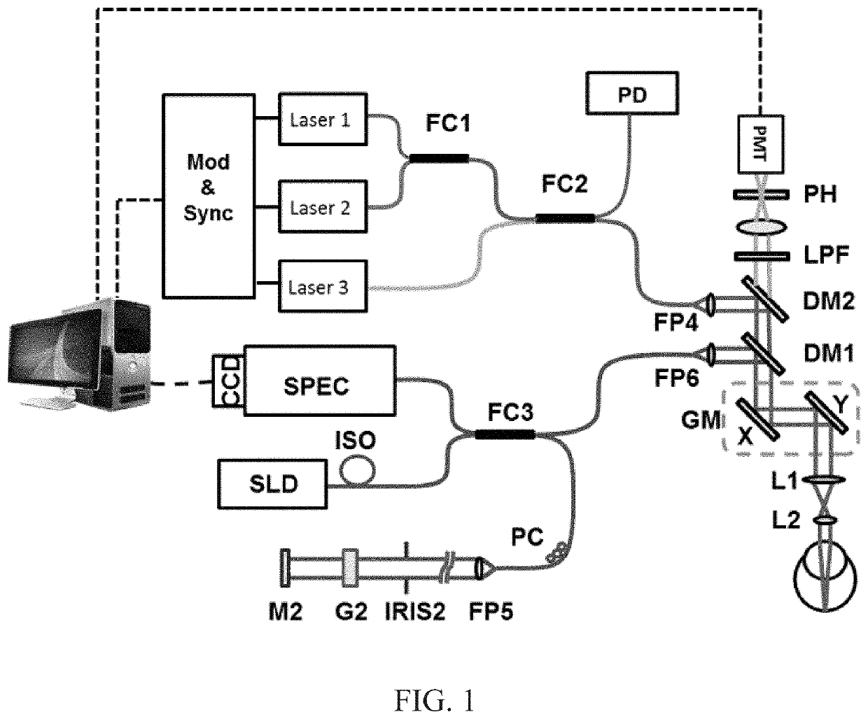
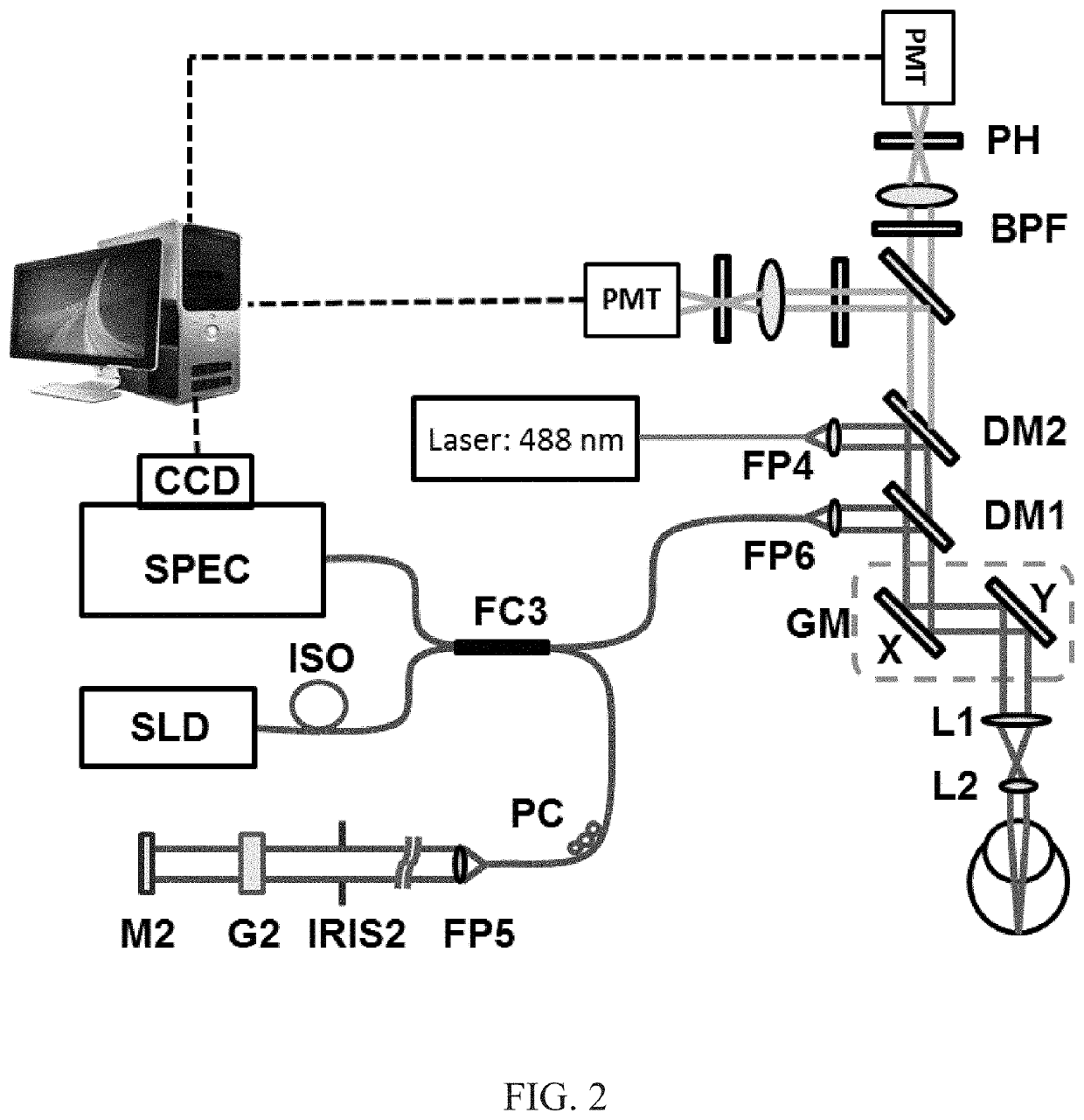
[0041]The phantom was scanned transversely with a system as described herein (see also, e.g., FIGS. 1 and 2). The light intensities from the center and outside of the tubes, which were labeled with Iin and Iout respectively, were acquired. Then measured optical density of a blood vessel (ODmeasured) was determined with the following formula,
[0042]OD(λ)measured=-log10Iin(λ)Iout(λ)
[0043]For comparison, the theoretical values of OD (ODcal) ...
example 2
[0046]A multi-wavelength experiment was performed. Using Blue 1 solution, the experiment of Example 1 was repeated with three wavelengths of 488 nanometers (nm), 506 nm, and 520 nm. The original dye solution was diluted to the concentrations of 1 / 80, 1 / 40, and 1 / 20, respectively. The measured OD(λ) is shown in FIG. 5.
[0047]Next, OD ratios were calculated for the three wavelengths (OD(488 nm) / OD(506 nm) and OD(520 nm) / OD(506 nm)). It was found that the OD ratios kept relatively constant as shown in FIG. 6. The highest error of OD ratio was only 1%, which demonstrated excellent accuracy of the methods and systems described herein. The oxygen saturation (SO2) is expected to have excellent accuracy because the oxygen saturation was determined with the OD ratios.
PUM
 Abstract
Description
Claims
Application Information
 - R&D Engineer
- R&D Manager
- IP Professional
- Industry Leading Data Capabilities
- Powerful AI technology
- Patent DNA Extraction
Browse by: Latest US Patents, China's latest patents, Technical Efficacy Thesaurus, Application Domain, Technology Topic, Popular Technical Reports.
© 2024 PatSnap. All rights reserved.Legal|Privacy policy|Modern Slavery Act Transparency Statement|Sitemap|About US| Contact US: help@patsnap.com