Eye implant devices and method and device for implanting such devices for treatment of glaucoma

a technology of eye implant and glaucoma, which is applied in the field of eye implant devices and methods and devices for treating glaucoma, can solve the problems of affecting the flow of fluid out of the eye, and affecting the vision of the ey

Active Publication Date: 2021-03-23
OMEGAD LLC
View PDF7 Cites 7 Cited by
  • Summary
  • Abstract
  • Description
  • Claims
  • Application Information

AI Technical Summary

Benefits of technology

[0013]The invention also comprises a drainage device which, when inserted into the sclera, permits the drainage of excess aqueous humor from within the anterior chamber of the eye and provides a remedy for the elevated pressure of glaucoma. The drainage device comprises a tube like structure through which aqueous humor is drained from the anterior chamber to the fluid collection channels beneath the conjunctiva. The drainage device may be conical in shape, being smaller in diameter at one end than the other. The drainage device may also be a mesh tube or drain or any other device that allows the flow of aqueous humor. The drainage device may contain a barb like structure at one end to secure the drainage device within the sclera and prevent the migration of the drainage device back in the direction of the anterior chamber.

Problems solved by technology

However, because of the nature of the endothelium cells, which are designed to pump fluid out of the cornea, the death of these cells can impair the vision of the eye.
If the device touches the endothelium, progressive endothelial cell loss will occur, causing the cornea to cloud, causing loss of vision and ultimately for the need for a corneal transplantation.
When the aqueous migration, described above, is insufficient to relieve the build-up of intra-ocular pressure, glaucoma results.
This pressure build-up is usually thought to be due to one or more obstructions in the trabecular meshwork through drainage areas behind the trabecular meshwork which can also impede flow of fluid out of the eye.
Unless controlled, the high pressures associated with glaucoma ultimately leads to permanent damage of the optic nerve, the nerve formed from the sensitive fibers of the retina.
The prior art shows many implantable devices and methods of implanting them, but the majority violate the wall of the sclera adjacent to the vascular tissue of the conjunctiva.
Such violation causes stimulation of vascular tissue.

Method used


Image

  • Eye implant devices and method and device for implanting such devices for treatment of glaucoma
  • Eye implant devices and method and device for implanting such devices for treatment of glaucoma
  • Eye implant devices and method and device for implanting such devices for treatment of glaucoma

Examples


Embodiment Construction

[0030]The present invention provides a device and a method for reducing intraocular pressure that overcomes the problems associated with prior devices and methods. The description below illustrates possible embodiments of the present invention and are in no way meant to be limiting. Other embodiments within the scope of the invention will be clear to those skilled in the art.

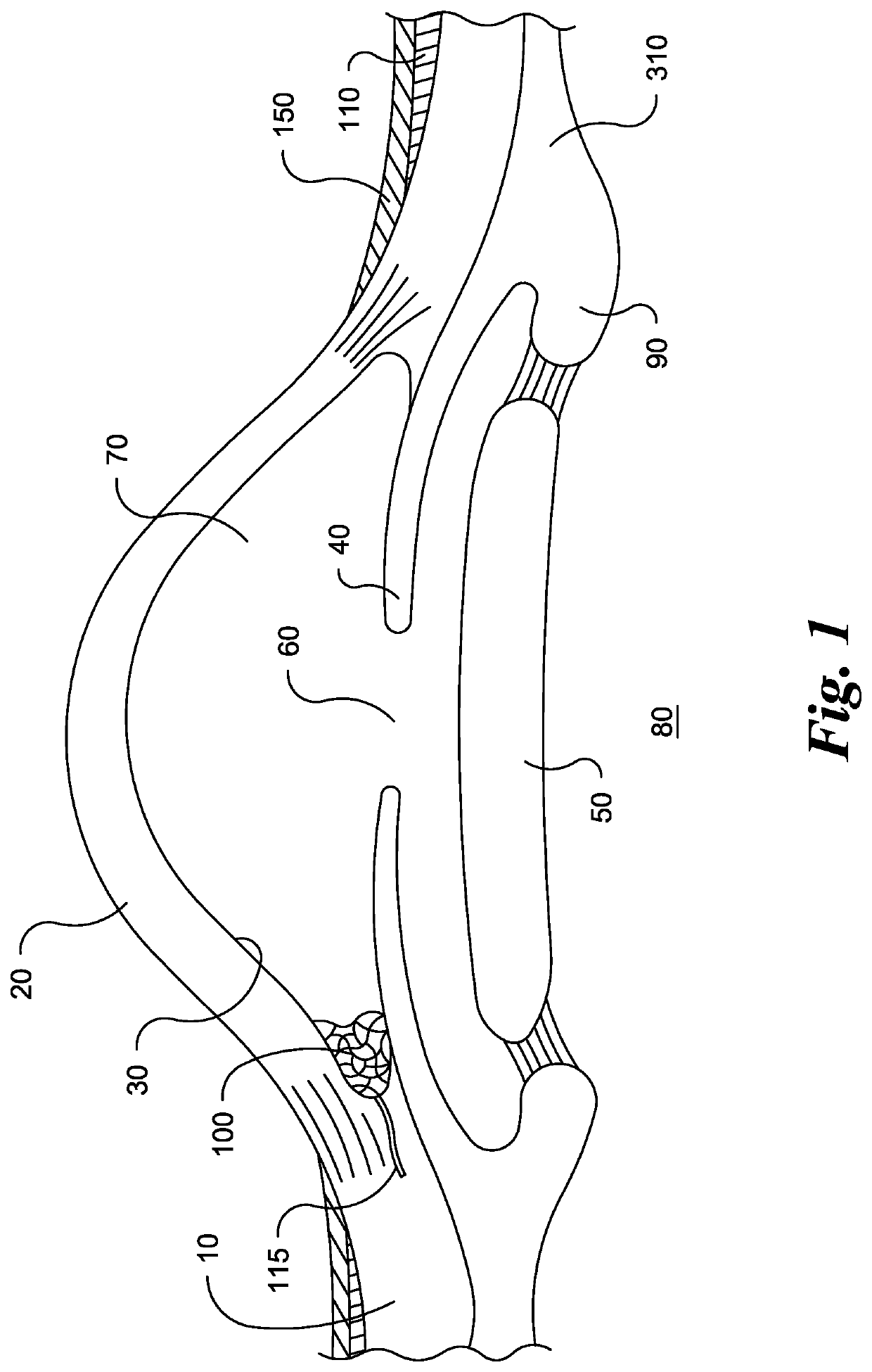
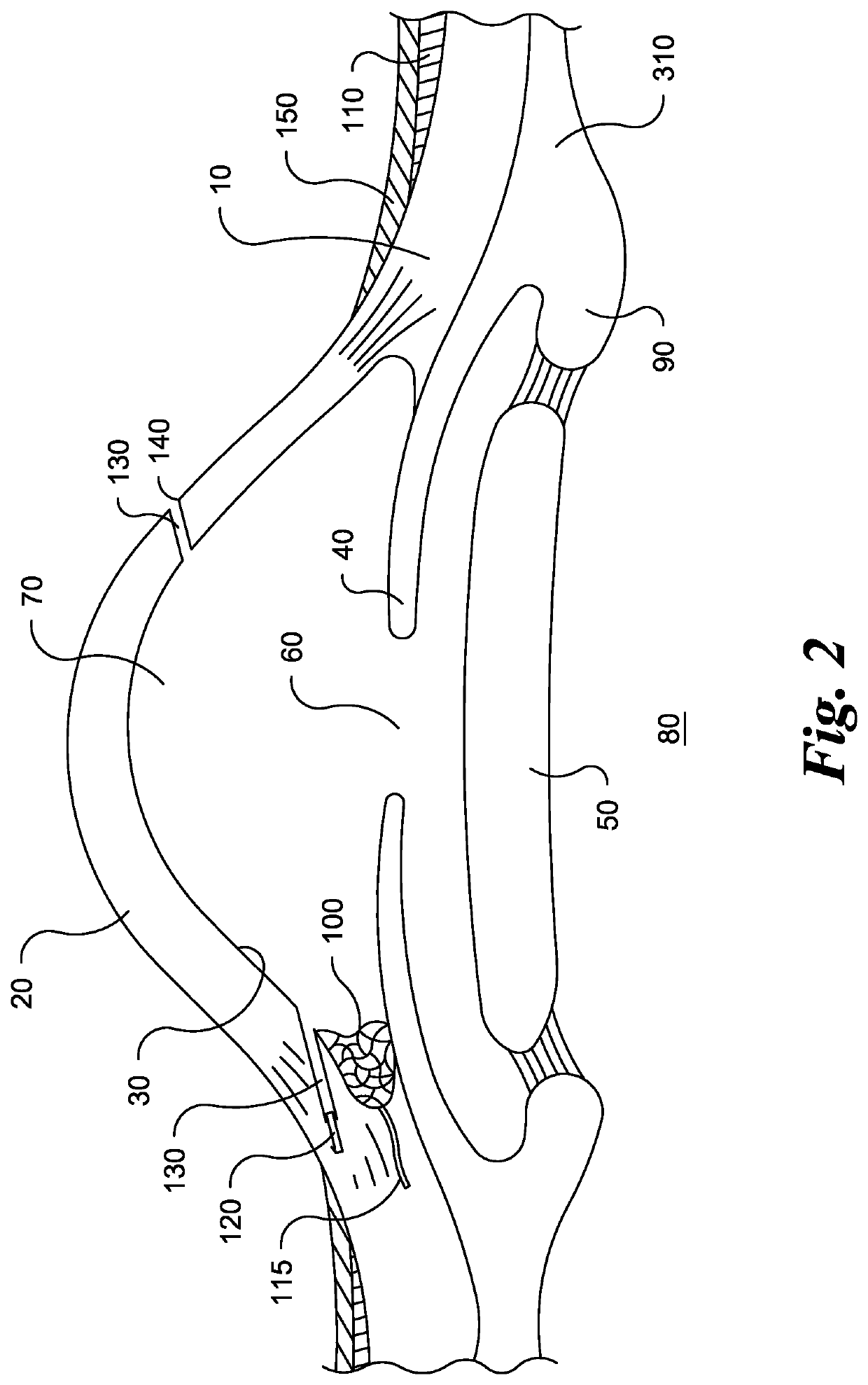
[0031]The subject invention involves a new method of treating glaucoma to insert one or more drainage devices into a location remote from the conjunctiva and tenons tissue into the sclera to facilitate drainage of aqueous humor from the eye. The incision may be remote from the area where the drainage device is to be inserted. The invention also comprises the drainage device and the delivery device, in particular, the delivery device for inserting the drainage device. The surgery for reducing intraocular pressure in the prior art typically involved inserting an implantable device through the conjunctiva. Often su...


PUM

PropertyMeasurementUnit
diameteraaaaaaaaaa
lengthaaaaaaaaaa
external diameteraaaaaaaaaa

Abstract

The present invention provides a method of relieving intraocular pressure by inserting a drainage device into the sclera near the trabecular meshwork from a remote location. The path of insertion is made from a corneal incision and avoids contact with the conjunctiva and tenons tissue. The drainage device comprises a tube like structure and may contain a barb at one end to secure the drainage device. A delivery device may be used to insert the drainage device. The delivery device may be inserted through a corneal incision and directed to the desired area for insertion in to the sclera. The delivery device may then be activated to shoot the drainage device into the sclera to promote increased drainage of aqueous humor and reduce the build up of intraocular pressure.

Description

BACKGROUND OF THE INVENTION[0001]This invention relates to the drainage of aqueous humor from eyes in the course of relieving eye disorders. Specifically, the invention relates to an implant which, when permanently affixed to or implanted in a specific area of the eye, will provide such drainage efficiently, for longer periods than heretofore accomplished, and, in short, will provide relief and prevent, or at least postpone, the adverse ultimate effects of glaucoma. The present invention also relates to a method for treating glaucoma, a method for inserting the implant, and a surgical device for inserting the implant.[0002]The eyeball is composed of three basic layers: (1) the sclera; (2) the middle layer or choroid; and (3) the retina. The sclera 10 is the outer layer of the eyeball. It consists of tough, white tissue consisting of meshwork fibers that serves as the supporting framework of the eye. At the front of the eye, the sclera is continuous with the clear, transparent cornea...

Claims


Application Information

Patent Timeline
Patent Type & Authority Patents(United States)
IPC IPC(8): A61F9/007A61M5/20A61M27/00A61F9/008
CPCA61F9/00781A61F9/00825A61M5/2053A61M27/002A61F2210/0004A61F2210/0076A61F9/007A61F9/008A61F2009/00865A61F2009/00868A61F2009/00891A61M5/2033A61M5/3134A61M5/315
Inventor SMITH, S. GREGORY
Owner OMEGAD LLC
Why Eureka
Try Eureka