Esophageal monitoring
a technology of esophageal monitoring and monitoring elements, applied in the field of esophageal monitoring, can solve the problems of causing esophageal fistula, adversely affecting tissues or organs adjacent to treated tissue or organs, damage to esophagus, etc., to prevent esophageal damage, facilitate the insertion of monitoring elements, and facilitate the withdrawal of monitoring elements.
- Summary
- Abstract
- Description
- Claims
- Application Information
AI Technical Summary
Benefits of technology
Problems solved by technology
Method used
Image

Examples
Embodiment Construction
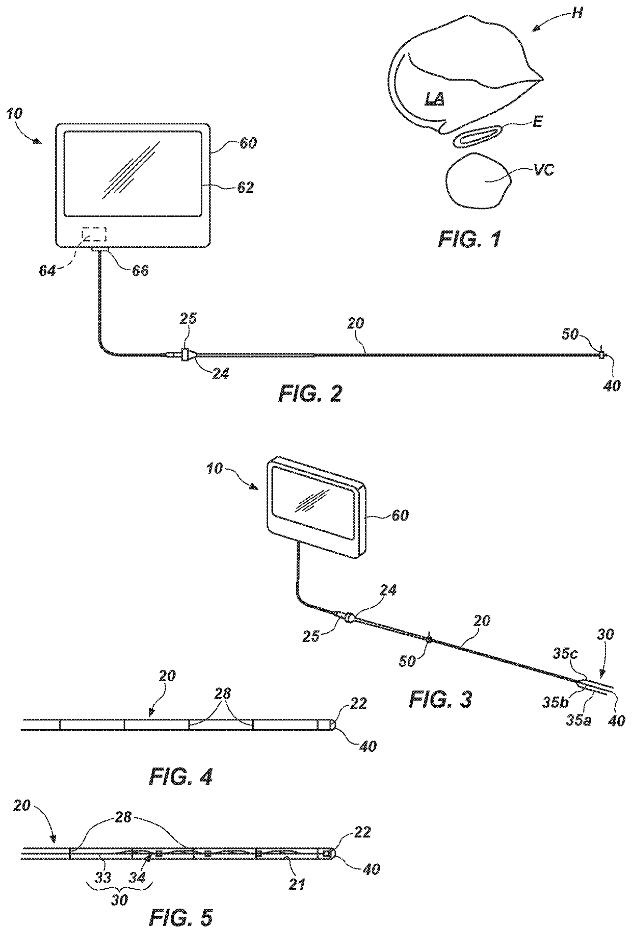
[0052]FIGS. 2 and 3 illustrate an embodiment of a system 10 according to this disclosure, which system 10 may also be referred to as an “esophageal monitoring system.” The system 10 is capable of monitoring a condition of a subject's esophagus while an ablation procedure is being performed on the posterior wall of the left atrium of the subject's heart (i.e., during a left atrial ablation procedure). As illustrated, the system 10 includes an insertion component 20, an esophageal monitoring device 30, a camera 40, a position sensor 50, and a monitor 60.
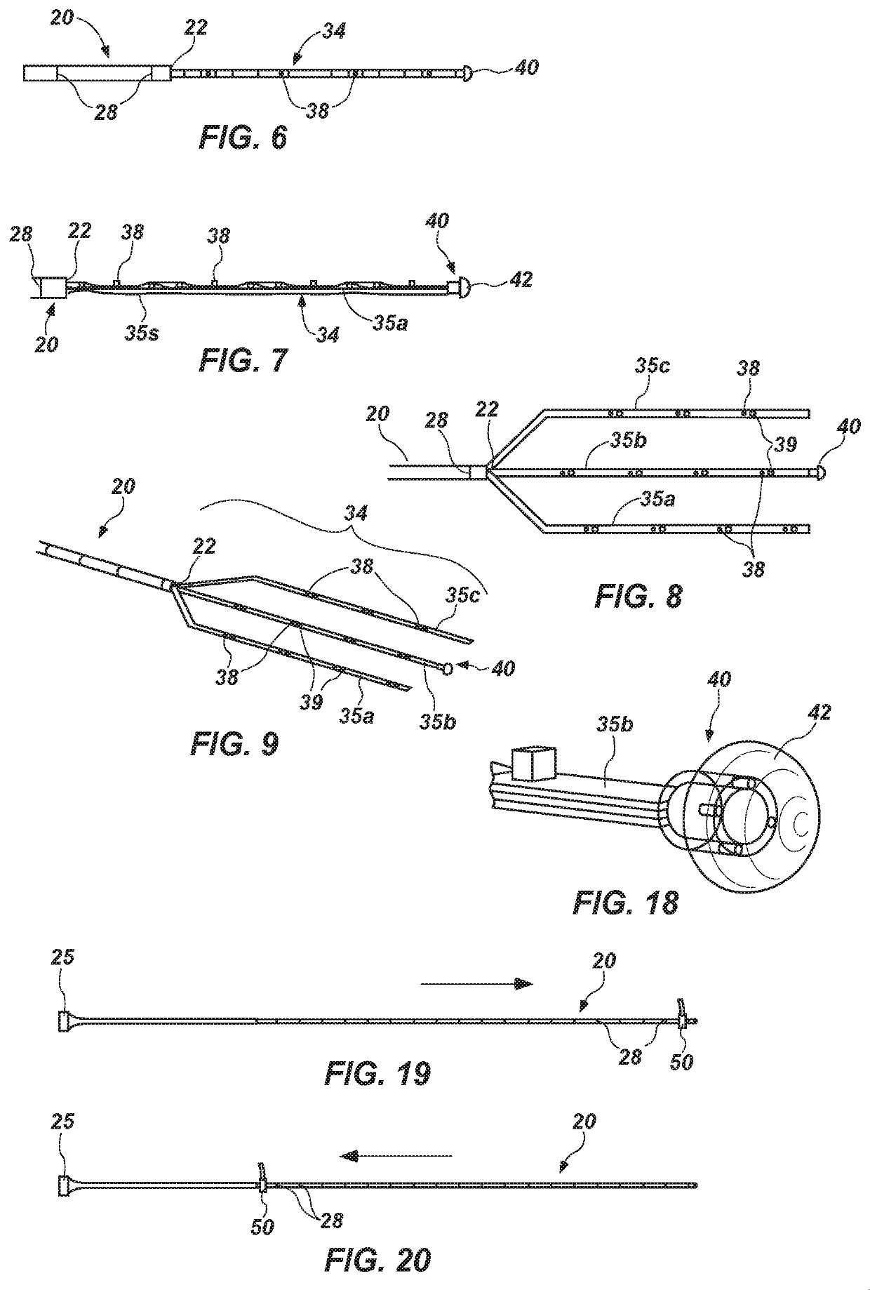
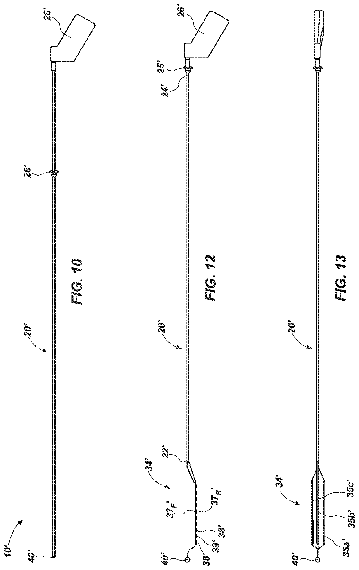
[0053]In FIG. 2, the esophageal monitoring device 30 is located within a lumen 21 (FIG. 5) of the insertion component 20, but the camera 40, which is located on a distal end 32 (FIG. 6) of the esophageal monitoring device 30, may be located at or just outside of (i.e., distal to) the distal end 22 of the insertion component 20.
[0054]In FIG. 3, a monitoring element 34 of the esophageal monitoring device 30 has been deployed from a dista...
PUM
 Abstract
Description
Claims
Application Information
 - R&D
- Intellectual Property
- Life Sciences
- Materials
- Tech Scout
- Unparalleled Data Quality
- Higher Quality Content
- 60% Fewer Hallucinations
Browse by: Latest US Patents, China's latest patents, Technical Efficacy Thesaurus, Application Domain, Technology Topic, Popular Technical Reports.
© 2025 PatSnap. All rights reserved.Legal|Privacy policy|Modern Slavery Act Transparency Statement|Sitemap|About US| Contact US: help@patsnap.com