
System and method for the objective evaluation of sympathetic nerve dysfunction

a sympathetic nerve and objective evaluation technology, applied in the field of apparatus, system and method for the non-invasive sudomotor assessment of corporeal pain, can solve problems such as ensuring that the instrument is properly aligned, providing an error reading, and causing problems such as problems

Active Publication Date: 2021-08-17
EPISCAN GLOBAL LLC
View PDF17 Cites 0 Cited by
  • Summary
  • Abstract
  • Description
  • Claims
  • Application Information

AI Technical Summary

Benefits of technology

[0015]Prior art instruments that utilize permanently installed sensor heads can be problematic as, during use, the permanent electrode can become contaminated with skin oils, moisture, or other contaminates that case errors in the skin conductance measurements. In addition, some clinical procedures require cleaning and disinfecting of the electrodes prior to use on the patient. Cleaning and disinfecting using normal aqueous or alcohol base solutions add additional surface conductivity unless the electrode is properly dried to remove the residual moisture (which also can cause erroneous measurements). Accordingly, to address the drawbacks of the permanently installed sensor head, it is an object of the present invention to manufacture the sensor head as a replaceable, detachable biopolar electrode assembly of sufficiently low cost that may be used either as an interchangeable electrode (allowing for proper sterilization and drying). Alternatively, the sensor head may be manufactured as a disposable product.
[0016]In certain embodiments of the present invention, the sensor head can be readily and securely attached to (and detached from) the device housing with each procedure and / or test subject, for example via a screw-in type mounting with spring loaded contact pins to complete the measurement circuit. Alternatively, the present invention contemplates a sensor head that can be physically separated from the device housing, thereby allowing it to be worn by a test subject over a period of time and configured for single and / or continuous monitoring and measurement. In this latter embodiment, the sensor is optionally provided with means to record measurements, such as an integral or on-board memory chip or memory card, and / or means to transmit recorded measurements to the hand-held housing, or, alternatively, directly to a local or remote computer system, database or physician. It may further be optionally configured to directly or indirectly coordinate with or connect to one or more preset ports on a standard laptop computer, smartphone, or tablet.
[0017]Many selective tissue conductance meters of the prior art use a direct (DC) current method wherein a small DC voltage is applied between the outer ring and center core of the concentric bipolar electrode assembly. As noted above, the amount of current measured between the two parts of the electrode is proportional to the skin conductivity. However, a problem with the DC current method is that, as the DC potential is applied to the skin or other body tissue, an ionophoresis effect is produced in which the current flow between the electrodes increases over time, thus producing progressively increased measurement values based upon the duration of the application of the DC currently. Consequently, in the context of DC-based measurements, controlling the time interval is critical for obtaining consistent results. However, the present invention eliminates this problem in certain preferred embodiments by utilizing alternating current (AC), more particularly a high frequency (1 to 100 kHz) AC signal to measure the current between the respective components of the bipolar electrode assembly. This eliminates the ionophoresis effect caused by the use of a DC current. Accordingly, in the context of the present invention, time of measurement is not a critical variable and thus measurements obtained are more consistent and accurate.
[0018]One objective of the present invention is to provide a multifunctional diagnostic device in which the sensor head is outfitted with multiple sensors for measuring multiple physiological parameters. For example, in one preferred embodiment, the sensor head and / or device housing optionally include other sensors such as (a) thermography cameras and optical infrared scanners to assess the heat and temperature of the affected region; (b) sweat-based glucose, lactate and theophylline biosensors that enable non-invasive transdermal scoring of analyte concentration in tissue, particularly muscle tissue; (c) pulse-oximeters to allows for measurement of oxygen saturation levels and assess pre- and post-flow to the affected region; (d) ultrasonic sensors and transducers that allow a medical practitioner to assess viability and recovery of muscle tissue.
[0019]In certain embodiments, the device housing may outfitted with liquid crystal display and optionally an onboard microprocessor and / or memory card or other storage means to allow for collected data to be temporarily stored, or recalled / displayed, until it can be downloaded (or uploaded) to a remote system. Alternatively, the connection between diagnostic device and associated microprocessor device can be wireless, using either short-range signal (such as a Bluetooth® or LAN network) or a long-range digital or cellular network to transfer data, thereby allowing the computer and the treating physician to be either local or remote. The onboard, local or remote microprocessor can record, transcribe and / or analyze specific measurements and / or enable local or remote analysis and diagnosis.
[0020]As noted above, in certain embodiments, the device housing can be “smart”, i.e., outfitted with a programmable computer chip or other complex microprocessing components that enables on-board programming and analysis. Alternatively, the device may be a simplified “dummy” device that receives all its programming instructions from a remote microprocessor, such as a laptop computer, tablet or Smartphone. So as to reduce user error, in a preferred embodiment, the device housing is outfitted with a display screen that allows the user to cycle through a menu of pre-programmed operating modes and modules, more preferably options that walk the medical practitioner through the requisite set-up, initialization, measurements and / or recordation processes.

Problems solved by technology

Consequently, if not aligned properly, the instrument will provide an erroneous reading.
Moreover, ensuring that the instrument is properly aligned when placing the electrode on parts of the body that are curved or contoured, or in places difficult to reach, is particularly problematic.

Method used


Image

  • System and method for the objective evaluation of sympathetic nerve dysfunction
  • System and method for the objective evaluation of sympathetic nerve dysfunction
  • System and method for the objective evaluation of sympathetic nerve dysfunction

Examples


examples

[0117]The present invention provides a comprehensive system for the quantitative assessment of regional sympathetic sudomotor dysfunction which is useful in the objective assessment of sympathetically maintained pain syndromes; a painless electrodiagnostic method requiring no sensory stimulation or subjective reports by patients; a handheld, self-powered device with an LCD display; a rapid and simple test procedure with automatic report generation; a HHSIFDA Regulatory Class II non-invasive device; and a sympathetic skin assessment approved by Medicare and most insurance companies for procedure reimbursement.

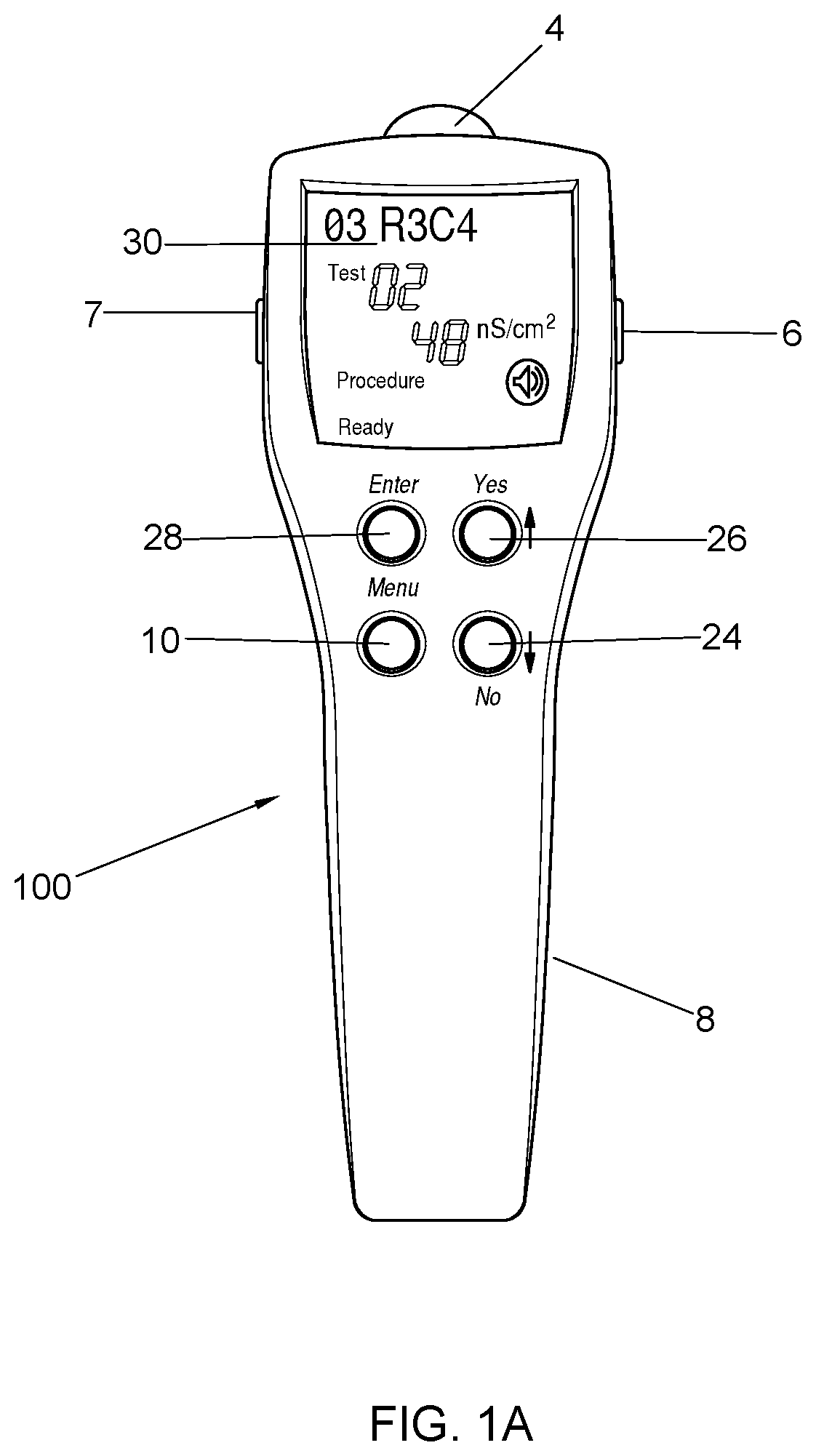
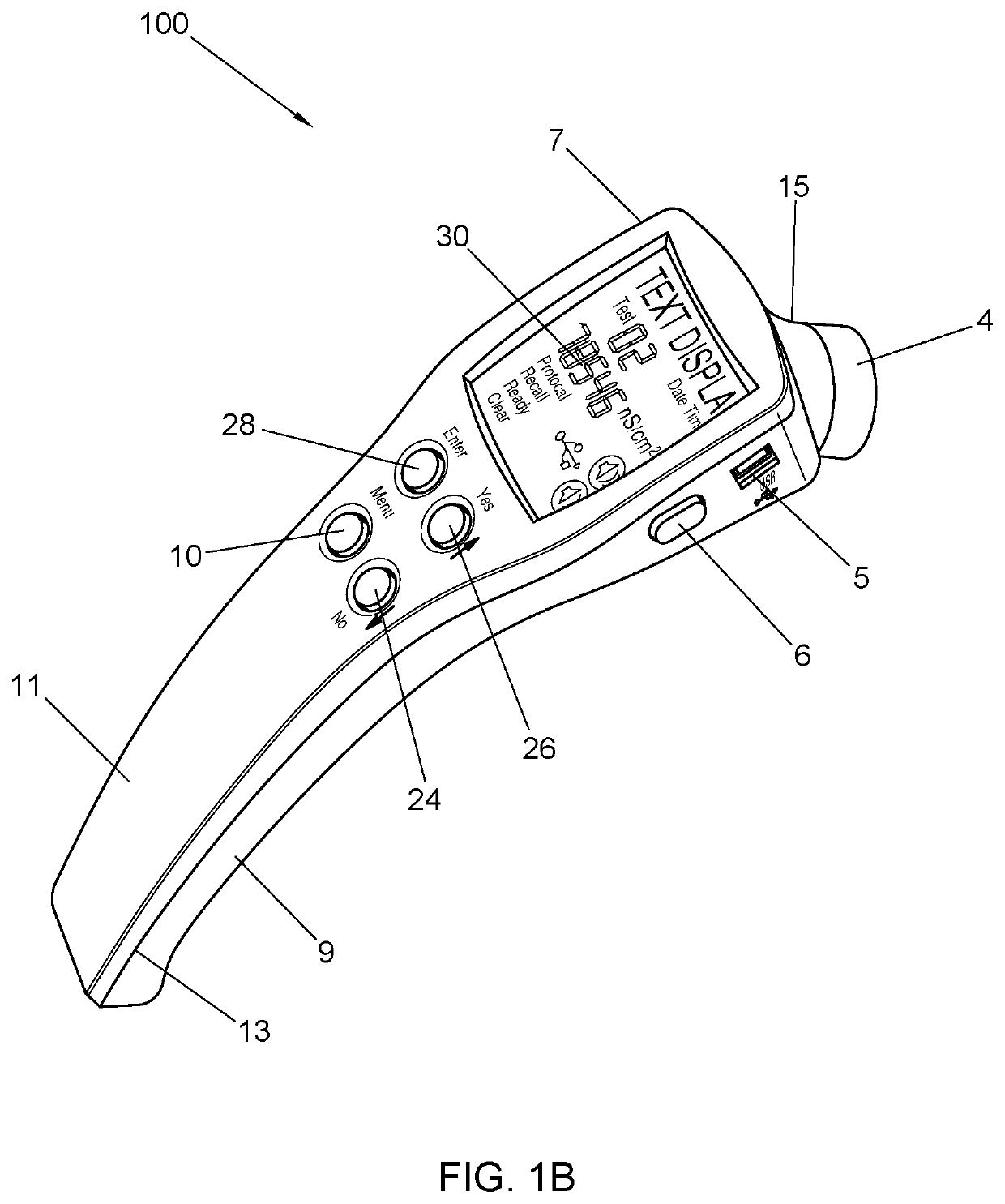
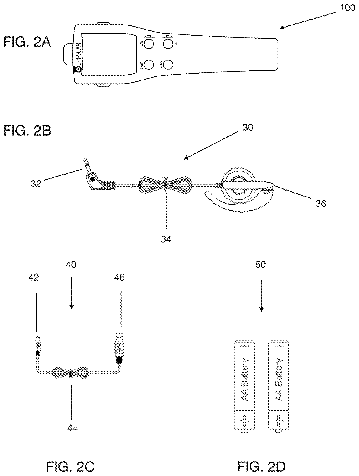
[0118]In illustrative embodiment an improved meter and monitoring system of the present invention of the inventive is presented in FIGS. 1-4. FIGS. 1A, 1B, and 2A respectively depict top-down, perspective, plan views of a diagnostic device (100) designed in accordance with the principles of the present invention. Device (100) includes a housing (8) fabricated of upper (11) and l...


PUM


Abstract

Various methods and machines have been used in the past to measure electrical characteristics of living tissue for purpose of locating an area of abnormal nervous system activity. However, whereas prior art methodologies merely allow for the detection of pain, the apparatus, system and method of the present invention allow for the objective assessment pain severity that finds utility not only the initial diagnosis but also the on-going treatment of any disease, disorder or injury associated therewith. To that end, the apparatus, system and method of the present invention allows medical practitioners to non-invasively and quantitatively distinguish organic pain from psychosomatic pain and legitimate pain patients from drug seekers and opiod addicts, as well as to directly and objectively compare the efficacy of different drug regimens and therapy protocols.

Description

PRIORITY[0001]This application claims the benefit of U.S. Provisional Application Ser. No. 62 / 434,169 filed Dec. 14, 2016, the contents of which are hereby incorporated by reference in their entirety.TECHNICAL FIELD OF THE INVENTION[0002]The present invention relates generally to the correlation between electrical tissue parameters, such as skin conductance, with sympathetic and parasympathetic nerve function (and dysfunction). More particularly, the present invention relates to an apparatus, system and method for the non-invasive sudomotor assessment of corporeal pain that allows for the objective measurement of pain severity that, in turn, finds utility not only in the diagnosis and treatment of any underlying disorder, disease or injury associated with the measured pain levels but also in determining the appropriate drug and dosage regimen, in distinguishing organic pain from psychosomatic pain and legitimate pain patients from drug seekers and opiod addicts, and in directly and ...

Claims


Application Information

Patent Timeline
Patent Type & Authority Patents(United States)
IPC IPC(8): A61B5/00A61B5/053G16H15/00G16H10/60G16H10/40G16H50/70G16H20/40G16H20/10G16H20/30A61B5/01A61B5/145A61B8/08A61B5/1455
CPCA61B5/4824A61B5/053A61B5/4041G16H10/40G16H10/60G16H15/00G16H20/10G16H20/30G16H20/40G16H50/70A61B5/0022A61B5/01A61B5/14532A61B5/14546A61B5/14551A61B5/4266A61B8/08A61B2503/40A61B2560/0214
Inventor SITTON, DAVID MICHAELDOUGHERTY, CHRISTOPHER P.ARNDT, III, SAMUEL F.
Owner EPISCAN GLOBAL LLC
Learn More