Hardness deriving device, medical imaging system, hardness deriving method and hardness deriving program storage medium

a hardness deriving and program technology, applied in applications, ultrasonic/sonic/infrasonic image/data processing, ultrasonic/sonic/infrasonic diagnostics, etc., can solve problems such as the inability to obtain detection results of breast hardness

Active Publication Date: 2021-09-28
FUJIFILM CORP
View PDF20 Cites 1 Cited by
  • Summary
  • Abstract
  • Description
  • Claims
  • Application Information

AI Technical Summary

Benefits of technology

[0023]Thus, the present disclosure may provide a hardness deriving device, a medical imaging system, a hardness deriving method, and a storage medium stored with a hardness deriving program, which are capable of consistently measuring the hardness of breast tissue.

Problems solved by technology

In elastography the degree of pressing of the breast generally differs according to the operator and the skill of the operator, such that consistent detection results for elastography, namely detection results for breast hardness, may not be obtained.

Method used


Image

  • Hardness deriving device, medical imaging system, hardness deriving method and hardness deriving program storage medium
  • Hardness deriving device, medical imaging system, hardness deriving method and hardness deriving program storage medium
  • Hardness deriving device, medical imaging system, hardness deriving method and hardness deriving program storage medium

Examples


first exemplary embodiment

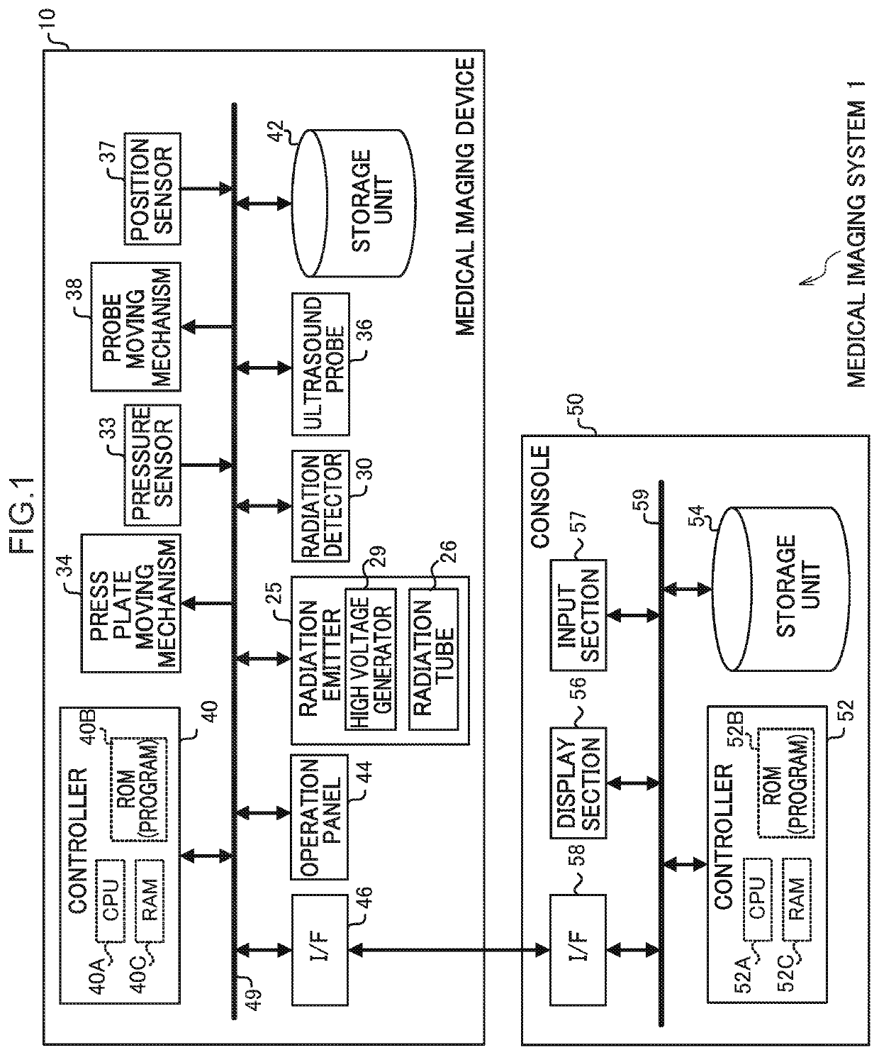
[0041]As illustrated in FIG. 1, a medical imaging system 1 of the first exemplary embodiment includes a medical imaging device 10 and a console 50.

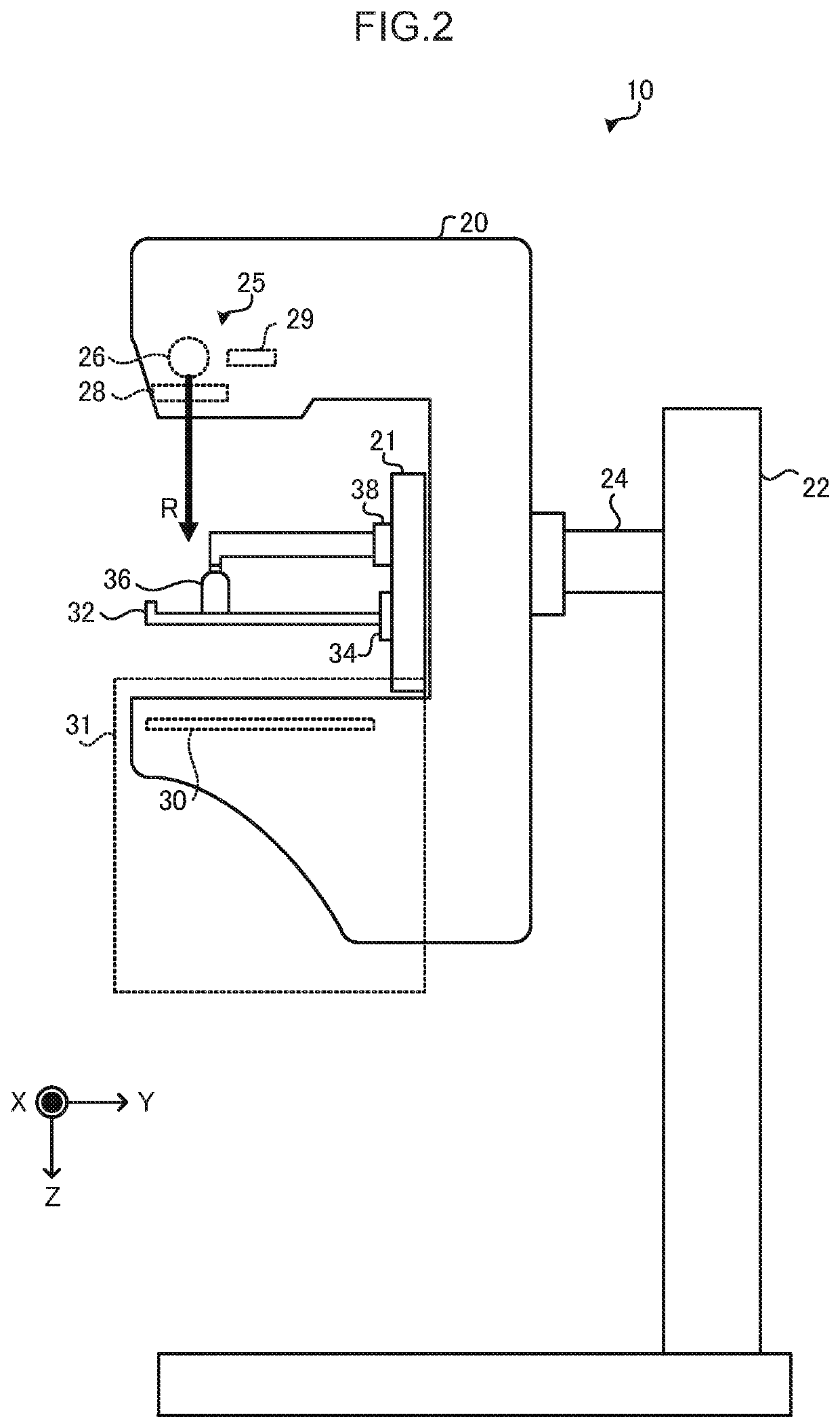
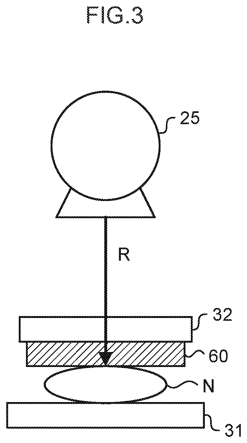
[0042]The medical imaging device 10 combines functionality of a mammography machine that performs radiographic imaging by radiating radiation R onto a breast of a subject and detecting the radiation R that has passed through the breast, and functionality of a ultrasound imaging device that performs ultrasound imaging by transmitting ultrasound waves through the breast, and receiving an ultrasound echo reflected by the interior of the breast.

[0043]There are two types of ultrasound imaging performed by the medical imaging device 10 of the present exemplary embodiment: normal ultrasound imaging (described in detail later), and ultrasound imaging for elastography. In the exemplary embodiments, “elastography” refers to detecting the hardness of breast tissue, rather than the overall hardness of the breast, as breast hardness. In the medical im...

second exemplary embodiment

[0136]Explanation follows regarding a second exemplary embodiment. Components similar to those of the medical imaging system 1 according to the first exemplary embodiment are appended with the same reference numerals, and detailed explanation thereof is omitted.

[0137]Configuration of a medical imaging system 1 is similar to that of the medical imaging system 1 of the first exemplary embodiment (see FIGS. 1 and 2), so explanation thereof is omitted.

[0138]In the present exemplary embodiment, the method of imaging the first ultrasound image and the second ultrasound image differs from that of the first exemplary embodiment. In the medical imaging device 10 of the first exemplary embodiment, the first ultrasound image and the second ultrasound image are imaged by scanning the ultrasound probe 36 across the surface of the press plate 32 at a constant (constant within a margin of error) speed using the probe moving mechanism 38. In contrast thereto, in the medical imaging device 10 of the...


PUM


Abstract

A hardness deriving device includes: an acquisition section configured to acquire plural ultrasound images obtained by ultrasound imaging a breast in a state in which the breast is pressed by a pressing member at a plurality of different pressures; and a deriving section configured to find an amount of change at corresponding points between the plural ultrasound images acquired by the acquisition section, and to derive information indicating a hardness of tissue of the breast based on the amount of change at the corresponding points.

Description

CROSS-REFERENCE TO RELATED APPLICATION[0001]This application claims priority under 35 USC 119 from Japanese Patent Application No. 2016-069187 filed on Mar. 30, 2016, the disclosure of which is incorporated herein by reference in its entirety.BACKGROUNDTechnical Field[0002]The present disclosure relates to a hardness deriving device, a medical imaging system, a hardness deriving method, and a storage medium stored with a hardness deriving program.Related Art[0003]Ultrasound imaging devices for use in diagnosis and observation of breast tissue are known that perform ultrasound imaging of a breast by scanning the breast with ultrasound waves. Elastography is known as technology to detect the hardness of breast tissue using such ultrasound images.[0004]Generally, when ultrasound images of a breast are imaged with an ultrasound imaging device, an operator moves an ultrasound probe over the surface of the breast of a subject, and performs imaging while pressing the breast with the ultras...

Claims


Application Information

Patent Timeline
Patent Type & Authority Patents(United States)
IPC IPC(8): A61B8/00A61B8/08A61B6/00
CPCA61B8/485A61B6/502A61B8/0825A61B8/403A61B8/4416A61B8/461A61B8/5223A61B8/5261A61B8/469A61B8/54G16H50/30
Inventor SENDAI, TOMONARI
Owner FUJIFILM CORP
Why Eureka
Try Eureka