
Myocardial CT perfusion image synthesis

a computed tomography and image technology, applied in the field of digital image processing, can solve the problems of less suitable imaging technologies for enabling combined detailed, inability to accurately assess the hemodynamic significance of coronary lesions, and inability to fully integrate the combined detailed,

Active Publication Date: 2021-12-28
KONINKLJIJKE PHILIPS NV
View PDF14 Cites 0 Cited by
  • Summary
  • Abstract
  • Description
  • Claims
  • Application Information

AI Technical Summary

Benefits of technology

This patent is about using computed tomography technology to analyze, synthesize, and visualize the function and structure of the heart. The goal is to provide a better and more efficient way to test and diagnose heart health.

Problems solved by technology

In coronary artery disease, accumulation of plaque material in the coronary arteries limits the supply of oxygen to the cardiac muscle, resulting in myocardial ischemia.
However, it may be a disadvantage of such prior-art approaches that these imaging technologies may be less suitable for enabling a combined detailed anatomic assessment of the coronary arteries and a functional evaluation of the downstream myocardial territory.
Therefore, CCTA methods known in the art may disadvantageously be limited in enabling a detailed assessment of hemodynamic significance of coronary lesions.
Therefore, the user is limited to an assessment of the coronary lesion, rather than being provided with a means for directly assessing the viability of the cardiac muscle.
However, it is a disadvantage that such views may be limited to a two-dimensional representation of the left ventricle, rather than allowing a three-dimensional assessment of perfusion over the entire CT volume.
While cardiac muscle viability can be visualized and assessed directly by such approach, it is a disadvantage of performing at least one additional CT perfusion scan that this implies an additional exposure of the patient to ionized radiation and a toxic contrast agent, e.g. in addition to the radiation exposure and the amount of contrast agent used for performing a CCTA scan.
Furthermore, between a CCTA scan and a CT perfusion scan, a time delay may be required to sufficiently reduce the residual contrast agent used for the CCTA scan in the body, which may limit the implementation in clinical practice.
It may be another disadvantage that the quantitative maps derived from physically acquired data may be noisy and unreliable due to a limited number of time points, e.g. caused by physical limitations of the data acquisition and / or in view of radiation dose management, compared to the complexity of the perfusion models used to generate the maps.

Method used

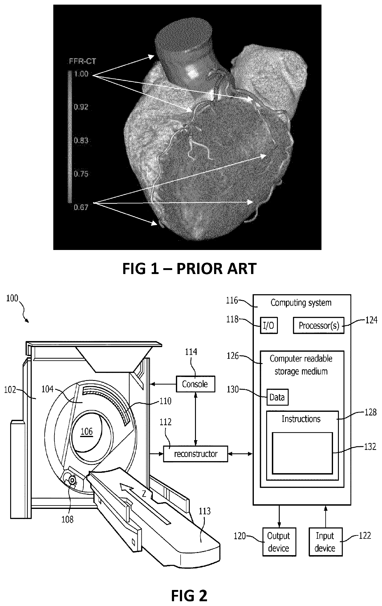

Image

  • Myocardial CT perfusion image synthesis
  • Myocardial CT perfusion image synthesis

Examples


Embodiment Construction

[0046]The present invention will be described with respect to particular embodiments and with reference to certain drawings but the invention is not limited thereto but only by the claims. The drawings described are only schematic and are non-limiting. In the drawings, the size of some of the elements may be exaggerated and not drawn on scale for illustrative purposes. The dimensions and the relative dimensions do not correspond to actual reductions to practice of the invention.

[0047]Furthermore, the terms first, second and the like in the description and in the claims, are used for distinguishing between similar elements and not necessarily for describing a sequence, either temporally, spatially, in ranking or in any other manner. It is to be understood that the terms so used are interchangeable under appropriate circumstances and that the embodiments of the invention described herein are capable of operation in other sequences than described or illustrated herein.

[0048]Moreover, t...


PUM


Abstract

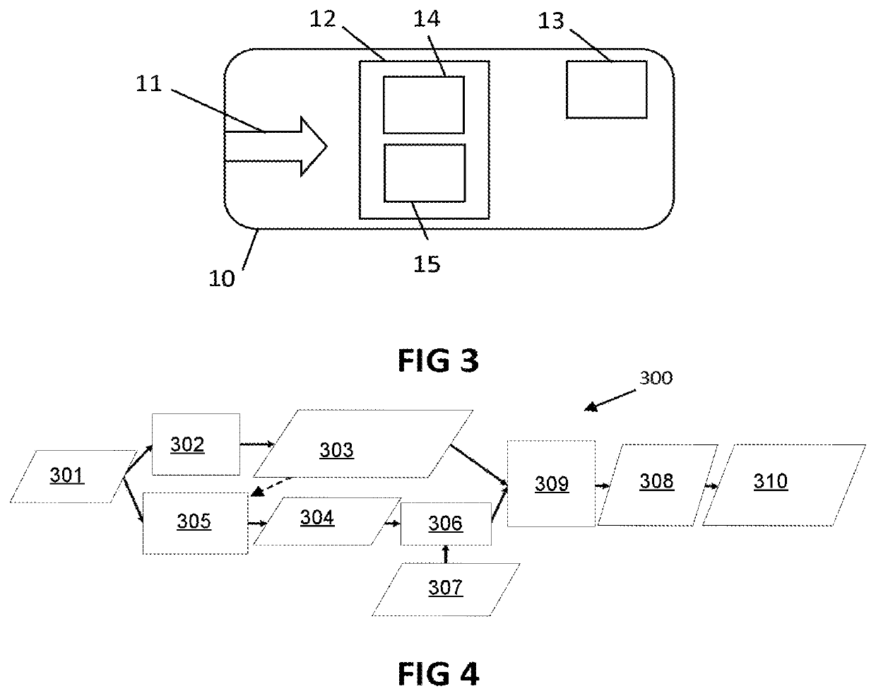
The present invention relates to image processing devices and related methods. The image processing device (10) comprises a data input (11) for receiving spectral computed tomography volumetric image data organized in voxels. The image data comprises a contrast-enhanced volumetric image of a cardiac region in a subject's body and a baseline volumetric image of that cardiac region, e.g. a virtual non-contrast image, wherein the contrast-enhanced volumetric image conveys anatomical information regarding coronary artery anatomy of the subject. The device comprises a flow simulator (12) for generating, or receiving as input, a three-dimensional coronary tree model based on the volumetric image data and for simulating a coronary flow based on the three-dimensional coronary tree model. The device comprises a perfusion synthesis unit (13) for generating a perfusion image representative of a blood distribution in tissue at at least one instant in time taking at least the baseline volumetric image and said coronary flow simulation into account.

Description

FIELD OF THE INVENTION[0001]The invention relates to the field of digital image processing. More specifically it relates to myocardial computed tomography (CT) perfusion image synthesis based on spectral CT data, e.g. coronary CT angiography data.BACKGROUND OF THE INVENTION[0002]Coronary artery disease (CAD) is an important cause of death worldwide. In coronary artery disease, accumulation of plaque material in the coronary arteries limits the supply of oxygen to the cardiac muscle, resulting in myocardial ischemia.[0003]Non-invasive imaging techniques known in the art for assessing the cardiac muscle viability include Myocardial Perfusion Imaging (MPI), in which nuclear imaging modalities such as SPECT and PET are used, and Cardiac Magnetic Resonance Imaging (CMR). However, it may be a disadvantage of such prior-art approaches that these imaging technologies may be less suitable for enabling a combined detailed anatomic assessment of the coronary arteries and a functional evaluatio...

Claims


Application Information

Patent Timeline
IPC IPC(8): A61B6/00G16H30/40G06T7/00G06T7/55
CPCA61B6/507A61B6/466A61B6/504A61B6/5205G06T7/0012G06T7/0014G06T7/0016G06T7/55G06T7/97G16H30/40A61B6/4241A61B6/486A61B6/503A61B6/5217A61B5/02007A61B5/026A61B5/0285G06T19/00A61B2576/023G16H50/50G06T13/20G06T2210/24G06T2210/41A61B5/7264A61B5/0033G06T11/008G06T2211/404G01V5/226
Inventor FREIMAN, MORDECHAY PINCHASGOSHEN, LIRAN
Owner KONINKLJIJKE PHILIPS NV
Learn More