Tissue classification using image intensities and anatomical positions

a tissue type and image intensity technology, applied in the field of medical image data processing, can solve the problems of inability to provide the anatomical positions over the complete image volume required for 3d tissue type classification, inability to determine internal structural information of segmented area, etc., and achieve the effect of reducing the development and maintenance effor

Active Publication Date: 2022-03-08
KONINKLJIJKE PHILIPS NV
View PDF8 Cites 0 Cited by
  • Summary
  • Abstract
  • Description
  • Claims
  • Application Information

AI Technical Summary

Benefits of technology

[0042]Thus, embodiments may be of particular interest for MR-only radiotherapy (RT) applications. An incorporation of spatial information according to embodiments may allow such separation and hence a generation of appropriate Pseudo-CT images. Thus, embodiments may combine a robust shape-based segmentation approach with a data-driven ML approach, which in combination may drastically reduce the development and maintenance effort required. For example, different internal bone structures of different patient populations may share the same anatomical model, while differently trained classifiers may be used, which only require representative training data.

Problems solved by technology

For the usage of medical image data, image segmentation and classification of voxels comprised by image data are an important but challenging task in order to identify and analyze anatomical structures of interest.
However, such model-based segmentation may be unable to determine internal structural information of the segmented area, since only the boundaries are modeled.
In other words, model-based segmentation only provides the anatomical positions at the organ boundaries but the anatomical positions over the complete image volume required for 3D tissue classification cannot be provided.

Method used


Image

  • Tissue classification using image intensities and anatomical positions
  • Tissue classification using image intensities and anatomical positions
  • Tissue classification using image intensities and anatomical positions

Examples


Embodiment Construction

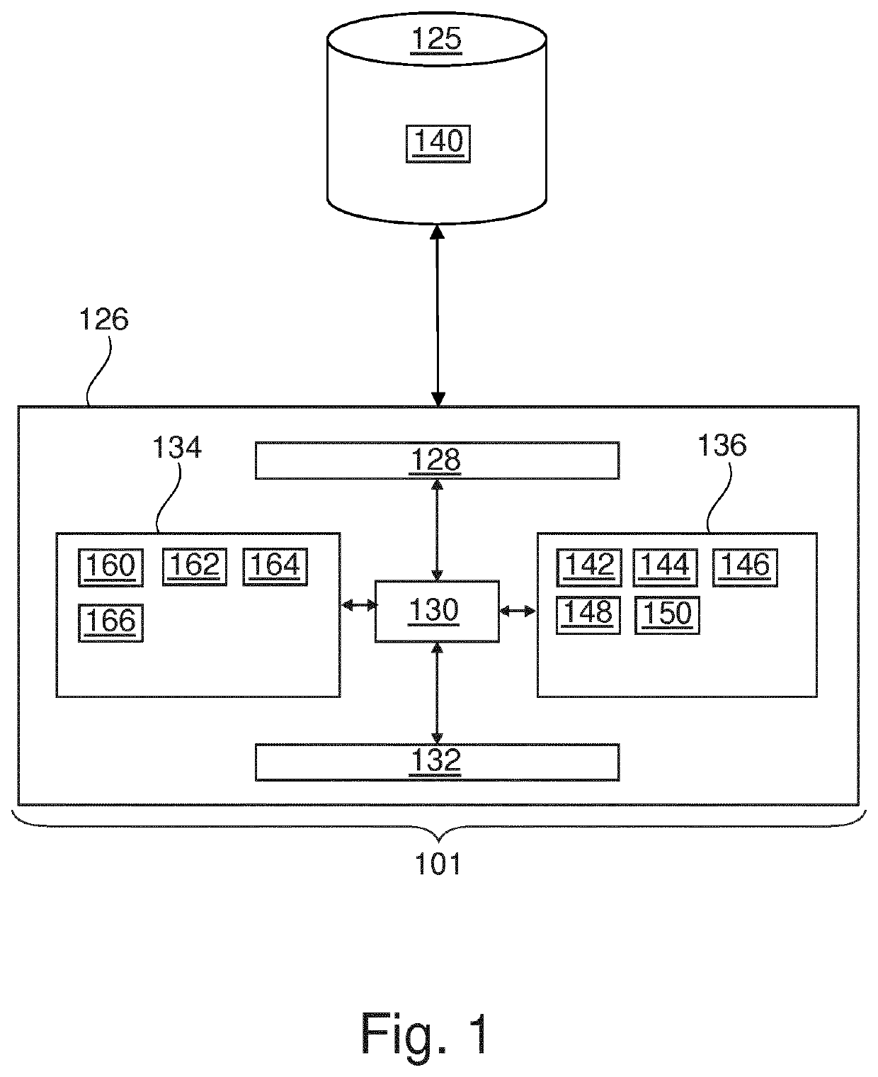
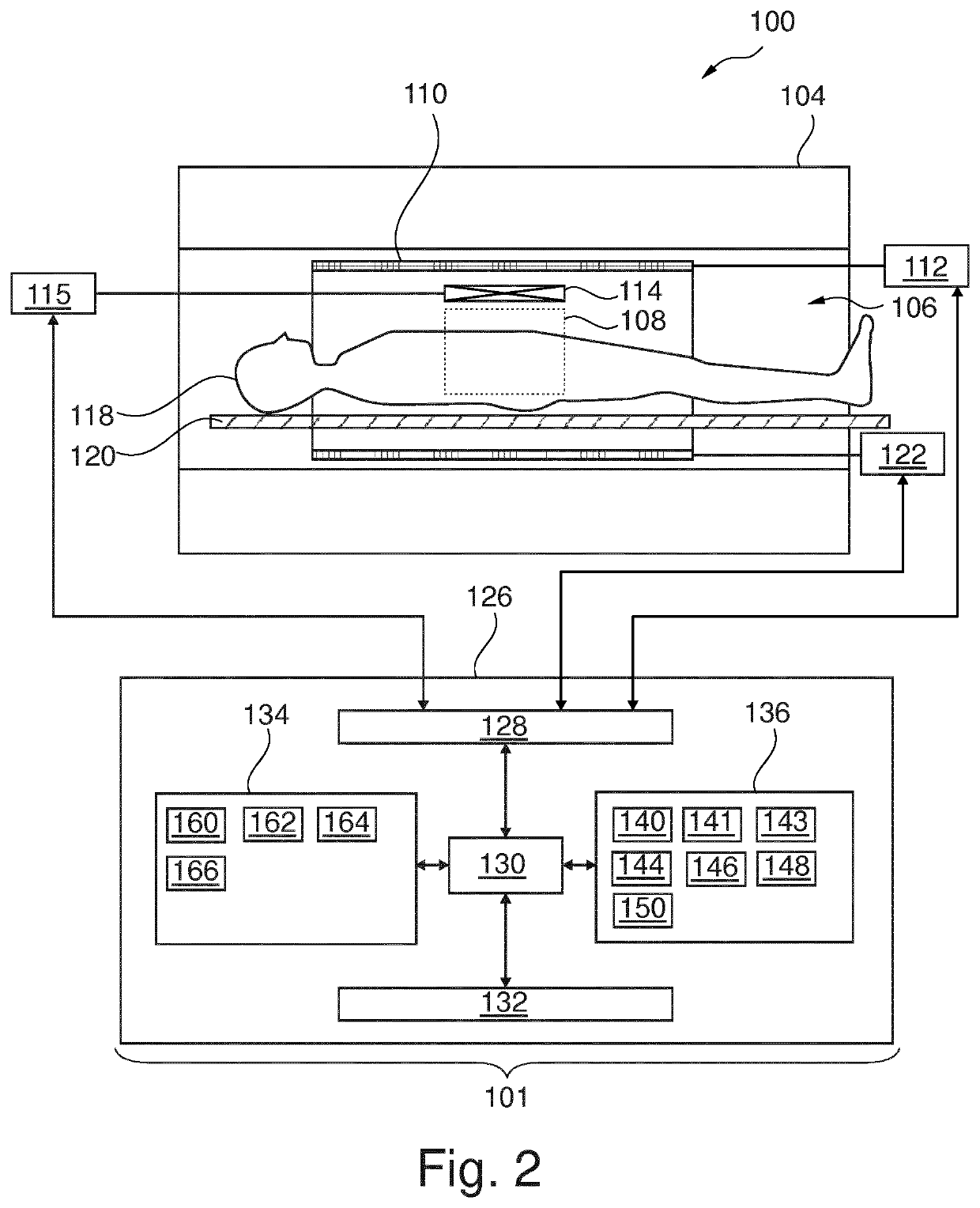
[0073]Like numbered elements in these figures are either equivalent elements or perform the same function. Elements which have been discussed previously will not necessarily be discussed in later figures if the function is equivalent.

[0074]FIG. 1 shows an example of a medical image data processing system 101 comprising a computer 126. The computer 126 is shown as containing a processor 130 which is operable for executing machine-readable instructions. The computer 126 is further shown as comprising a user interface 132, computer storage 134 and computer memory 136 which are all accessible and connected to the processor 130. Furthermore, the computer 126 may communicatively be connected with a database 125. The computer 126 may be configured for requesting data, like medical image data 140 from the database 125 via the communication interface 128. According to embodiments, the database 125 may be provided by an external system and accessible for the computer 126 via a communication n...


PUM


Abstract

The invention relates to a medical image data processing system (101) for image segmentation. The medical image data processing system (101) comprises a machine learning framework trained to receive an anatomical position of a voxel and to provide a tissue type classification. An execution of machine executable instructions by a processor (130) of the medical image data processing system (101) causes the processor (130) to control the medical image data processing system (101) to: —receive medical image data (140) comprising an anatomical structure of interest, —fit an anatomical frame of reference (302, 402) to the medical image data (140) using model-based segmentation, —classify tissue types represented by voxels of the medical image data (140) using the machine learning framework, wherein anatomical positions of the voxels with respect to the anatomical frame of reference (302, 402) are used as the input to the machine learning framework.

Description

CROSS REFERENCE TO RELATED APPLICATIONS[0001]This application is a U.S. national phase application of International Application No. PCT / EP2019 / 050358 filed on Jan. 9, 2019, which claims the benefit of EP Application Serial No. 18151866.3 filed on Jan. 16, 2018 and is incorporated herein by reference.FIELD OF THE INVENTION[0002]The invention relates to processing medical image data, in particular it relates to methods and apparatuses for classifying tissue types using medical image data.BACKGROUND OF THE INVENTION[0003]For the usage of medical image data, image segmentation and classification of voxels comprised by image data are an important but challenging task in order to identify and analyze anatomical structures of interest.[0004]In order to analyze an anatomical structure of interest, not only its boundary contours, but also its internal structure may be important. It may in particular be important to determine which tissue types are comprised by the respective anatomical struc...

Claims


Application Information

Patent Timeline
Patent Type & Authority Patents(United States)
IPC IPC(8): G06K9/00G06T7/00G16H30/40G01R33/48G01R33/56G06K9/62G06V10/24
CPCG06T7/0014G01R33/4828G01R33/5608G06K9/6256G06K9/6262G16H30/40G06K2209/05G06T2207/10088G06T2207/20081G06T2207/30004G06T7/0012G06T2207/10081G06T2207/20084G06T2207/30016G06T7/12G06V10/24G06V2201/03G06F18/214G06F18/217
Inventor BUERGER, CHRISTIANRENISCH, STEFFEN
Owner KONINKLJIJKE PHILIPS NV
Why Eureka
Try Eureka