
Bioactive soft tissue implant and methods of manufacture and use thereof

a soft tissue implant and bioactive technology, applied in the field of bone suture anchors, can solve the problems of inability to use standard sutures as cores, high cost, and often insufficient effect of suture and coating combinations

Active Publication Date: 2022-05-17
HOWMEDICA OSTEONICS CORP
View PDF191 Cites 0 Cited by
  • Summary
  • Abstract
  • Description
  • Claims
  • Application Information

AI Technical Summary

Benefits of technology

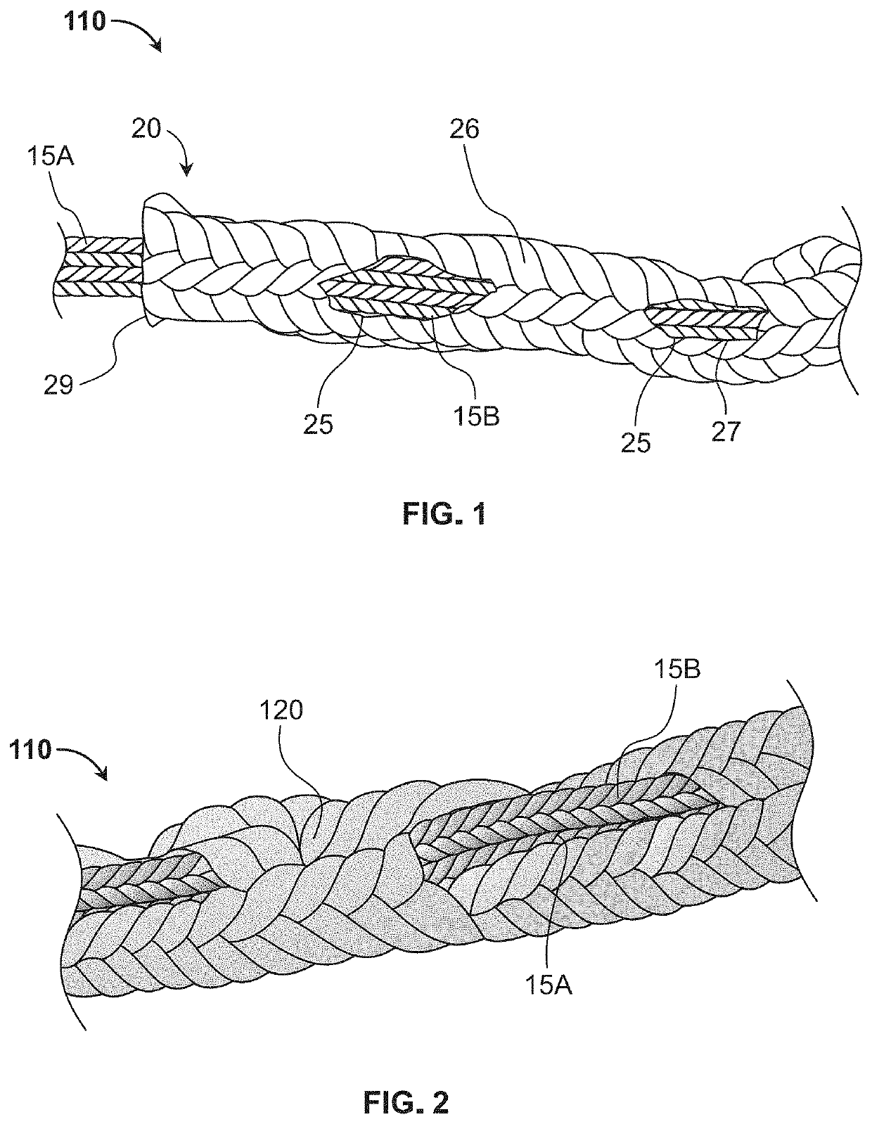
The patent describes a bioactive filamentary structure that includes a sheath coated with a mixture of synthetic bone graft particles and a polymer forming a scaffold structure. The synthetic bone graft particles may be made of calcium phosphate and a bioactive additive like bone chips or collagen. The polymer may be precipitated from a polymer solution and applied to the filamentary structure. The filamentary structure may be used in a surgical repair to promote bone growth and ingrowth. The patent also describes a method for making the bioactive filamentary structure by applying a polymer solution around a filamentary structure and allowing the polymer to precipitate. The polymer solution may contain polycaprolactone and glacial acetic acid. The technical effects of this patent include providing a bioactive filamentary structure that promotes bone growth and ingrowth, and a method for making the structure using a polymer solution.

Problems solved by technology

These suture and coating combinations often do not satisfy their desired efficacy as coatings are either removed during insertion of coated sutures due to abrasive contact with neighboring tissue or are rapidly degraded and diffused into the body before the coating can exert significant beneficial effects at the target site.
Supplying high concentrations of bioactive material to the target site is not an effective solution as such concentrations may negate the benefits and indeed could be harmful, and further are relatively costly.
These designs require a concentration of bioactive material such as living cells and therapeutic agents throughout the cores and thus cannot be used with standard sutures as cores.
However, the use of autogenous bone exposes the patient to a second surgery, pain, and morbidity at the donor site.
Allograft devices, which are processed from donor bone, also carry the risk of disease transmission.
These devices are restricted in terms of variations on shape and size and have sub-optimal strength properties that decrease after implantation.
Also, since companies that provide allograft implants obtain their supply from donor tissue banks, there tend to be limitations on supply.
Unfortunately, those synthetic scaffolds lack the foam-like consistency, and are of a higher density and stiffer, and lack several desirable properties, such as porosity to soak up and retain biological fluids, moldability and compressibility to fit into a hole or defect in bone.

Method used


Image

  • Bioactive soft tissue implant and methods of manufacture and use thereof
  • Bioactive soft tissue implant and methods of manufacture and use thereof
  • Bioactive soft tissue implant and methods of manufacture and use thereof

Examples


example 1-1

CL Solution

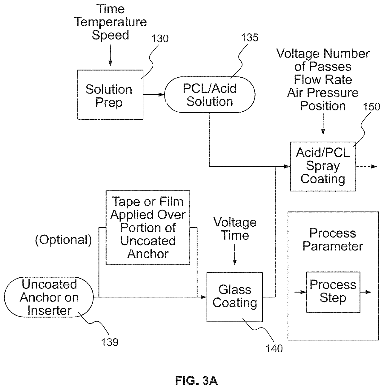
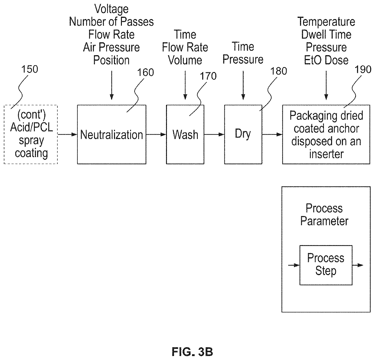
[0193]About 1.5 gram of PCL was added to about 10 g of glacial acetic acid (GAA) in a 50 ml plastic sample tube and the mixture was agitated over the course of several hours by intermittently vortexing using a Fisher Scientific Vortex Mixer to form a PCL solution. Other forms of mixing or sample agitation may be contemplated to solubilize the PCL. The PCL-GAA solution has a concentration of PCL greater than about 10 wt % and less than about 15 wt %. Concurrently, about 3.4 g of calcium phosphate (Vitoss® micromorsels, 1-2 mm, available from Stryker Orthobiologics) were placed into a mold (Aluminum, rectangular shaped, about 1.0 inch wide×4.0 inches long× 7 / 18 inches deep), where the mold was sprayed with cooking oil and excess oil wiped away with a kimwipe prior to adding the calcium phosphate. About 12 g of the 10 wt %-15 wt % PCL-GAA solution was added to the mold containing the calcium phosphate, and the mixture of PCL, GAA, and calcium phosphate was allowed to sit for...

example 1-2

ution

[0194]Example 1-2 is made using the same procedure described for Example 1-1, except about 1 g of PCL is added to about 10 g of GAA to form a 10 wt % PCL-GAA solution. The resulting scaffold is depicted in FIG. 8B.

example 1-3

lution

[0195]Example 1-3 is made using the same procedure described for Example 1-1, except about 0.75 g of PCL is added to about 10 g of GAA to form a 7.5 wt % PCL_GAA solution. The resulting scaffold is depicted in FIG. 8C.


PUM

PropertyMeasurementUnit
sizeaaaaaaaaaa
sizeaaaaaaaaaa
timeaaaaaaaaaa

Abstract

A bioactive filamentary structure includes a sheath coated with a mixture of synthetic bone graft particles and a polymer solution forming a scaffold structure. In forming such a structure, synthetic bone graft particles and a polymer solution are applied around a filamentary structure. A polymer is precipitated from the polymer solution such that the synthetic bone graft particles and the polymer coat the filamentary structure and the polymer is adhered to the synthetic bone graft particles to retain the graft particles.

Description

CROSS-REFERENCE TO RELATED APPLICATIONS[0001]The present application is a divisional application of U.S. patent application Ser. No. 15 / 584,620 filed May 2, 2017, now U.S. Pat. No. 10,729,548, which claims the benefit of the filing date of U.S. Provisional Patent Application No. 62 / 330,584 filed May 2, 2016, and is a continuation-in-part of U.S. patent application Ser. No. 15 / 234,239 filed Aug. 11, 2016, published as U.S. Patent Application Publication No. 2017 / 0043052 A1, which claims the benefit of the filing date of U.S. Provisional Patent Application No. 62 / 204,119, filed Aug. 12, 2015, the disclosures of all of which are hereby incorporated herein by reference.FIELD OF THE INVENTION[0002]The present invention relates to bone suture anchors, and more particularly, to bioactive suture anchors and the preparation thereof.BACKGROUND OF THE INVENTION[0003]Surgical sutures and suture anchors are used to close or hold together various types of soft tissue, including but not limited to...

Claims


Application Information

Patent Timeline
Patent Type & Authority Patents(United States)
IPC IPC(8): A61F2/28A61L31/16A61L31/12A61L31/00A61F2/30A61F2/46A61L27/02A61L27/36A61L27/40A61L27/26
CPCA61F2/28A61F2/30767A61F2/4601A61L27/02A61L27/025A61L27/365A61L27/3645A61L27/40A61L31/005A61L31/127A61L31/16A61F2/30965A61F2002/2835A61F2002/3006A61F2002/30062A61F2002/3093A61L27/26A61L2420/02A61L2420/06C12N2533/18A61F2002/30985A61F2002/30064A61F2310/00796A61B17/0401A61B2017/0406A61B2017/00526A61B2017/00004A61B2017/00893C08L67/04
Inventor SHETH, SONNYARAZAWA, DAVIDBURLEY, J. BROOKSMITH, SARAH ELIZABETHHAVENER, MATTHEW B.SAN ANTONIO, JAMES
Owner HOWMEDICA OSTEONICS CORP
Learn More