Protein delivery with porous metallic structure
a porous metallic and protein technology, applied in the field of medical devices and biologic therapies, can solve the problems of limited supply of autografts, less ideal materials than autografts, and efficient incorporation of osteoinductive materials, and achieve the effect of improving the elution of osteoinductive proteins
- Summary
- Abstract
- Description
- Claims
- Application Information
AI Technical Summary
Benefits of technology
Problems solved by technology
Method used
Image
Examples
example constructs
[0029]The present invention encompasses a number of composite constructs that meet the design criteria discussed above. Table 1 sets forth several constructs according to various embodiments of the present invention, with “Design 1” including granules as discussed above, and “Design 2” being free from such granules. Furthermore, “Design 3” also includes a number of 1-2 mm diameter openings through the matrix. These designs provide embodiments suitable for loading of osteoinductive proteins, as described herein. This listing is exemplary rather than comprehensive, and it will be appreciated that other constructs which meet the design criteria above are within the scope of the present invention.
[0030]
TABLE 1EXEMPLARY CONSTRUCTSDesign 1Design 2Design 3BiocompatibleTi-NiTi-NiTi-NiMatrixintermetallicintermetallicintermetallicmaterial—material—material— 50-500 μm 50-500 μm50-500 μmpore sizepore sizepore sizeOpenings / NoneNone1-2 mmFenestrationsdiameterThrough theMatrixGranule Size225-800 μ...
example 1
[0031]Samples of Ti—Ni matrices representative of Phusion Metal™ Cervical Cage (PorOsteon Inc., Menlo Park, Calif.) were examined and loaded with BMP-GER-NR described in U.S. Pat. No. 8,952,131 and incorporated herein in its entirety.
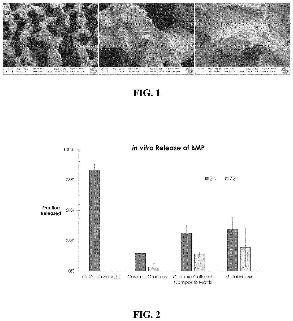
[0032]Scanning electron micrographs of the matrices are shown in FIG. 1, and demonstrate a highly rough and textured surface with features as small as 100 nm in size. BMP-GER-NR was loaded into the matrix by applying a protein solution (0.3 mg / mL in a pH 4 buffer solution) directly to the entire matrix in a volume equal to approximately 40-60% of the pore space within the matrix. The loaded matrices were allowed to stand for at least 15 minutes to enable the solution to fully penetrate the matrix, and then were placed into release buffer mimicking biological fluids comprised of saline and serum. Benchtop retention assays using ELISA were used to detect BMP-GER-NR in the release buffer at various time points. Approximately 34% of the initially loaded BMP...
example 2
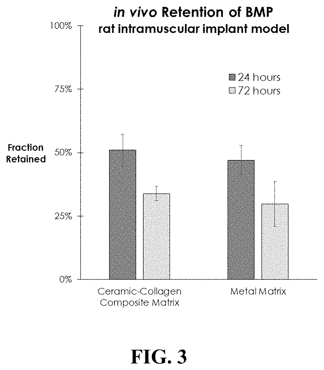
[0033]An in vivo study in the rat intramuscular implant model was conducted to compare the retention of BMP-GER-NR by the Ti—Ni matrices with plant-based collagen matrices loaded with CDHA granules (as described in U.S. Ser. No. 62 / 333,571). Equal quantities of radiolabeled BMP-GER-NR solution were applied to samples of the Ti—Ni and collagen matrices for a time sufficient to allow absorption into the matrices, and then implanted into a quadriceps muscle pouch in rats. Quantitative scintigraphy was performed immediately after surgery, and then subsequently after 24 and 72 hours. BMP retention was similar at both 24 and 72 hours for the collagen sponges and Ti—Ni metal matrices, as shown in FIG. 3. These data indicate that the nature of the pore structure and surface characteristics of the Ti—Ni matrix are effective in retaining BMP-GER-NR on or near the matrix for a clinically-effective time period in vivo.
PUM
| Property | Measurement | Unit |
|---|---|---|
| pore size | aaaaa | aaaaa |
| pore size | aaaaa | aaaaa |
| diameter | aaaaa | aaaaa |
Abstract
Description
Claims
Application Information
 - R&D
- Intellectual Property
- Life Sciences
- Materials
- Tech Scout
- Unparalleled Data Quality
- Higher Quality Content
- 60% Fewer Hallucinations
Browse by: Latest US Patents, China's latest patents, Technical Efficacy Thesaurus, Application Domain, Technology Topic, Popular Technical Reports.
© 2025 PatSnap. All rights reserved.Legal|Privacy policy|Modern Slavery Act Transparency Statement|Sitemap|About US| Contact US: help@patsnap.com


