Minimally invasive cardiac surgery procedure

a minimally invasive, cardiac surgery technology, applied in the field of surgical procedures, can solve the problems of significant trauma to the patient, particularly traumatic procedures, and long recovery tim

Inactive Publication Date: 2001-06-21
THE CLEVELAND CLINIC FOUND +1
View PDF0 Cites 75 Cited by
  • Summary
  • Abstract
  • Description
  • Claims
  • Application Information

AI Technical Summary

Problems solved by technology

Such procedures cause significant trauma to the patient, and recovery time.
Such procedures are particularly traumatic.
Incisional pain tends to require significant postoperative analgesia and postoperative discomfort tends to result in significant patient morbidity and lengthy hospital stays.
In addition, because the pericardial sack is opened underlying the sternum, after the procedure the heart has a tendency to become adherent to the sternum.
This can be problematical in the event of subsequent procedures.
However, such techniques require special instrumentation and special skills to perform, and may extend the time the heart is arrested and the duration of the procedure.

Method used


Image

  • Minimally invasive cardiac surgery procedure
  • Minimally invasive cardiac surgery procedure
  • Minimally invasive cardiac surgery procedure

Examples


Embodiment Construction

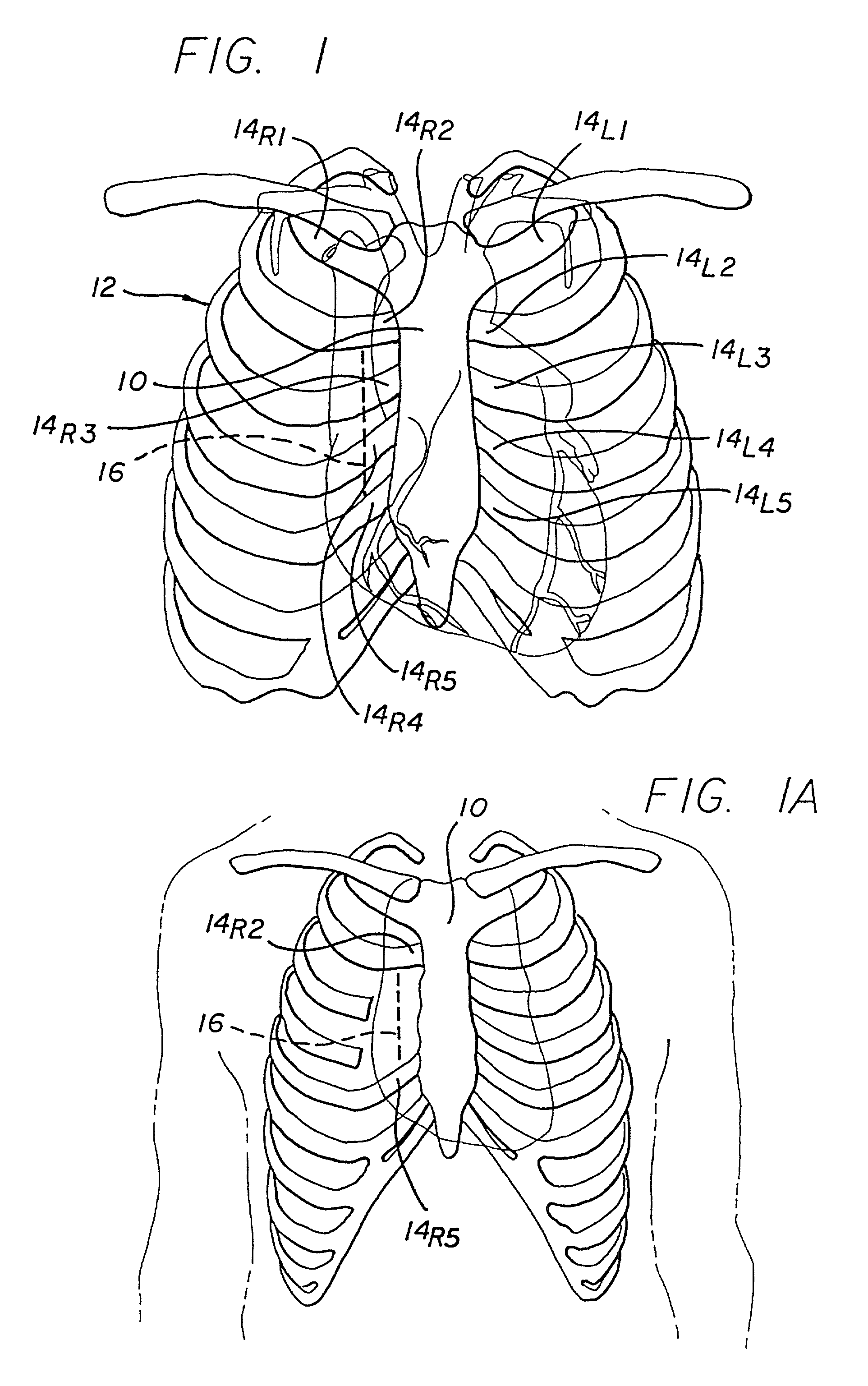
[0041] Referring now to FIG. 1, in a typical human, a sternum 10, a planary bone structure centrally disposed in the chest, is connected to a plurality of ribs 12 by respective costal cartilages 14.sub.R1, 14.sub.R2, 14.sub.R3, 14.sub.R4, 14.sub.R5, and 14.sub.L1, 14.sub.L2, 14.sub.L3, 14.sub.L4, 14.sub.L5. The heart and great vessels are located within a tissue sack (pericardium), located beneath the sternum, extending laterally under the costal cartilages and ribs, with the aorta disposed in part underlying the second and third right costal cartilages 14.sub.R2 and 14.sub.R3 and a portion of the right coronary artery located generally underlying the vicinity of the fourth and fifth right costal cartilages 14.sub.R4 and 14.sub.R5.

[0042] In accordance with one aspect of the present invention, it has been determined that a surgery on portions of the heart and great vessels located between a point approximately three centimeters above the supra annular ridge and the mid-ventricular ca...


PUM


Abstract

A minimally invasive approach for surgery on portions of the heart and great vessels. A parasternal incision is made extending across a predetermined number of costal cartilages, e.g., a right parasternal incision extending from the lower edge of the second costal cartilage to the superior edge of the fifth costal cartilage. One or more costal cartilages, e.g., the third and fourth, are then excised to provide access to the portion of the heart or great vessels of interest, for example between a point approximately three centimeters above supra annular ridge and the mid ventricular cavity, and a desired procedure completed. A minimally invasive procedure for repair or replacement of the aortic valve is disclosed that includes making a transverse incision of about 10 cm in length over the second or third intercostal space in the thorax of the patient, dividing the sternum transversely following the incision, retracting the transversely divided sternum, exposing the ascending aorta, and incising the ascending aorta to provide access to an area adjacent the aortic valve.

Description

[0001] This application is a continuation of U.S. Application Ser. No. 08 / 801,494, filed Feb. 18, 1997, which is a continuation-in-part of U.S. Application Ser. No. 08 / 603,313, filed Feb. 19, 1996, now issued as U.S. Pat. No. 5,752,526.[0002] 1. Field of the Invention[0003] The present invention relates to surgical procedures and, more specifically, to minimally invasive procedures for mitral and aortic valve repair or replacement surgeries including making small access incisions in the sternal area.[0004] 2. Description of the Related Art[0005] Various types of surgical procedures are performed on the heart and the great vessels. Many of such procedures, particularly those involving the aorta, and aortic valve employ a gross thoracotomy, e.g., a median sternotomy, in order to gain access to the involved portion of the heart or vessel. In other words, the procedures entail splitting open the patient's chest. Such procedures cause significant trauma to the patient, and recovery time....

Claims


Application Information

Patent Timeline
Patent Type & Authority Applications(United States)
IPC IPC(8): A61B17/00A61B17/02A61F2/24
CPCA61B17/00234A61B17/02A61B17/0218A61B2017/00243A61F2/2427A61F2/2445A61F2/2466Y10S623/902Y10S623/914Y10S623/922Y10S623/918
Inventor COSGROVE, DELOS M.
Owner THE CLEVELAND CLINIC FOUND
Why Eureka
Try Eureka