Stereoscopic microscope

a microscope and microscope technology, applied in the field of stereoscopic microscopes, can solve the problems of difficult to distinguish minute tissues of intricate organs, other staff, nurses, interns,

Inactive Publication Date: 2002-06-27
ASAHI KOGAKU KOGYO KK
View PDF1 Cites 25 Cited by
  • Summary
  • Abstract
  • Description
  • Claims
  • Application Information

AI Technical Summary

Benefits of technology

[0016] With this construction, the object light rays incident on the close-up optical system form the primary images having predetermined parallax at the field stops through the zoom optical systems. The close-up optical system is adjusted so that the front focal point thereof coincides with the object. Thus, the close-up optical system has a function of a collimator lens that converts the divergent light rays from the object into the parallel light rays. The primary images are transmitted by the relay optical systems. The inter-axis distance reducing element reduces the inter-axis distance of the right and left light rays. The primary images are re-imaged by the relay optical systems as the secondary images on the adjacent regions on the single image taking surface of the image taking device, respectively. The captured images are displayed on a display device such as an LCD panel or a CRT. The lead surgeon and the other stuffs can observe the magnified stereoscopic images on the display devices through stereoscopic viewers.

Problems solved by technology

Since it is difficult to distinguish minute tissues of an intricate organ such as a brain by the naked eye, the surgical microscope is required to proceed surgery on such an organ.
However, with the conventional optical stereoscopic microscope, although a lead surgeon or his / her assistant can observe the microscopic image, other staffs such as anesthetists, nurses, interns, and advisers who works at some remote locations cannot observe the same microscopic image.
Therefore, they could not pursue their share of tasks with sufficient accuracy and promptness.
Similarly, the adviser could not provide timely and proper advice from the remote locations.
Accordingly, the lead surgeon D and other surgical staffs who are observing these images can easily identify the diseased part, which would be difficult to be identified with actual images only.
Further, if a mirror is used as the optical path deflecting element, a setting angle error of the mirror largely deviates the direction of the reflected light.
However, if the aberrations have the opposite directional properties, the right and left images have different pictures in the specific part of the object, which significantly interferes with the stereoscopic observation.
The spherical aberration can be reduced using a large number of lenses, while it increases cost, weight and space of the close-up optical system.
However, the higher the magnification of the zoom optical system is, the longer the focal length thereof is, which enlarges the total size and weight of the microscope.
When the magnification M.sub.R is larger than -1, the focal length of the zoom optical system should be longer to obtain the predetermined total magnification of the microscope, which disturbs the reduction in size of the zoom optical systems.

Method used


Image

  • Stereoscopic microscope
  • Stereoscopic microscope
  • Stereoscopic microscope

Examples


second example

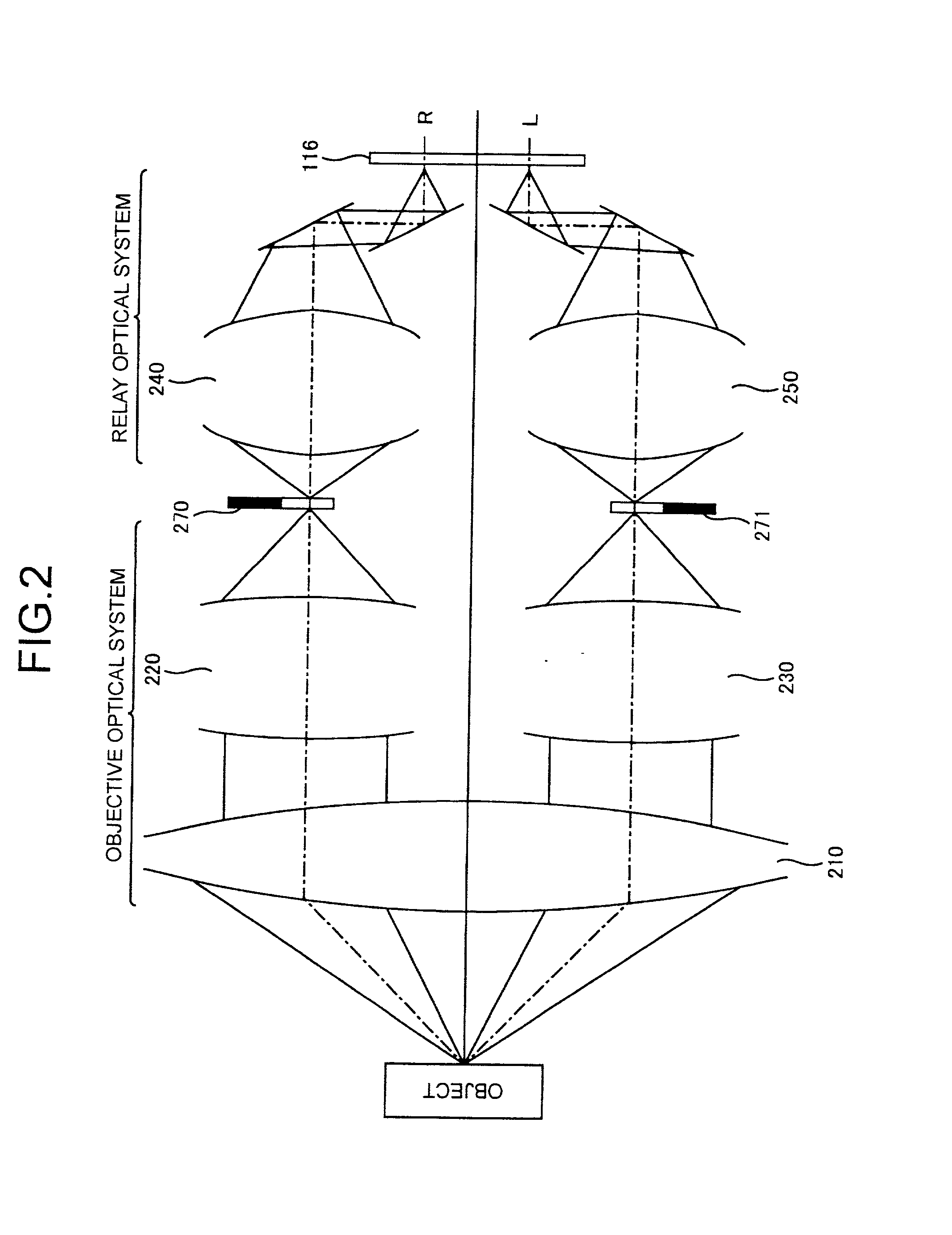
[0113] FIG. 11 shows the image taking optical system 200 of the second example in the developed fashion. The numerical constructions thereof are described in TABLE 2. The elements are indicated by the same surface numbers as the first example.

[0114] In the second example, the focal length of the close-up optical system 210 is variable in the range of 532.3 mm through 645.3 mm, and the imaging magnification M.sub.R of the relay optical systems 240, 250 is -1.5.

4TABLE 2 Surface Number r d n .nu.d 1 -440.000 6.50 1.51633 64.1 2 164.547 9.20 1.62004 36.3 3 472.000 30.34 4 403.219 5.40 1.80518 25.4 5 141.500 16.50 1.67003 47.3 6 -292.600 35.66 7 96.650 2.00 1.67270 32.1 8 32.889 5.50 1.61800 63.4 9 321.250 0.20 10 42.000 4.00 1.61800 63.4 11 154.980 8.12 12 -199.500 1.50 1.83400 37.2 13 6.892 3.70 1.84666 23.8 14 18.878 30.41 15 -15.142 1.20 1.48749 70.2 16 .infin. 18.30 17 19.083 3.80 1.49700 81.6 18 -19.083 0.50 19 26.525 4.50 1.48749 70.2 20 -11.258 1.00 1.83481 42.7 21 3-78.000 21.52...

third example

[0115] FIG. 12 shows the image taking optical system 200 of the third example in the developed fashion. The numerical constructions thereof are described in TABLE 3. The elements are indicated by the same surface numbers as the first example.

[0116] In the third example, the focal length of the close-up optical system 210 is variable in the range of 532.3 mm through 645.3 mm, and the imaging magnification M.sub.R of the relay optical systems 240, 250 is -1.875.

5TABLE 3 Surface Number r d n .nu.d 1 -440.000 6.50 1.51633 64.1 2 164.547 9.20 1.62004 36.3 3 472.000 30.34 4 403.219 5.40 1.80518 25.4 5 141.500 16.50 1.67003 47.3 6 -292.600 35.66 7 77.320 1.60 1.67270 32.1 8 26.311 4.40 1.61800 63.4 9 257.000 0.16 10 33.600 3.20 1.61800 63.4 11 123.984 6.49 12 -159.600 1.20 1.83400 37.2 13 5.514 2.96 1.84666 23.8 14 15.102 24.34 15 -12.114 0.96 1.48749 70.2 16 .infin. 14.64 17 15.266 3.04 1.49700 81.6 18 -15.266 0.40 19 21.220 3.60 1.48749 70.2 20 -9.006 0.80 1.83481 42.7 21 -302.400 17.22 ...

fourth example

[0117] FIG. 13 shows the image taking optical system 200 of the fourth example in the developed fashion. The numerical constructions thereof are described in TABLE 4. The elements are indicated by the same surface numbers as the first example.

[0118] In the fourth example, the focal length of the close-up optical system 210 is variable in the range of 532.3 mm through 645.3 mm, and the imaging magnification M.sub.R of the relay optical systems 240, 250 is -2.0.

6TABLE 4 Surface Number r d n .nu.d 1 -440.000 6.50 1.51633 64.1 2 164.547 9.20 1.62004 36.3 3 472.000 30.34 4 403.219 5.40 1.80518 25.4 5 141.500 16.50 1.67003 47.3 6 -292.600 35.66 7 72.487 1.50 1.67270 32.1 8 24.667 4.12 1.61800 63.4 9 240.937 0.15 10 31.500 3.00 1.61800 63.4 11 116.235 6.08 12 -149.625 1.12 1.83400 37.2 13 5.169 2.78 1.84666 23.8 14 14.159 22.82 15 -11.357 0.90 1.48749 70.2 16 .infin. 13.72 17 14.312 2.85 1.49700 81.6 18 -14.312 0.38 19 19.894 3.38 1.48749 70.2 20 -8.444 0.75 1.83481 42.7 21 -283.500 16.14 ...


PUM


Abstract

The stereoscopic microscope includes a common close-up optical system that faces an object, a pair of zoom optical systems that form a pair of primary image, a pair of field stops, a pair of relay optical systems that relay the primary images to form a pair of secondary images, an inter-axis distance reducing element, an image taking device and an illuminating optical system. The object light rays incident on the close-up optical system form the primary images having predetermined parallax at the field stops through the zoom optical systems. The inter-axis distance reducing element reduces the inter-axis distance of the right and left light rays. The primary images are re-imaged by the relay optical systems as the secondary images on the adjacent regions on the single image taking surface of the image taking device, respectively.

Description

[0001] 1. Field of the invention[0002] The present invention relates to a stereoscopic microscope for magnifying an object, and more particularly, to a stereoscopic microscope in which an image of the object is electronically taken by an image taking device such as a CCD.[0003] 2. Description of the Related Art[0004] A stereoscopic microscope is used as a surgical microscope for magnifying minute tissues such as brain cells during surgery.[0005] Since it is difficult to distinguish minute tissues of an intricate organ such as a brain by the naked eye, the surgical microscope is required to proceed surgery on such an organ. Besides, since it is impossible to observe the three-dimensional structure of a tissue with a monocular microscope, a stereoscopic microscope has been used to enable three-dimensional magnifying observation of the tissue in order to perform accurate operations.[0006] However, with the conventional optical stereoscopic microscope, although a lead surgeon or his / her...

Claims


Application Information

Patent Timeline
Patent Type & Authority Applications(United States)
IPC IPC(8): G02B21/22
CPCG02B21/22
Inventor TACHIHARA, SATORUTANAKA, CHINARI
Owner ASAHI KOGAKU KOGYO KK
Why Eureka
Try Eureka