
Markers and screens

a technology applied in the field of markers and screens, can solve the problems of mismatch in timing and have not been identified to date, and achieve the effects of enhancing the detectability of disease phenotype, increasing the detectability of synapse on activation, and improving the detection efficiency of disease phenotyp

Inactive Publication Date: 2005-03-03
ACKERMANN MANUEL +1
View PDF0 Cites 0 Cited by
  • Summary
  • Abstract
  • Description
  • Claims
  • Application Information

AI Technical Summary

Benefits of technology

[0063] It is most preferred that the exogenous genetic material be added to the male DNA complement of the zygote prior to its being processed by the ovum nucleus or the zygote female pronucleus. It is thought that the ovum nucleus or female pronucleus release molecules which affect the male DNA complement, perhaps by replacing the protamines of the male DNA with histones, thereby facilitating the combination of the female and male DNA complements to form the diploid zygote.
[0067] The number of copies of the DNA sequences which are added to the zygote is dependent upon the total amount of exogenous genetic material added and will be the amount which enables the genetic transformation to occur. Theoretically only one copy is required; however, generally, numerous copies are utilized, for example, 1,000-20,000 copies of a gene, in order to insure that one copy is functional. As regards the present invention, there is generally an advantage to having more than one functioning copy of each of the inserted exogenous DNA sequences to enhance the phenotypic expression of the exogenous DNA sequences.
[0121] Thus the invention provides methods of diagnosis and use of the materials disclosed herein in such methods, particularly in respect of neuropathologies. The invention further provides methods of enhancing the detectability of the disease phenotype of a transgenic animal model wherein the detectability of a synapse on activation, is increased by use of detectable cellular component as described above.

Problems solved by technology

It is known in the art that there is a mismatch in timing between the electrophysiological effects of LTP, which appear immediately, and the associated morphological changes, which require some 30 min to develop.
However, although such a distinctive tag has been mooted (41) it has not been identified to date.
However the fact that the AMPA receptors turn over at the synapse suggests that they are not durable markers of activation and may in any case be present in even non-activated mature synapses.

Method used


Image

  • Markers and screens
  • Markers and screens
  • Markers and screens

Examples


example 1

Profilin I and Profilin II in Cultured Hippocampal Neurons

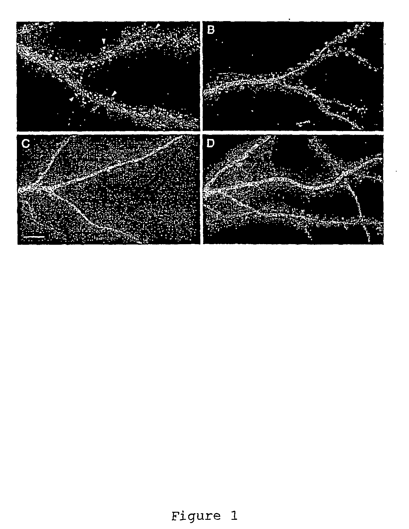
[0136] Expressing GFP-tagged profilin I and profilin II in cultured hippocampal neurons revealed differences in their distribution within dendrites (FIG. 1 and Table 1). In 4% of cells expressing profilin II (n=239) the GFP-tagged protein was highly enriched in heads of dendritic spines (FIG. 1A) whereas in others cells spine enrichment was weak or undetectable (FIG. 1B). GFP-tagged profilin I showed only weak or no enrichment in spines (FIG. 1C). For comparison the distribution of unmodified GFP in control cells is shown in FIG. 1D.

[0137] Corresponding results with gelsolin are shown in Table 5. ‘G2-6-GFP’ is a based on a gelsolin mutant which lacks the domain 1 (and hence the severing function) of whole gelsolin.

example 2

NMDA Receptors Regulate Profilin II Accumulation in Spine Heads

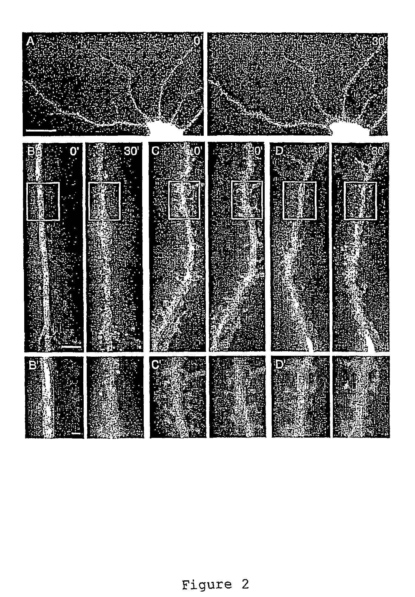
[0138] The strong enrichment of profilin II in spines of some cells but not others suggested that its cytoplasmic localization might be activity dependent. To test this hypothesis we exposed hippocampal neurons expressing GFP-tagged profilin II or profilin I or unmodified GFP to 10 μM glutamate (FIG. 2). Images of living cells captured before and after treatment showed a strong redistribution of profilin II-GFP into the heads of spines that started 5 to 8 minutes after glutamate addition and was maximal after 30 minutes (FIGS. 2A and B). Glutamate treatment also produced a weaker shift of GFP tagged profilin I into spines (FIG. 2D) which, however, never approached the high levels seen for profilin II. The same treatment did not change the distribution of unmodified GFP (FIG. 2C).

[0139] To determine which sorts of glutamate receptor might be involved in profilin targeting we treated profilin II-GFP transfected cells wit...

example 3

Increased Calcium Levels are Required for Spine Targeting of Profilin II

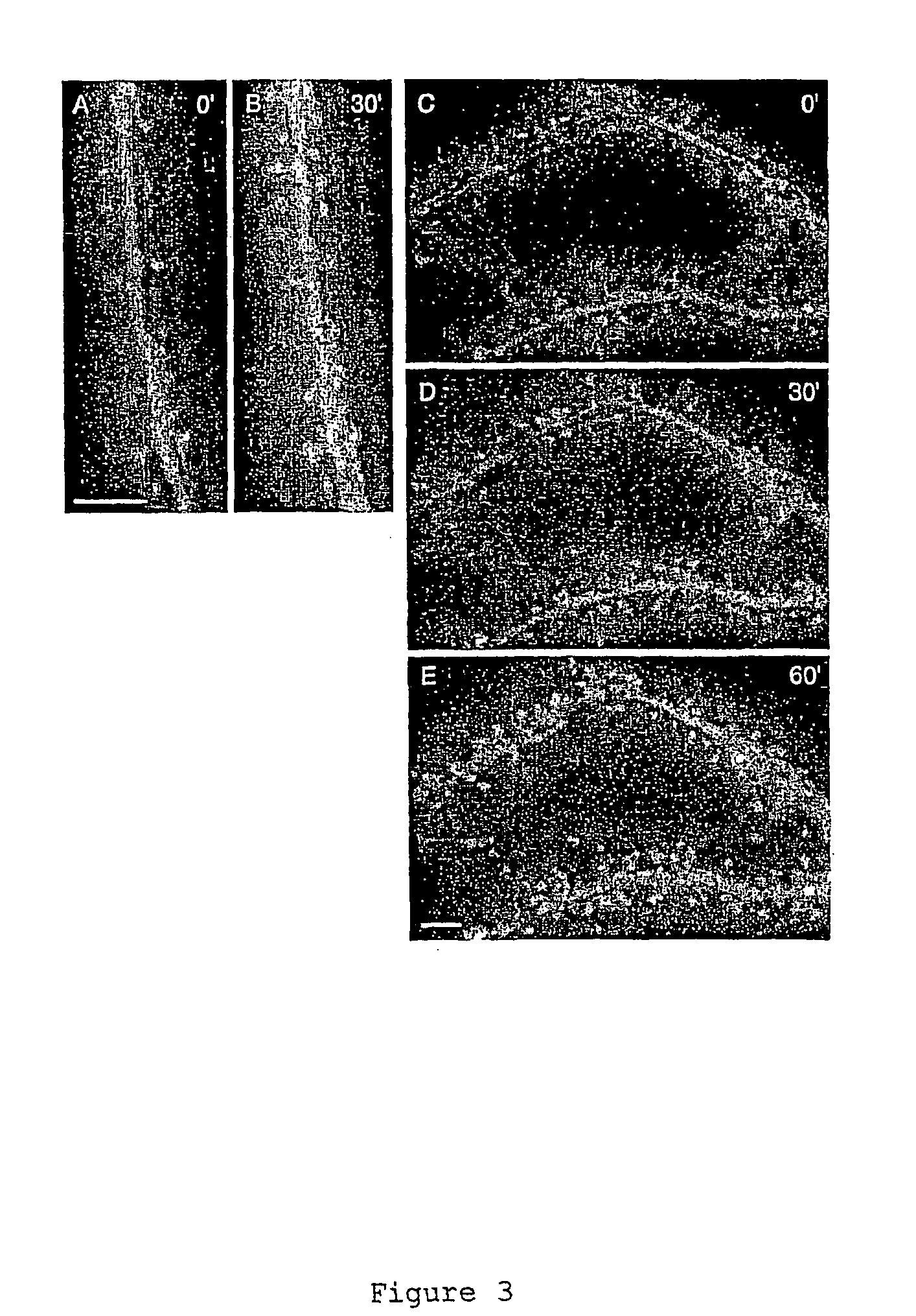
[0141] The involvement of NMDA receptors in the accumulation of profilin II in spines suggested that increases in cytoplasmic Ca levels may be required to trigger profilin II targeting. Consistent with this idea, profilin II levels in spines failed to increase when cells were treated with glutamate in the absence of extracellular Ca2+ (Table 3, see also supplementary figure). To determine whether elevation of intracellular Ca2+ is alone sufficient to trigger the targeting process, we treated neurons with thapsigargin (1 μM) which results in the release of Ca2+ from internal stores and leads to influx of extracellular Ca2+ (21, 22). Following thapsigargin treatment 10 out of 15 cells tested showed accumulation of profilin II in spines (Table 3). When cells were treated with thapsigargin in the absence of extracellular Ca2+ there was only weak redistribution of profilin II in a minority of cells (Table 3a) sugges...


PUM

PropertyMeasurementUnit
volumeaaaaaaaaaa
pHaaaaaaaaaa
pHaaaaaaaaaa

Abstract

Provided are in vivo and in vitro methods for identifying or detecting a synapse which has been activated, or assessing the level of activation of a synapse, which method comprises: (i) determining the presence and\or amount, in a morphologically specialised postsynaptic site in the synapse (e.g. a dendritic spine), of a detectable cellular component associated with the activation (which is a ‘tag’ or ‘marker’ for the activation e.g. an actin-cytoskeleton interacting protein such as profilin II or gelsolin), and (ii) correlating the result of the determination with synaptic activation. Such assays can be useful in identifying processes involved in LTP, and also more generally in identifying modulators of synaptic activation or transmission, and hence cognitive function.

Description

TECHNICAL FIELD [0001] The present invention relates generally to methods and materials for use in markers of synaptic activity and use of these for identifying modulators of such activity. BACKGROUND ART [0002] It is known that experimental manipulations of experience produce changes in the shape and number of dendritic spines that form the postsynaptic contact sites for excitatory synapses in the brain (1). [0003] A wide variety of ideas about the function of dendritic spines have been proposed. These include that they exist solely to increase the surface area of the neuron available for receiving synaptic contacts, that their role is to protect neurons from excessive excitation, that they act as electrophysiological compartments independent of the dendrite or as biochemical compartments. Evidence that the numbers or shapes of dendritic spines can change according to the sensory input of animals during development or with an animal's behavioural status has led to the proposal that...

Claims


Application Information

Patent Timeline
Patent Type & Authority Applications(United States)
IPC IPC(8): C12Q1/02G01N33/15G01N33/50G01N33/58G01N33/68
CPCG01N33/5076G01N33/6896G01N33/582
Inventor ACKERMANN, MANUELMATUS, ANDREW IAN
Owner ACKERMANN MANUEL
Learn More