Capsule endoscope

a capsule endoscope and endoscope technology, applied in the field of endoscopes, can solve the problem of excessive length of the capsule endoscope, and achieve the effect of improving the ease of use of the capsule and favorable correction of astigmatism

Active Publication Date: 2005-03-10
OLYMPUS CORP
View PDF15 Cites 89 Cited by
  • Summary
  • Abstract
  • Description
  • Claims
  • Application Information

AI Technical Summary

Benefits of technology

The present invention relates to a capsule endoscope that may be swallowed by a patient for the purpose of examining inside portions of a living body. More particularly, the present invention discloses various conditions to be met by a capsule endoscope in order to improve the ease with which the capsule may be swallowed by miniaturization of the capsule size while assuring an optimum depth of field and favorable correction of astigmatism over the entire field of view of the capsule endoscope.

Problems solved by technology

However, when the radius of curvature of the transparent cover in a central portion of the field of view is the same as the radius of curvature near the periphery of the field of view, due to the fact that the radius of curvature of the transparent cover is determined according to the external diameter of the capsule, there is a problem in that the length of the capsule endoscope becomes excessive.

Method used


Image

  • Capsule endoscope
  • Capsule endoscope
  • Capsule endoscope

Examples


embodiment 1

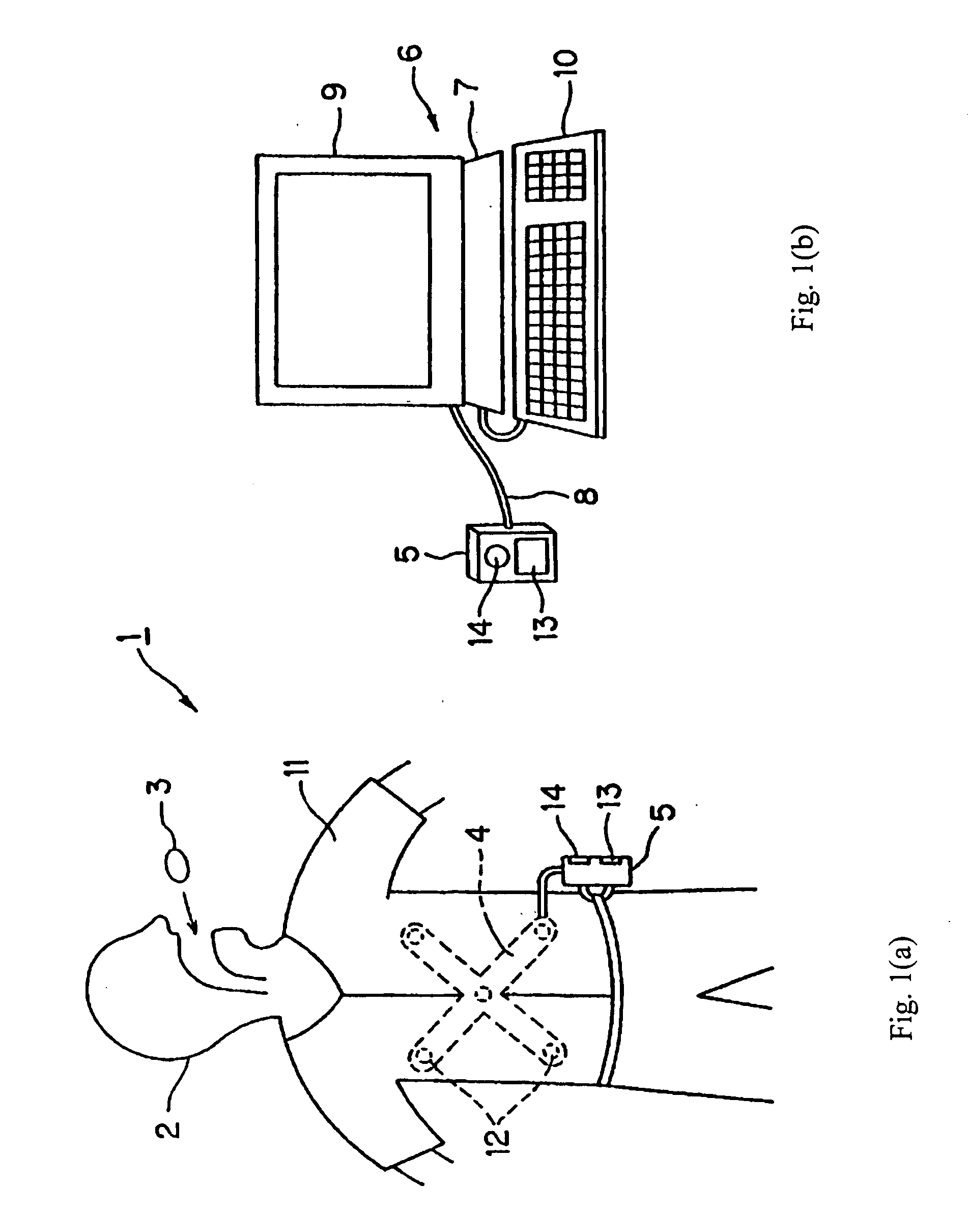
FIGS. 1(a), 1(b) and 2 relate to Embodiment 1 of the present invention. As shown in FIG. 1(a), a capsule endoscope device 1 is provided with a capsule endoscope 3 and an external unit 5. A patient 2 swallows the capsule endoscope 3 and, when the capsule endoscope 3 passes through a body passage within the patient's body cavity, an internal wall surface of the body passage is optically imaged and image data signals are transmitted from the capsule endoscope 3 via radio waves. The transmitted image data signals are received by an antenna unit 4 that is provided outside the body of the patient 2, and the image data signals are stored in an external unit 5. The external unit 5 has built-in memory, for example, a COMPACT FLASH MEMORY® having a storage capacity of 1 GB, for the purpose of storing the image data signals.

During the examination or after the examination, as shown in FIG. 1(b), the external unit 5 may be connected to a display system 6, so that the stored image data can be ...

embodiment 2

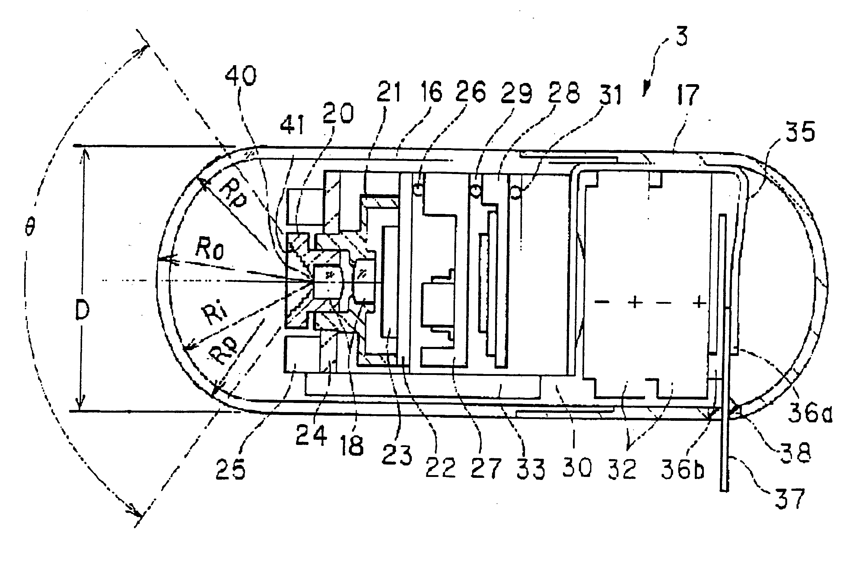
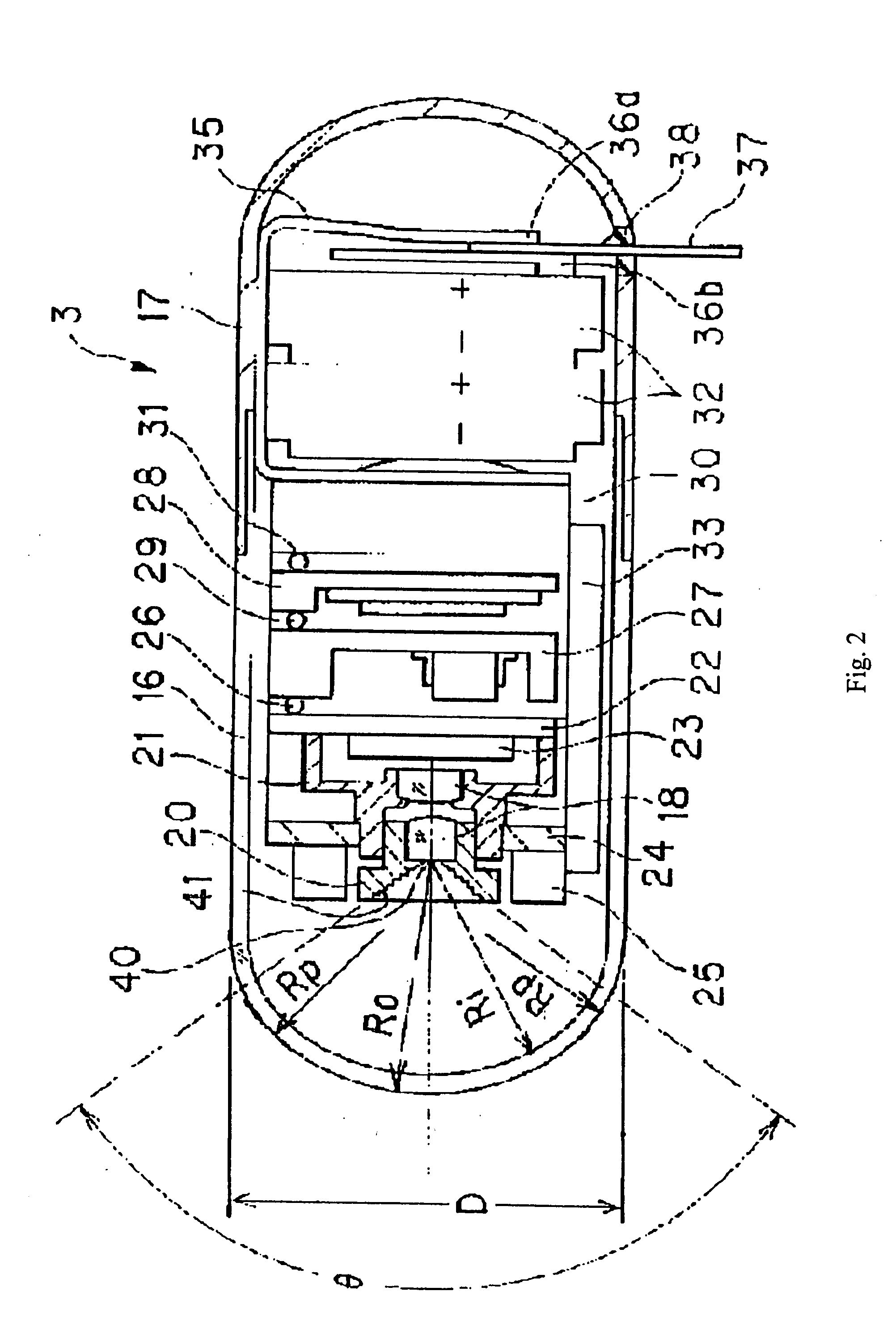
FIG. 3 shows Embodiment 2 of the present invention. In the capsule endoscope 51, a transparent front cover 53 engages with the front end of an exterior cover 52, which is a cylinder having a closed rear end that is rounded at the periphery and occluded. The transparent front cover is adhered to the exterior cover 52 so the inside is a watertight structure, and an objective optical system 54 is positioned inside the capsule so as to receive light that transmits through the front cover.

The objective optical system 54 is formed by attaching a first lens and a second lens to a first lens frame 55 and a second lens frame 56, respectively, and is arranged in a center position to the rear of the transparent cover 53. A CMOS image detector 58 is arranged at the image surface of the objective optical system 54. The CMOS image detector 58 is mounted in a centrally located recess that is established in the front surface of a substrate 57, and the second lens frame 56 is engaged with the firs...

embodiment 3

In the capsule endoscope 71 shown in FIG. 4(a), a roughly hemispherical transparent cover 73 is connected and secured to an exterior case 72, which comprises a cylinder having an occluded rear portion that is substantially hemispherical and is sealed to the transparent cover 73 so as to be watertight. In the capsule endoscope shown in FIG. 4(b), the configuration of the transparent cover 73 has been modified such that the radius of curvature of a portion where a light ray from the periphery of the field of view of the objective optical system 76 passes through is smaller than the radius of curvature of a portion where a light beam from the center of the field of view passes through. In each of the capsule endoscopes shown in FIGS. 4(a) and 4(b), there is provided within the sealed capsule the below-mentioned components.

An objective optical system 76 that is attached to a lens frame 75 is centrally positioned about the cylinder axis within the capsule so as to receive light that p...


PUM


Abstract

A capsule endoscope is disclosed that includes a capsule and a transparent cover having a central axis and that is located in front of an objective optical system within the capsule. The objective optical system has a depth of field, a field of view, and an image surface that has curvature of field, and is arranged on the central axis of the transparent cover and the capsule. The field of view of the objective optical system includes a spherical surface that is tangent to the transparent cover at a periphery of the field of view, and the near point of the depth of field on the optical axis of the objective optical system is positioned farther from the objective optical system than is the transparent cover. Various conditions are satisfied so as to provide high quality imaging.

Description

This application claims the benefit of priority of Japanese Patent Application 2003-314708 filed Sep. 5, 2003, the disclosure of which is hereby incorporated by reference. BACKGROUND OF THE INVENTION In recent years, endoscopes have been widely used in both the medical and industrial fields. Recently, a capsule endoscope has been introduced for use in medical applications that can be swallowed by a patient; see for example, FIG. 1 of Japanese Laid-Open Patent Application 2001-91860 and FIG. 1 of International Patent Publication WO 01 / 65995 A2. Such endoscopes are advantageous in that they greatly reduce the pain associated with an endoscopic examination by avoiding the pain associated with inserting the insertion portion of a conventional endoscope. Japanese Laid-Open Patent Application 2001-91860 discloses a conventional capsule endoscope wherein an objective lens and an illuminator, made from light emitting diodes that are symmetrically positioned about the objective lens, are ...

Claims


Application Information

Patent Timeline
Patent Type & Authority Applications(United States)
IPC IPC(8): A61B1/00A61B1/05A61B1/273A61B1/31G02B13/00G02B23/24
CPCA61B1/00096A61B1/00188G02B23/2423A61B1/2733A61B1/31A61B1/041
Inventor YOSHINO, KOICHIRO
Owner OLYMPUS CORP
Why Eureka
Try Eureka