
Fluid emitting suture needle

Active Publication Date: 2005-03-31
ETHICON INC
View PDF19 Cites 96 Cited by
  • Summary
  • Abstract
  • Description
  • Claims
  • Application Information

AI Technical Summary

Problems solved by technology

Although this time-tested method of delivering medication to the body serves its singular purpose effectively, it is not readily adapted to serve the alternate function of closing wounds, since multiple passes of the suture needle through the tissue surrounding the wound and knotting of the suture are typically involved in the wound closure process.
Likewise, because suture needles are not designed to transport a fluid and are not easily connected directly to an external reservoir of fluid without severely impairing their primary function as a tool for wound closure, suture needles are not used for drug delivery.
Although suture needles have been improved over the years to exhibit an exemplary combination of handling and performance properties, including but not limited to strength, stiffness, ductility, and lubricity, a number of problems are associated with the use of surgical needles.
For example, the transmission of blood born pathogens occurring from accidental needle sticks poses a risk to the medical professionals conducting the wound closure procedure.
However, since most tissue is not easily penetrated with blunt point needles, the additional level of safety from accidental needle sticks is achieved only by sacrificing handling characteristics and performance of the needle.
Additionally, blunt point needles in many cases will cause a higher level of tissue trauma than their sharp point counterparts.
Besides posing a nuisance to the surgeon, in certain surgeries such as those involving blood vessel anastomosis and certain organ surgeries, or in the cases where patients are suffering from hemophilia or consuming blood thinning medicines, hemostasis may be quite difficult to achieve.
As a further example, surgical site infections are a source of many post-operative complications and deaths each year.
However, the types of sutures that may be effectively combined with antimicrobial agents, as well as the duration and zone of efficacy are limited.

Method used


Image

  • Fluid emitting suture needle
  • Fluid emitting suture needle
  • Fluid emitting suture needle

Examples


example

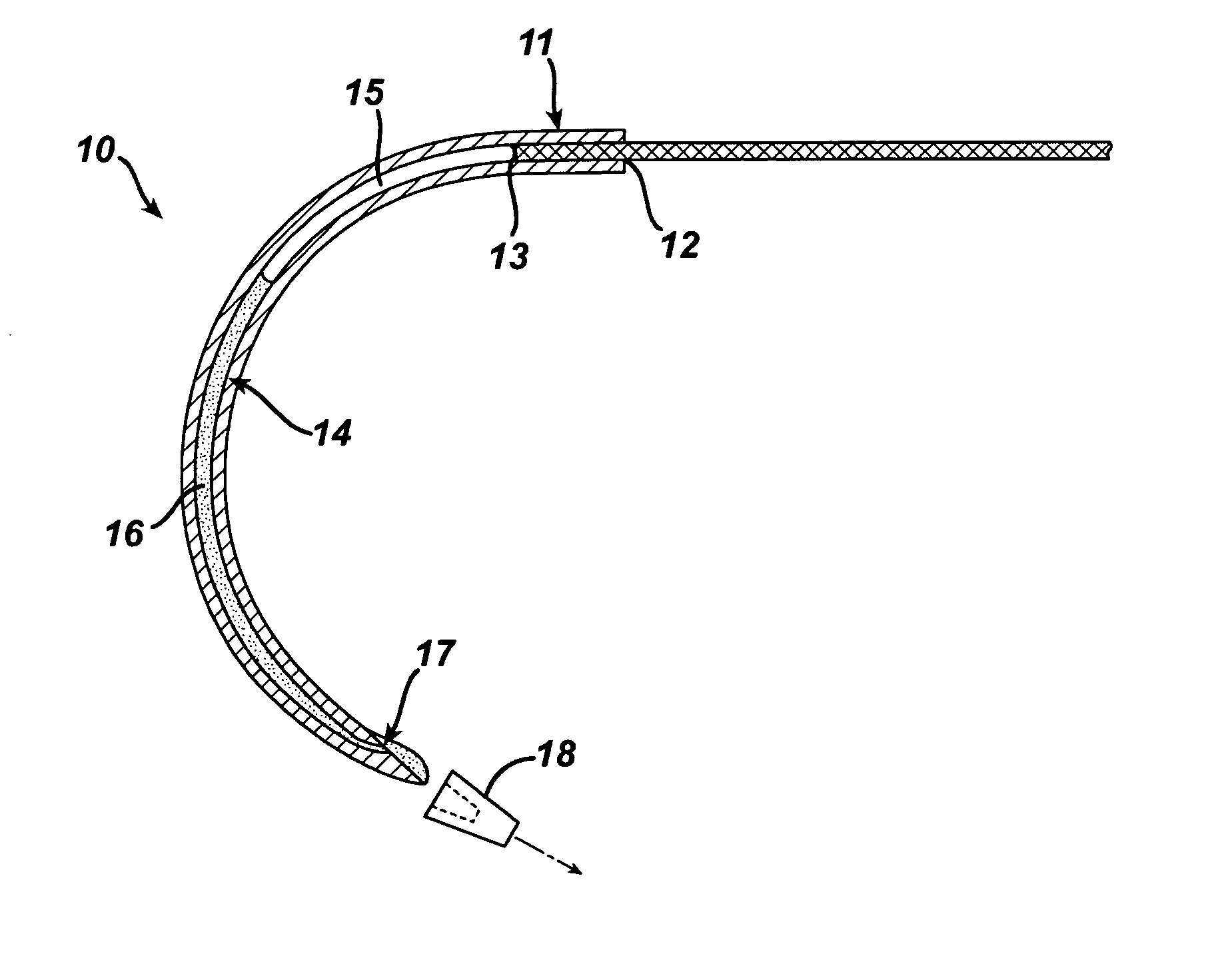
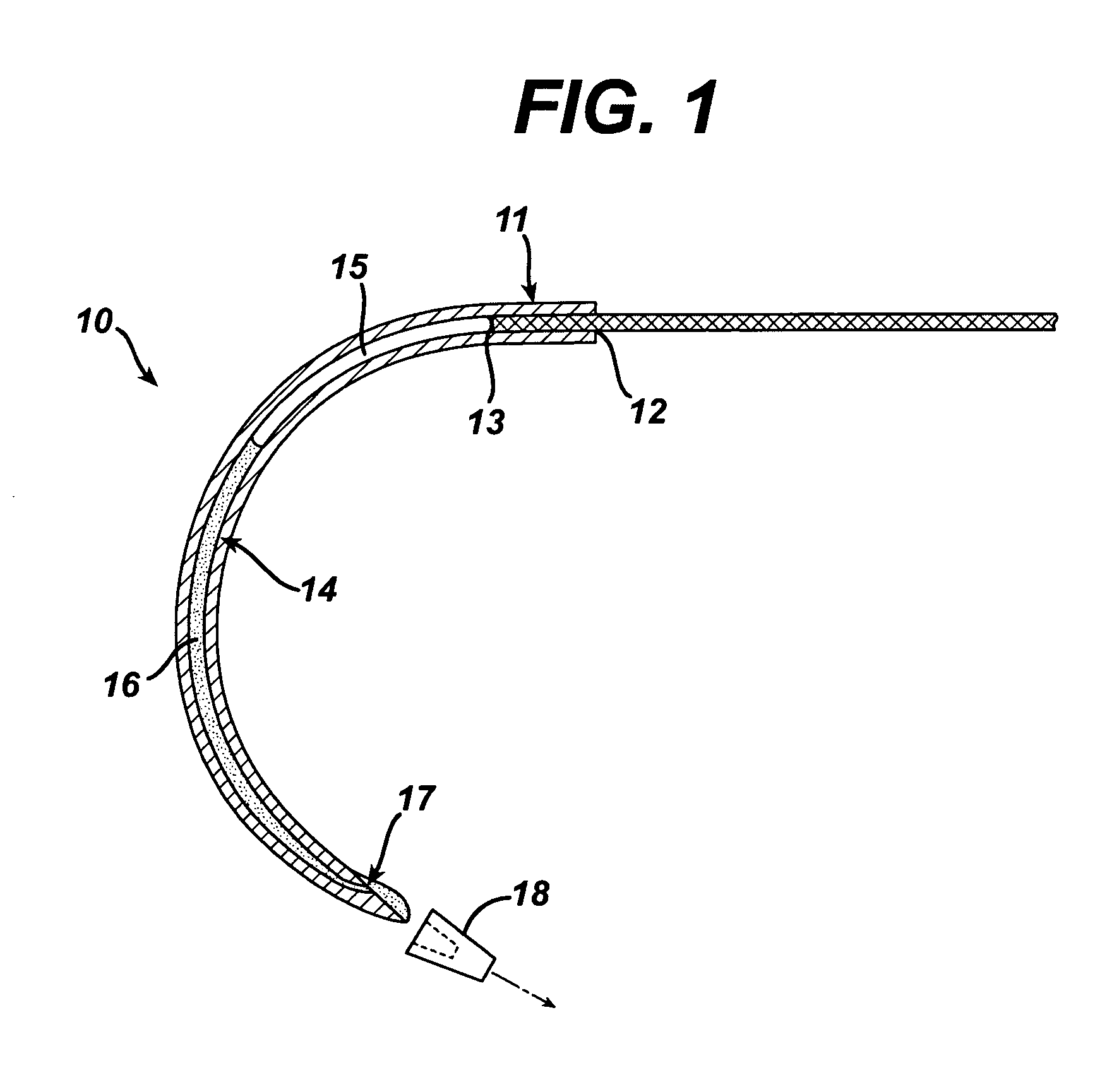
In vitro trials were conducted to evaluate the therapeutic efficacy of delivering an antimicrobial agent from the suture needle described in FIG. 1. Fluid emitting suture needles with a nominal outside diameter of 0.032″ and an inside diameter of 0.019″ were filled with a Triclosan bearing solution and pressurized to 2 atmospheres pressure. The liquid vehicle was a mixture of 75% propylene glycol and 25% Ethanol. Triclosan was added at a concentration of 0.1 g / ml solution. The total volume of fluid contained within the needle was ˜3 microliters. A control set of needles that were not loaded with an active agent were produced as well for comparison. Multifilament PET sutures and monofilament polypropylene sutures were attached to the needles. Devices were activated by removing the cap 18 shown in FIG. 1 and passed through agar plates containing various bacteria commonly found in surgical site infections, including: Staphylococcus Aureus, Escherichia. Coli and Enterococcus Facili. A ...


PUM


Abstract

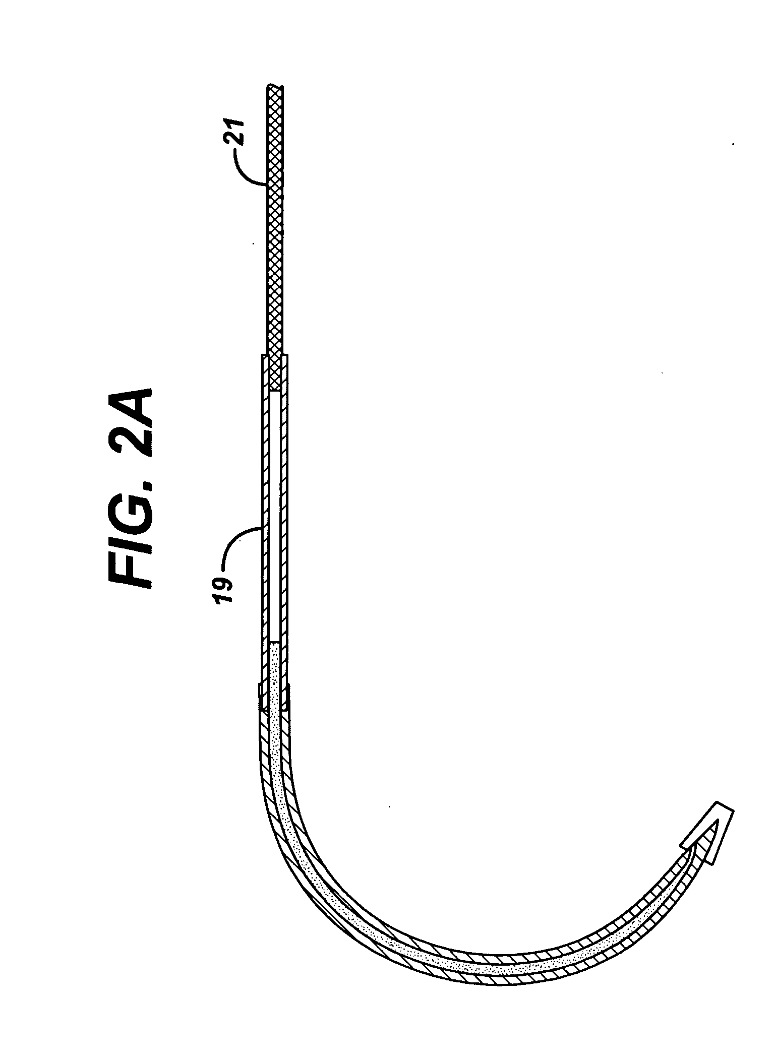
The invention provides a multifunctional suture needle that may be used to draw a suture through tissue surrounding a wound while simultaneously delivering a bioactive fluid through the needle tip. The suture needle possesses an internal cavity capable of containing a fluid, and a fine aperture adjacent to the point of the needle through which the fluid may egress. The fluid may be driven from the needle through the needle tip with a compressed gas that is sealed within the cavity adjacent to the fluid. Alternatively a fluid conducting suture may be employed to deliver fluid through the internal passage of the suture needle and out the aperture adjacent to the tip of the needle. The rate at which the fluid is emitted from the suture needle may be controlled by carefully selecting the fluid viscosity, design of the needle or suture passages, and pressure applied to the fluid.

Description

FIELD OF INVENTION The present invention relates to multifunctional devices that may be used to close surgical wounds. More particularly the invention relates to functional suture needles that may be used to emit therapeutic or bioactive agents or fluids during the wound closure procedure. In particular, the invention relates to a device that incorporates a suture needle having an internal passage that is in turn connected to a fine orifice adjacent to the needle tip, wherein the fluid is emitted through said orifice. BACKGROUND OF INVENTION Suture needles have long been used to guide and draw sutures through the tissue surrounding a wound. Even today, the function of commercially available suture needles continues to be singular in nature, namely to guide and position the suture to close wounds. Unlike hypodermic needles commonly employed to deliver fluids subcutaneously, suture needles must serve as a tool to guide and draw a suture into position along the path of a wound. Hypo...

Claims


Application Information

Patent Timeline
IPC IPC(8): A61B17/06
CPCA61B17/06066A61B2017/06052A61B2017/06185A61B2017/061A61B2017/0608
Inventor CICHOCKI, FRANK RICHARD JR.
Owner ETHICON INC
Learn More