
Method for slice position planning of tomographic measurements, using statistical images

a tomographic and statistical image technology, applied in tomography, instruments, applications, etc., can solve problems such as difficulty in comparing images obtained from respective scans

Active Publication Date: 2005-04-28
THE GENERAL HOSPITAL CORP +1
View PDF8 Cites 91 Cited by
  • Summary
  • Abstract
  • Description
  • Claims
  • Application Information

AI Technical Summary

Benefits of technology

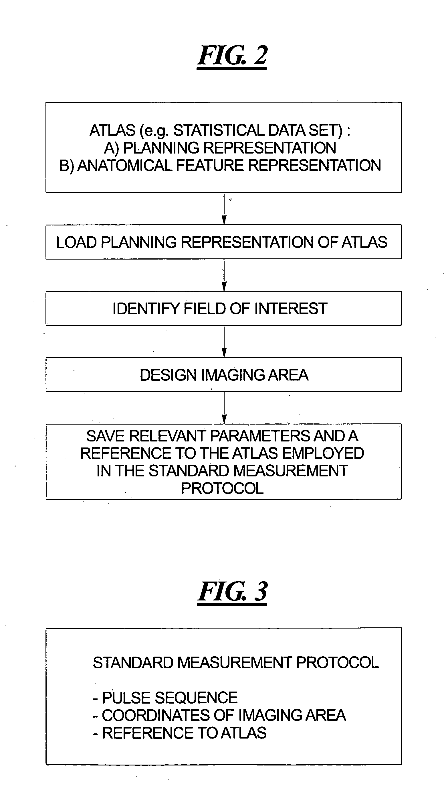
[0009] It is an object of the present invention to provide a method for slice position planning of tomographic measurements which avoids the above-described manual slice re-alignment. A further object of the present invention is to provide such a method which avoids the need for a localizer protocol of the type described above. This object is achieved in accordance with the principles of the present invention in a method for slice position planning of MR measurements wherein, instead of planning the slices for each individual patient for each individual scan, the slice or slices for a particular scan is / are planned using a statistical dataset which represents the geometrical details of the organ of interest in the scan. The statistical dataset represents a “standard” image of the organ of interest. The dataset can be obtained from a standard organ atlas, many of which are known and accessible, or can be produced from a data acquisition system by averaging several measured datasets that have previously been obtained from other patients, and that have been stored. The statistical dataset, or atlas, is displayed as a planning representation in a global slice-positioning environment. The measurement for an imaging area (geometrical parameters, sequence parameters, etc.) is planned using this statistical dataset, and is stored as a standard measurement protocol for the particular “standard” human organ in question. The standard measurement protocol includes information regarding, for example, the position of the imaging area in the dataset and the position of the imaging area regarding the “standard” human organ. The standard measurement protocol also includes information as to the number of slices, the orientation of the slices, the number of pixels per slice, the size of the pixels, etc. in the imaging area. The standard measurement protocol may allow the measurement of a series of imaging areas and / or may contain information regarding saturation areas, etc.
[0012] A significant advantage of the inventive method is the high degree of reproducibility that can be obtained in the examination. Because the imaging area is each time automatically determined by starting from the standard image area, that is adjusted according to the actual anatomical features of the patient (that in general do not change), the same patient can be scanned a number of times, separated by relatively long time durations, so that the respective images from the time-separated scans can be compared in a meaningful manner. This is particularly advantageous when the scans are for the purpose of following up treatment for a particular pathological condition, such as monitoring the size of a cancerous tumor during the course of radiation therapy or chemotherapy. When a patient is scanned at times that are separated from each other, it sometimes can be difficult to compare the images obtained from the respective scans because it cannot be reliably determined that changes in the images detected as a result of the comparison have occurred because of an actual change in the size of the tumor, or because the orientation of the slice in one of the images was not identical to the orientation of the slice in the other of the images. Because of the high degree of reproducibility achieved with the inventive procedure, when changes are detected between time-separated images, it can be more reliably assumed that those changes represent actual anatomical changes, rather than changes resulting from inconsistent slice positioning.
[0013] A further advantage obtained with the inventive method is a higher patient throughput with regard to the imaging installation, achieved by the significant decrease in time for planning each tomographic measurement.

Problems solved by technology

When a patient is scanned at times that are separated from each other, it sometimes can be difficult to compare the images obtained from the respective scans because it cannot be reliably determined that changes in the images detected as a result of the comparison have occurred because of an actual change in the size of the tumor, or because the orientation of the slice in one of the images was not identical to the orientation of the slice in the other of the images.

Method used


Image

  • Method for slice position planning of tomographic measurements, using statistical images
  • Method for slice position planning of tomographic measurements, using statistical images
  • Method for slice position planning of tomographic measurements, using statistical images

Examples


Embodiment Construction

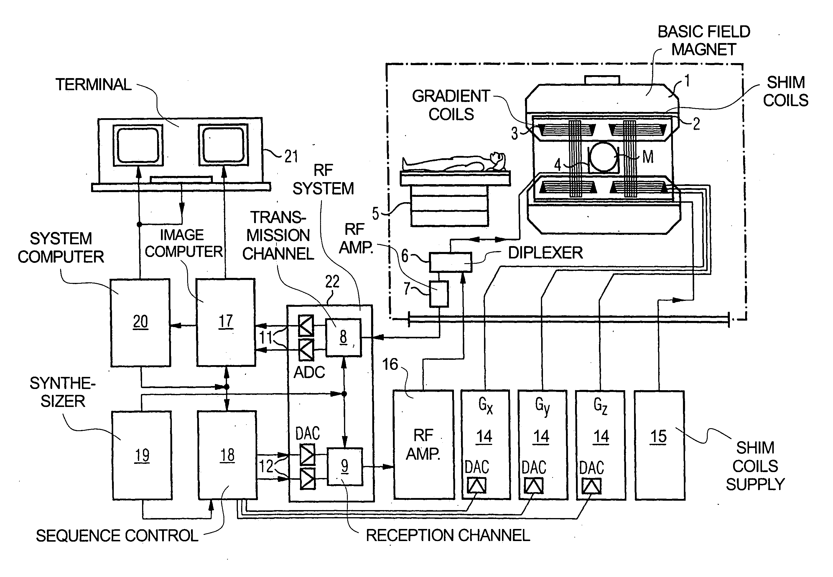
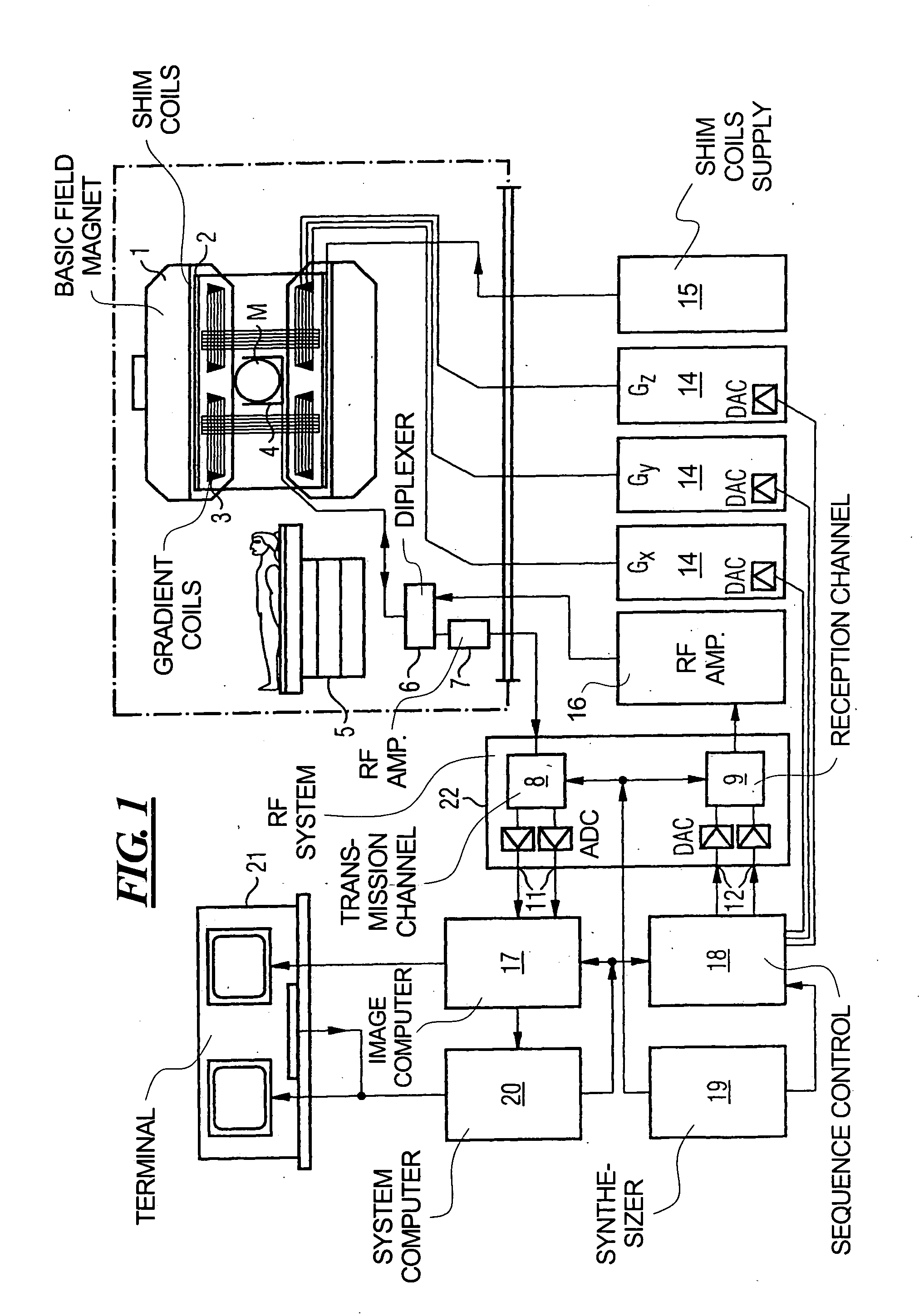
[0026]FIG. 1 schematically illustrates a magnetic resonance imaging (tomography) apparatus for generating a magnetic resonance image of a subject, as an example of a tomographic imaging modality operable according to the present invention. The components of the nuclear magnetic resonance tomography apparatus correspond to those of a conventional tomography apparatus, but it is controlled according to the invention. A basic field magnet 1 generates a time-constant, intense magnetic field for polarization (alignment) of the nuclear spins in the examination region of a subject such as, for example, a part of a human body to be examined. The high homogeneity of the basic magnetic field required for the nuclear magnetic resonance measurement is defined in a spherical measurement volume M in which the part of the human body to be examined is introduced. For supporting the homogeneity demands and, in particular, for eliminating time-invariable influences, shim plates of ferromagnetic mater...


PUM


Abstract

In a method and computer program product for operating a tomographic imaging apparatus, a standard measurement protocol is generated by displaying a planning representation of a standard object, defining a spatial position of a standard imaging area in the planning representation, and storing, as the standard measurement protocol for the standard object, a reference to the standard object and parameters of the standard imaging area. Such a standard measurement protocol can then be used in the slice position planning for an actual tomographic measurement, by obtaining data representing features of an examination object, corresponding to the standard object, determining a geometrical relation of the features of the examination object to features of the standard object, and generating an object-specific measurement protocol wherein the imaging area is positioned relative to the examination object by modification of the standard measurement protocol.

Description

BACKGROUND OF THE INVENTION [0001] 1. Field of the Invention [0002] The present invention is directed to a method for slice position planning of tomographic (magnetic resonance) measurements, including a protocol for operating a magnetic resonance imaging apparatus. [0003] 2. Description of the Prior Art [0004] Magnetic resonance imaging (MRI, also known as magnetic resonance tomography (MRT)) is based on the physical phenomenon of nuclear magnetic resonance and has been successfully utilized as an imaging modality in medicine and in biophysics for more than fifteen years. In this imaging modality, a subject, such as a living patient, is subjected to a strong, constant magnetic field. As a result, the nuclear spins of the atoms in the subject, that were previously irregularly oriented, are aligned. Radio-frequency energy emitted into the subject then excites these “ordered” nuclear spins to a specific resonance. This resonance generates the actual measurement signal, which is receiv...

Claims


Application Information

Patent Timeline
IPC IPC(8): A61B5/05A61B5/055A61B5/107A61B6/00A61B6/03
CPCA61B5/055A61B6/506A61B6/03
Inventor SCHRECK, OLIVERMULLER, MIKEHARDER, MARTINHOLLENBACH, HANS-PETERSCHMITT, FRANZNIMSKY, INESDALE, ANDERSVAN DER KOUWE, ANDRE
Owner THE GENERAL HOSPITAL CORP
Learn More