

Monoclonal antibody imaging and therapy of tumors that express met and bind hepatocyte growth factor
a tumor and met expression technology, applied in the field of immunology and cancer diagnosis and treatment, can solve the problems of poor patient outcome and high met expression in tumors, and achieve the effect of rapid and effective detection of tumors autocrin
- Summary
- Abstract
- Description
- Claims
- Application Information
AI Technical Summary
Benefits of technology
Problems solved by technology
Method used
Image

Examples
example 1
Materials and Methods
Reagents
[0183]125I was purchased as NaI (480-630 mBq (13-17 mCi) per μg iodine) from Amersham Corp. (Arlington Heights, Ill.). C-28 rabbit polyclonal antibody reactive with the C-terminal portion of human Met was purchased from Santa Cruz Biotechnology, Inc.
Cell Lines and Tumors
[0184] Imaging studies were initiated with a constituted mixture of S-114 cells (NIH 3T3 cells transformed with hHGF and hMet (Rong S et al., Cell Growth Differ. 1993;4:563-569) and M-114 cells (NIH 3T3 cells transformed with mHGF and mMet). Cells were grown in DMEM containing 8% calf serum. SK-LMS-1, a human leiomyosarcoma cell line autocrine for hMet and hHGF (Jeffers M et al., Mol Cell Biol. 1996;16:1115-1125), was maintained in Dulbecco's modified Eagle's medium (DMEM) supplemented with 10% FBS. DA3, a mouse mammary carcinoma cell line expressing mMet (Firon M et al., Oncogene 2000;19:2386-2397), was grown in DMEM supplemented with 10% FBS and antibiotics.
Production and Charac...
example 2
Characterization of Anti-Met mAb by Immunofluorescence
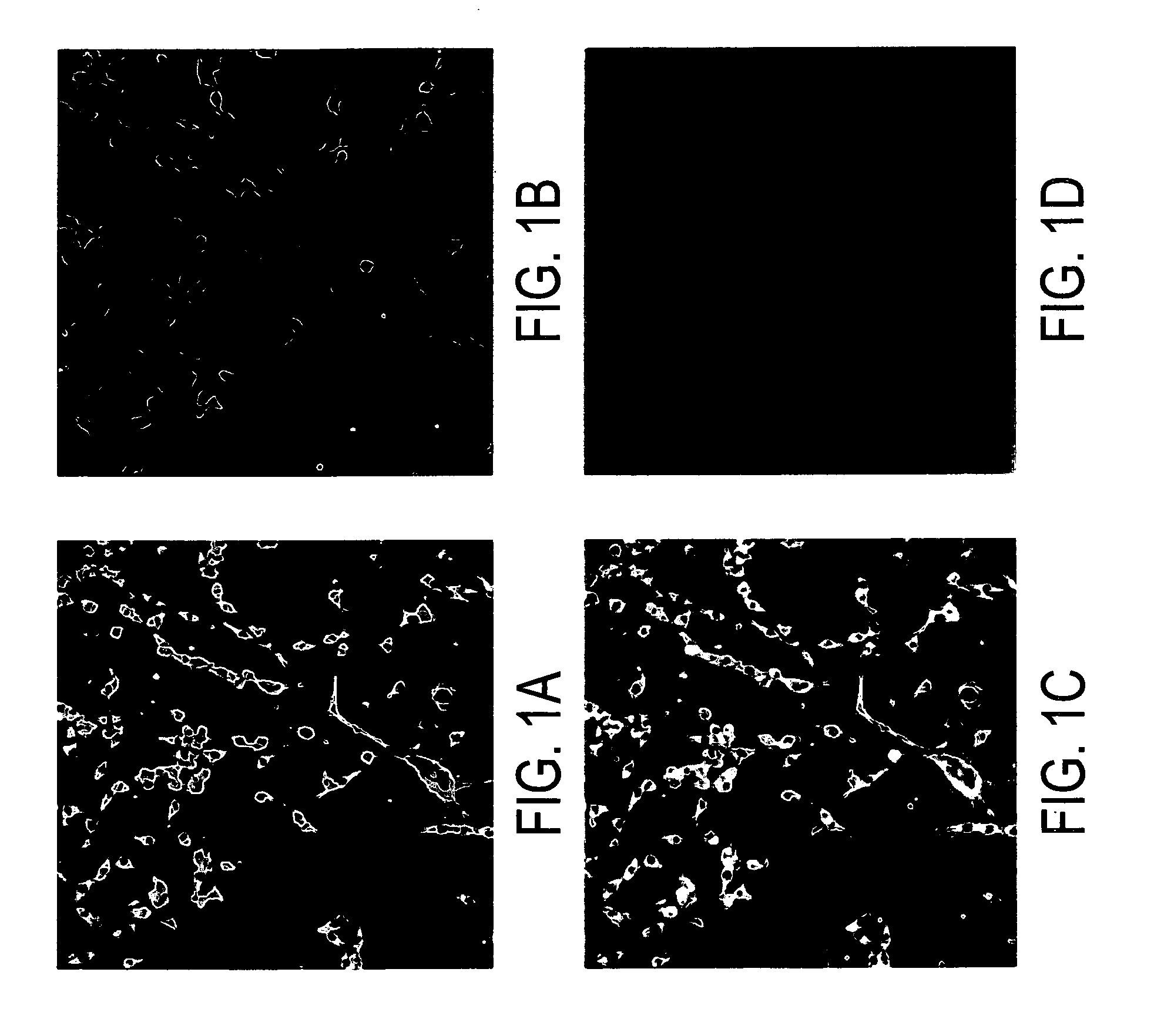
[0195] The mAb specific for the anti-hMet ECD (2F6) was characterized for IF with S-114 cells expressing hMet. Results are shown in FIG. 1. S-114 cells fixed in acetone / methanol were stained with both mAb 2F6 (=Met3) (in green, panel A) and the rabbit polyclonal antibody against the Met C-terminal peptide antibody C-28 (in red, panel B). Colocalization of staining (yellow) is evident in panel C. A Nomarski image is provided (panel D) to show the unstained location and characteristics of the cells in culture.
example 3
Image Analysis and Quantitation
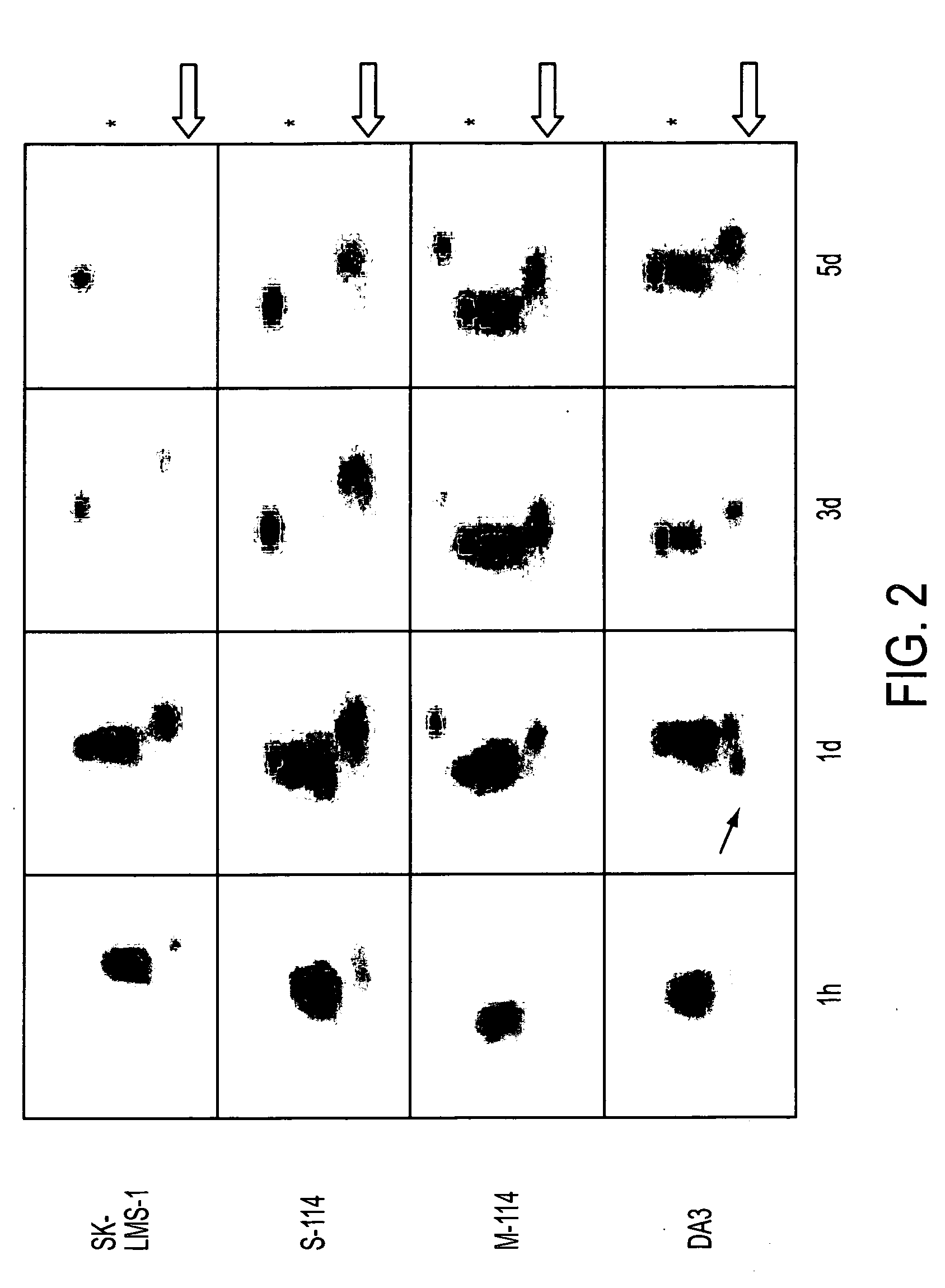
[0196] Serial total body gamma camera images of individual tumor-bearing mice were obtained between one hour and five days following i.v. injection of the 125I-mAb mixture reactive with hHGF and hMet. See FIG. 2. Activity was evident in the human tumors (SK-LMS-1 and S-114, both of which express hHGF and hMet) as early as one hour postinjection and prominently thereafter.
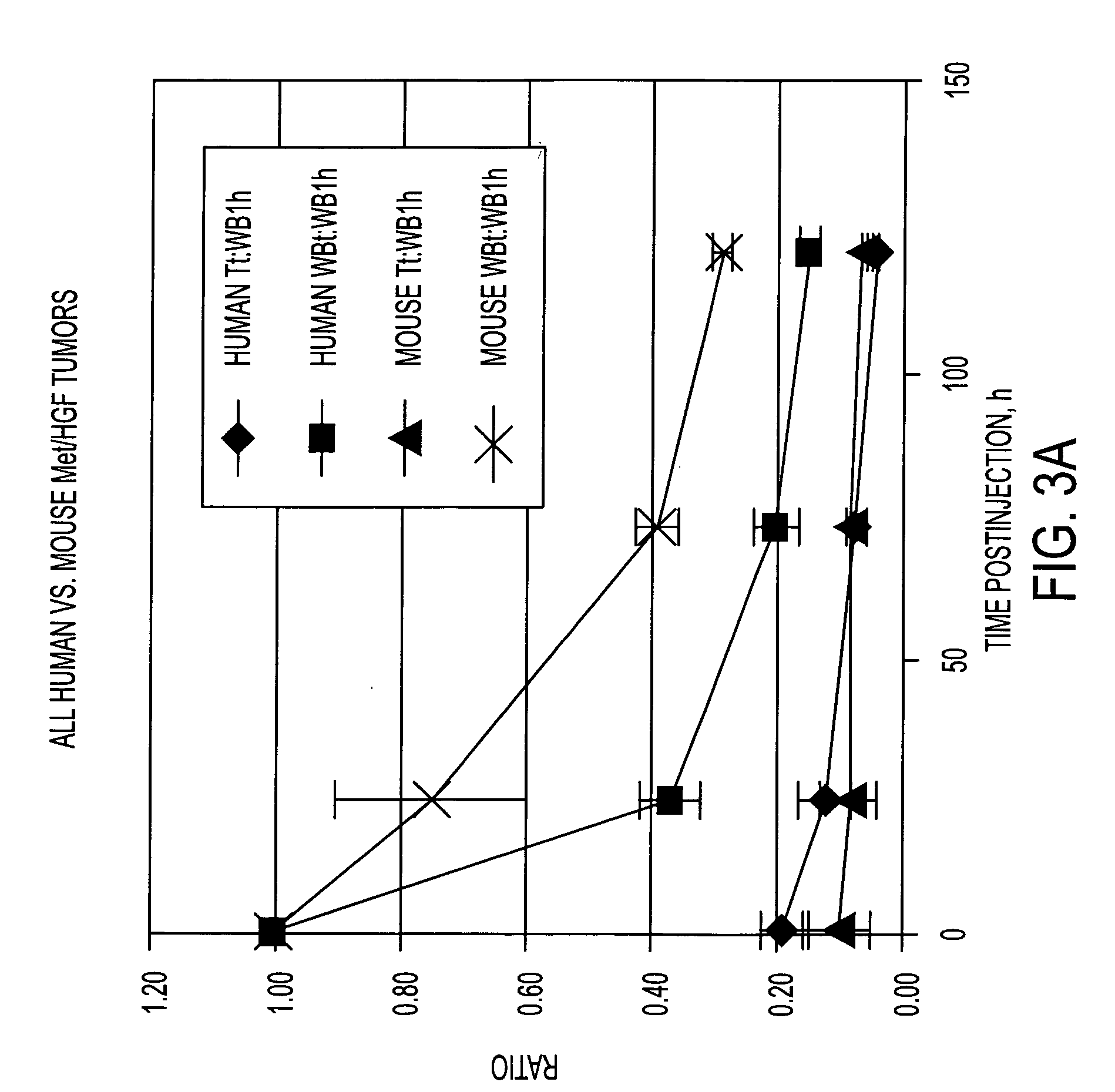
[0197] Activity was also clearly seen as early as one day postinjection in murine tumors (M-114, which expresses mHGF and mMet, and DA3, expressing mMet alone). Nevertheless, mice bearing human tumors cleared radioactivity more rapidly from the circulation than mice bearing murine tumors, as evidenced by their much lower levels of visceral radioactivity at three and five days postinjection and more conspicuous thyroid activity (reflecting uptake of free radioiodine released from labeled mAbs). Even though the absolute radioactivity levels in human and murine tumors generally appeared...
PUM
| Property | Measurement | Unit |
|---|---|---|
| composition | aaaaa | aaaaa |
| therapeutic composition | aaaaa | aaaaa |
| MRI | aaaaa | aaaaa |
Abstract
Description
Claims
Application Information