Engraftable neural progenitor and stem cells for brain tumor therapy

a neural progenitor and brain tumor technology, applied in the field of gene therapy, can solve the problems of large distribution of viral vector-mediated genes to large regions, and the number of cells typically in need is often disappointingly limited

Inactive Publication Date: 2005-08-04
CHILDRENS MEDICAL CENT CORP +2
View PDF12 Cites 34 Cited by
  • Summary
  • Abstract
  • Description
  • Claims
  • Application Information

AI Technical Summary

Problems solved by technology

Most gene therapy strategies to date are viral vector-based, yet extensive distributions of sufficient amounts of viral vector-mediated genes to large regions and numbers of cells typically in need has often been disappointingly limited.

Method used


Image

  • Engraftable neural progenitor and stem cells for brain tumor therapy
  • Engraftable neural progenitor and stem cells for brain tumor therapy
  • Engraftable neural progenitor and stem cells for brain tumor therapy

Examples


example 1

Migratory Capacity of NSCs in Culture

[0036] To determine properties of the NSCs in association with glioma cells, studies were initially preformed in culture comparing the relative migratory capacity of NSCs (clone C17.2) to fibroblasts (the lacZ-expressing TR-10 fibroblast cell line) when co-cultured with glioma cells. C17.2 and TR-10 cells were maintained in Dulbecco's modified Eagle's medium (DMEM; Mediatech, Washington, D.C.) supplemented with 10% fetal calf serum (FCS; Sigmiia, St. Louise, Mo.), 5% horse serum (HS;Gibco), 1% Glutamiiie (2 mM; Gibco), 1% penicillin / streptomycin (Sigma). CNS-1 cells were stably transduced with the PGK-GFP-IRES-NeoR retroviral vector construct to express green fluorescent protein (GFP) as previously described (ref. Aboody-Guterman et. al, 1997), and maintained in RPMI-1640 (Bio Whittaker) supplemented with 10% FCS and 1% penicillin / streptomycin (Sigma). Cell structure studies were performed in 100 mm petri dishes under standard conditions: humidi...

example 2

Transgene-Expressing NSCs Migrate Throughout and Beyond Invading Tumor Mass In Vivo

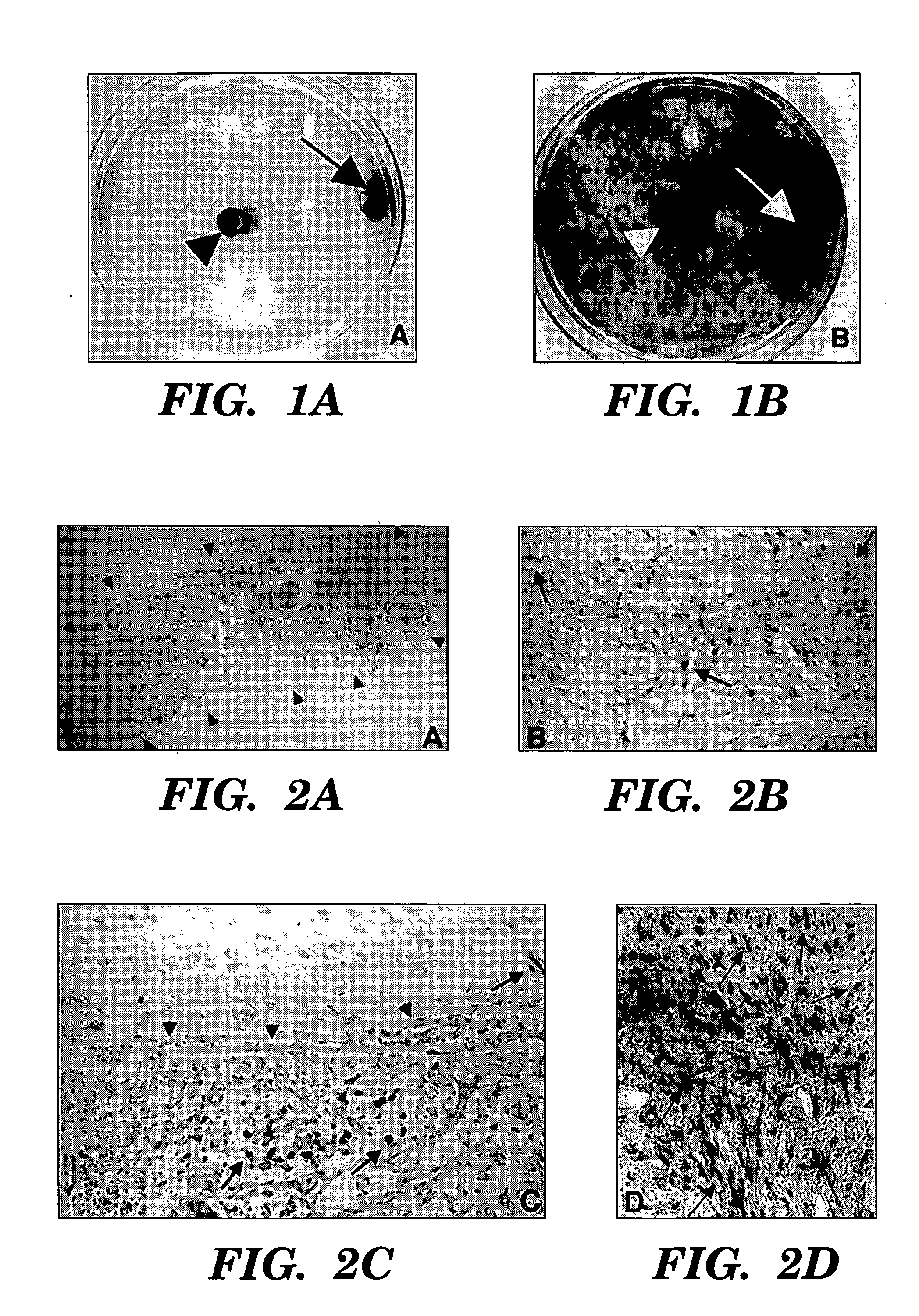
[0038] To determine the behavior of clone C17.2 NSCs introduced into brain tumors, experimental animals (syngeneic adult rats) first received an implant of 4×10⁴ D74 rat glioma cells in 1 μl injected into the right frontal lobe. Four days later, 1×10⁵ C17.2 NSCs in 1.5 μl PBS were injected at same coordinates directly into the D74 tumor bed. Animals were then sacrificed at days 2, 6, and 10 post-intratumoral injection and cryostat sections of the brains were processed with Xgal histochemistry for β-galactosidase (βgal) activity to detect donor-derived cells and counterstained with neutral red to detect tumor cells.

[0039] Donor C17.2 NSCs were found extensively dispersed throughout the tumor within a few days, spanning an 8 mm width of tumor as rapidly as 2 days after injection (FIGS. 2A, 2B). This is a much more extensive and rapid dispersion compared to previous reports of 3T3 fibroblasts grafted i...

example 3

NSCs “Track” Infiltrating Tumor Cells

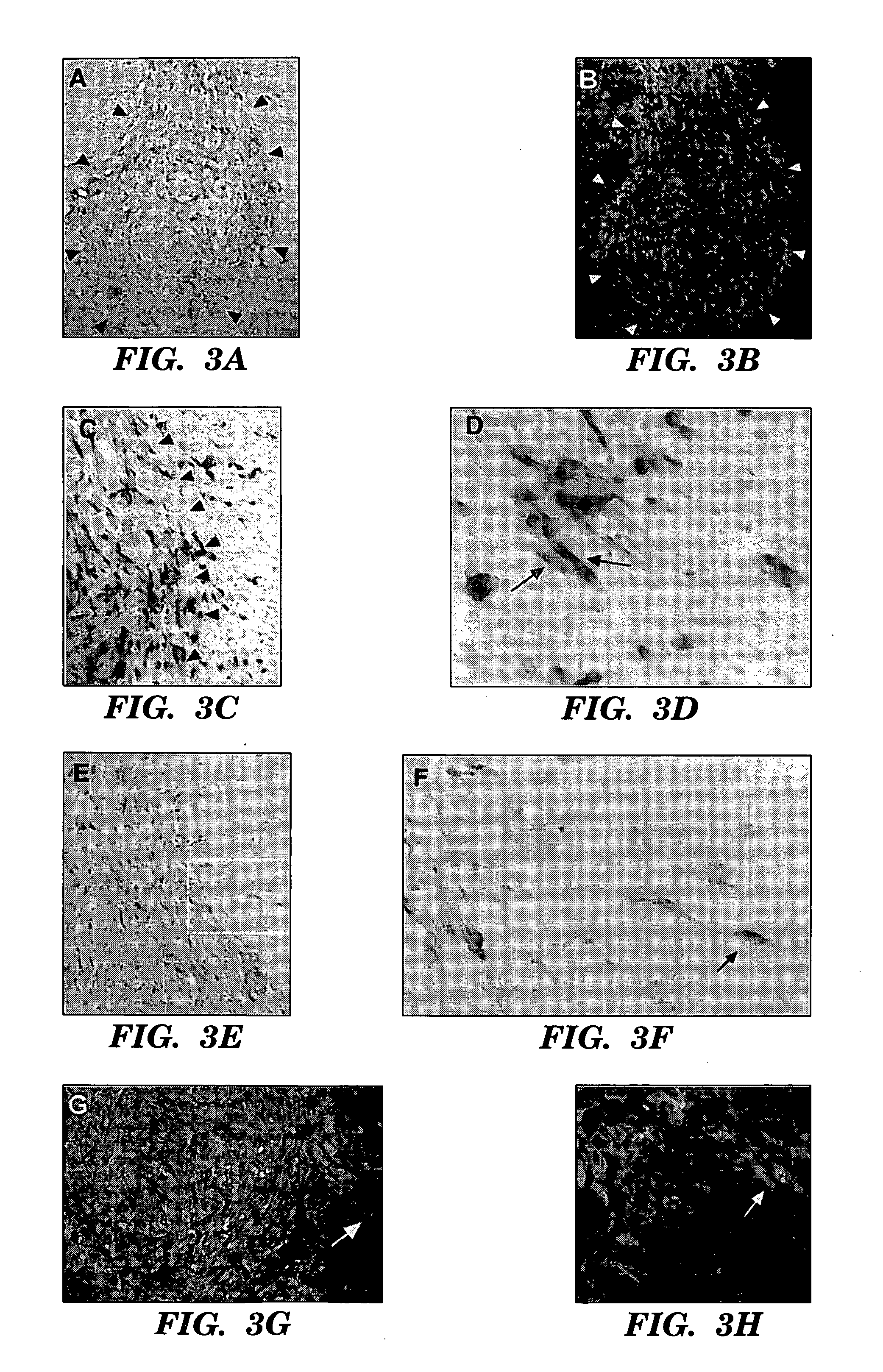
[0041] CNS-1 tumor cells were labelled by retroviral transduction with green fluorescent protein (GFP), prior to implantation, to better distinguish single cells away from the main tumor bed17. GFP-expressing CNS-1 glioma cells (3×10⁴) in 1 μl PBS injected into right frontal lobe at stereotaxic coordinates 2 mm lateral to bregma, on coronal suture, 3 mm depth from dura. 4×10⁴ C17.2 or TR-10 cells in 1 μl PBS injected at same coordinates directly into tumor bed on day 6. 3-4 C17.2 animals (2 BUdR labelled, 1 BUdR pulsed) and 1-2 TR-10 control animals (1 BUdR labelled). Animals were sacrificed on days 9,12, 16 and 21 post-tumor implantation. Cryostat sectioned, fixed brain tissue was stained either with β-galactosidase (C17.2 cells blue) and neutral red (tumor cells dark red) or double immunofluorescence with Texas red anti-β-galactosidase (C17.2 cells red) and FITC anti-GFP (tumor cells green).

[0042] (FIGS. 3A, 3B) parallel sections: low power o...


PUM

PropertyMeasurementUnit
concentrationsaaaaaaaaaa

Abstract

One of the impediments to the treatment of some human brain tumors (e.g. gliomas) has been the degree to which they expand, migrate widely, and infiltrate normal tissue. We demonstrate that a clone of multipotent neural progenitor stem cells, when implanted into an experimental glioma, will migrate along with and distribute themselves throughout the tumor in juxtaposition to widely expanding and aggressively advancing tumor cells, while continuing to express a foreign reporter gene. Furthermore, drawn somewhat by the degenerative environment created just beyond the infiltrating tumor edge, the neural progenitor cells migrate slightly beyond and surround the invading tumor border. When implanted at a distant sight from the tumor bed (e.g., into normal tissue, into the contralateral hemisphere, into the lateral ventricles) the donor neural progenitor/stem cells will migrate through normal tissue and specifically target the tumor cells. These results suggest the adjunctive use of neural progenitor/stem cells as a novel, effective delivery vehicle for helping to target therapeutic genes and vectors to invasive brain tumors that have been refractory to treatment.

Description

RELATED APPLICATIONS [0001] This application is a continuation-in-part of pending U.S. Ser. No. 09 / 133,873, filed on Aug. 14, 1998, which is incorporated herein by reference.GOVERNMENT SUPPORT [0002] This invention was made with support from the NIH under grant number P20-HD18655, and the United States government has certain rights in this invention.FIELD OF THE INVENTION [0003] This invention is in the field of gene therapy, more particularly the field of using neuronal cells to treat brain tumors. BACKGROUND [0004] An effective gene therapy for the treatment of brain tumors has been an elusive goal for many years. Glioblastoma multiforma, which is virtually untreatable, and the less malignant anaplastic astrocytoma account for about one-quarter of the 5,000 intracranial gliomas diagnosed yearly in the United States; 75 percent of gliomas in adults are of this category. Because of its profound and uniform morbidity, it contributes more to the cost of cancer on a per capita basis th...

Claims


Application Information

Patent Timeline
Patent Type & Authority Applications(United States)
IPC IPC(8): A61K35/12A61K38/12C12N5/0797C12N5/10
CPCA61K35/12A61K48/0008A61K48/0075C12N5/0623C12N7/00A61K38/00C12N2510/00C12N2710/16052C12N2710/16643C12N2740/10052C12N15/86A61P43/00Y02A50/30
Inventor SNYDER, EVAN Y.ABOODY, KAREN S.BREAKEFIELD, XANDRA O.LYNCH, WILLIAM P.
Owner CHILDRENS MEDICAL CENT CORP
Why Eureka
Try Eureka