Device for removing an elongated structure implanted in biological tissue

a biological tissue and elongated technology, applied in the field of methods and surgical devices, can solve the problems of difficult to remove the lead from the heart area without causing trauma to the area, severe damage to the vein, and difficulty in removing the elongated structure, etc., and achieve the effect of improving gripping

Inactive Publication Date: 2005-09-01
COOK VASCULAR
View PDF90 Cites 22 Cited by
  • Summary
  • Abstract
  • Description
  • Claims
  • Application Information

AI Technical Summary

Benefits of technology

[0017] In other aspects of the invention, the gripping member can comprise a double helix of opposite hands, a helical spring, a braided structure, a pattern of fenestrations, a woven structure, a polymeric tu

Problems solved by technology

However, problems can be encountered in attempting removal of an elongated structure implanted in biological tissue.
While cardiac electrical leads typically have a useful life of many years, over time pacemaker and defibrillator leads unfortunately become encapsulated by fibrotic tissue against the heart itself or the wall of the vein, or against other surrounding tissue.
The fibrotic tissue is tough and makes it difficult to remove the lead from the area of the heart without causing trauma to the area.
For example, when small diameter veins through which a pacemaker lead passes become occluded with fibrotic tissue, separating the lead from the vein can cause severe damage to the vein such as dissection or perforation of the vein.
Furthermore, separation of the lead from the vein is usually

Method used


Image

  • Device for removing an elongated structure implanted in biological tissue
  • Device for removing an elongated structure implanted in biological tissue
  • Device for removing an elongated structure implanted in biological tissue

Examples


Embodiment Construction

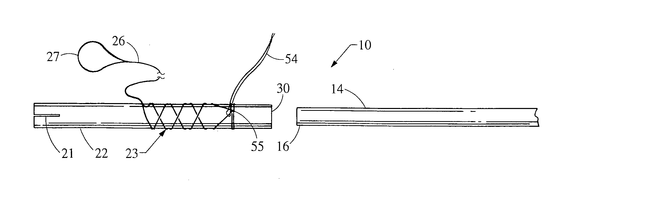
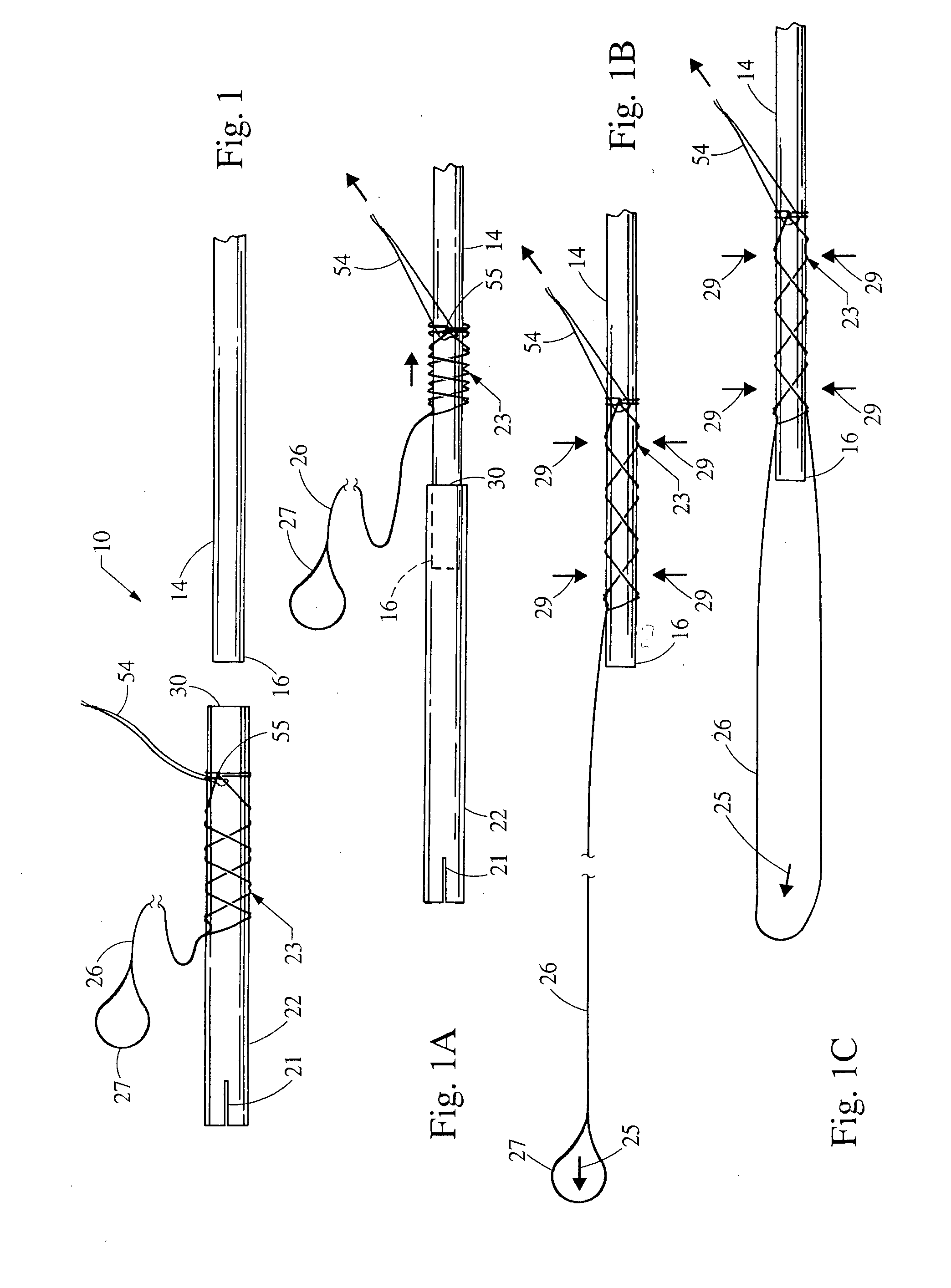
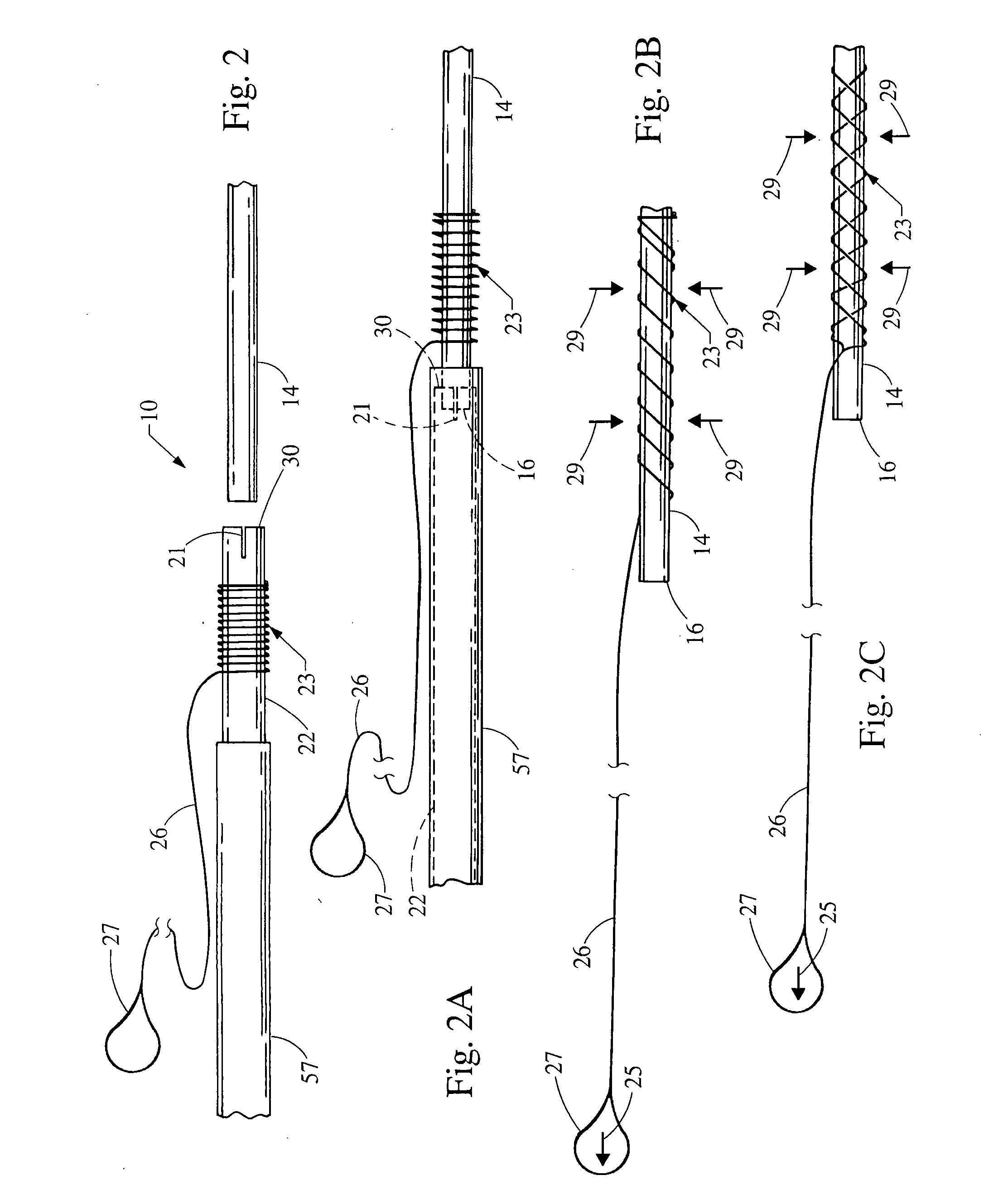
[0042] With reference first to FIGS. 1-16, a device 10 for removing or extracting a previously implanted elongated structure 14 from a patient is shown. The elongated structure 14 to be removed has an outside dimension or diameter, a proximal end 16 and a distal end located within the patient. The distal end of the elongated structure 14 may be located within or outside the vascular system of the patient. For example, when the elongated structure 14 is the cardiac pacemaker lead, the distal end will be located within the vascular system of the patient, and in particular, within a chamber of the patient's heart (such as in an atrium or ventricle of the heart). Alternatively, when the elongated structure 14 is a defibrillator lead, the distal end will be located either in or about the heart of the patient. The distal ends of other types of elongated structures 14 may not be and need not be near the heart at all; the device 10 will still be useful for removing them. The proximal end 16...


PUM


Abstract

A device for removing from a patient a previously implanted elongated structure, such as a pacemaker lead or a defibrillator lead, that may be encapsulated in biological tissue of the patient. In one form, the device comprises a sheath member having an inside dimension greater than the outside dimension of the elongated structure, and a gripping member positioned about the sheath member. The gripping member has a longitudinal passage extending substantially therethrough, which longitudinal passage is dimensioned to encircle at least a portion of the elongated structure. A proximal portion of the gripping member may extend outwardly beyond the proximal end of the elongated structure and outwardly of at least the vascular system of the patient. The gripping member may comprise a first configuration having a first diameter that is greater than the outside dimension of the elongated structure, and a second configuration having a second diameter that is substantially the same as the outside dimension of the elongated structure.

Description

RELATED APPLICATION [0001] The present patent document claims the benefit of the filing date under 35 U.S.C. §119(e) of Provisional U.S. Patent Application Ser. No. 60 / 548,754, filed Feb. 27, 2004, which is hereby incorporated by reference.BACKGROUND [0002] 1. Technical Field [0003] This invention relates generally to methods and surgical devices, and more particularly to a method and a device for separating encapsulating biological tissue from an implanted elongated structure (for example, an implanted cardiac electrical lead such as a pacemaker or defibrillator lead), and / or for removing such an elongated structure from a patient. [0004] 2. Background Information [0005] A variety of medical treatments and surgical methods entail implanting an elongated structure in the body of a human or veterinary patient. Examples of such elongated structures include catheters, sheaths and cardiac electrical leads (such as pacemaker leads and defibrillator leads), and a variety of other devices....

Claims


Application Information

Patent Timeline
IPC IPC(8): A61B17/22A61N1/05
CPCA61B17/22031A61N1/056A61B2017/2215A61B17/221
Inventor LUI, CHUN KEEGOODE, LOUIS B.NORLANDER, BARRY E.
Owner COOK VASCULAR
Why Eureka
Try Eureka