

Extended, ultrasound real time 3D image probe for insertion into the body
a real-time 3d imaging and ultrasound technology, applied in the field of ultrasound probes and instruments for real-time 3d imaging, can solve the problems of difficult to manufacture ultrasound matrix arrays at frequencies above 7 mhz, high cost and space consumption of probes inserted into the body, etc., and achieve the effect of reducing the number of wires
- Summary
- Abstract
- Description
- Claims
- Application Information
AI Technical Summary
Benefits of technology
Problems solved by technology
Method used
Image

Examples
Embodiment Construction
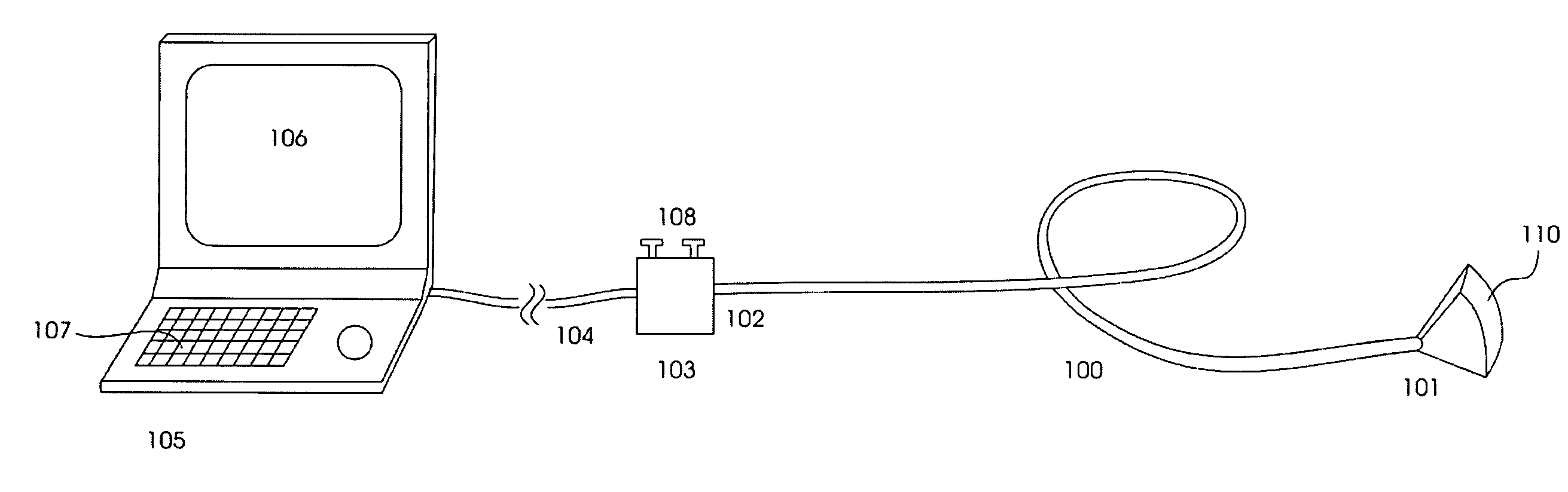
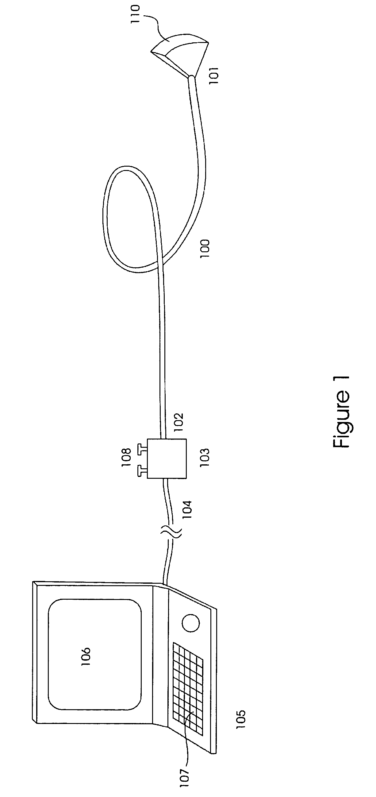
[0024] The invention relates to an ultrasound real time 3D imaging system, which in a typical embodiment is composed of the components shown in FIG. 1, where 100 shows an elongated imaging probe with a distal imaging tip 101 and a proximal end 102 that is connected to an utility console interface 103. The imaging ultrasound beam is transmitted from the distal tip of the probe enabled to be scanned within a three-dimensional (3D) region 110 to be imaged. The utility interface further connects via the cable 104 the probe signals to an ultrasound imaging instrument 105. The imaging instrument has an image display screen 106 for visualization of the images and also other information, and a key board interface 107 for user control of the instrument.
[0025] In this particular embodiment, the imaging probe 100 is a particularly flexible catheter probe for example allowing double curving of the probe, which has advantages for imaging inside tortuous vessels and the heart cavities. In other ...
PUM
 Abstract
Description
Claims
Application Information