Method for multiple site, right ventricular pacing with improved left ventricular function

a right ventricular and pacing technology, applied in electrotherapy, therapy, heart stimulators, etc., can solve the problems of congestive heart failure, cardiac contraction, not observable on an ecg, etc., and achieve the effect of improving the contraction of the rv and less tim

Inactive Publication Date: 2005-09-15
QUETZAL BIOMEDICAL
View PDF2 Cites 52 Cited by
  • Summary
  • Abstract
  • Description
  • Claims
  • Application Information

AI Technical Summary

Benefits of technology

[0032] The goal is to more closely mimic the Purkinje septal activation sequence in the left ventricle that would occur in a normal heart. Applications of this therapy include the treatment of Bradycardia (or any other disease requiring artificial cardiac pacing) and congestive heart failure. This therapy is also of value in heart transplants where artificial pacing is necessary and for the treatment of irregular cardiac contraction following thoracic surgery. The goal is to generate improved cardiac performance and also minimize myocardial wall strain in order to prevent, slow, or reverse the progression of heart failure.
[0037] An interesting consequence of PRT is to produce a shortened QRS complex. This is due to improving activation synchronicity over the entire heart, not just the left ventricle. However, it should be emphasized that narrowing the QRS complex is not the goal of PRT, rather it is a mere side effect, and that mere narrowing of the QRS has not been shown to correlate clinically with improved cardiac function.
[0042] More than five electrodes in a line oriented along the long axis of the RV septum, or a patch of electrodes deploying at least five electrodes along the RV septal long axis and some number of electrodes more anterior and posterior, will generate a fused action potential on the LV septum in less time. A preferred embodiment for a line of electrodes on the RV septum along the long axis is 9 or 10 electrodes spaced 6 or 7 mm apart. This allows for the possibility that some of the electrodes may not be in good contact with the myocardium. PRT will be delivered so long as at least four or five of these electrodes, spread approximately evenly apart, are in contact with the septum.
[0043] In addition to stimulating at a minimum of five locations along the RV septal long axis, additional electrodes can be located on the right ventricular free wall to improve contraction of the RV. This is not necessary for optimizing LV activation, but may be of value in optimizing RV activation and is a way of treating RV heart failure.

Problems solved by technology

This depolarization wave front passes across all the cells of both atria and results in atrial contraction.
The atrium also repolarizes but this event (the U wave) is masked by activity in the ventricle and consequently it is not observable on an ECG.
Congestive heart failure is a condition that causes many deaths annually.
These symptoms are associated with the inability of the heart to pump sufficient blood.
Cardiac output insufficiency may be caused by the failure of the heart to contract in an efficient way.
If the physiologic conduction system has broken down or due to scar tissue associated with myocardial infarctions, the chambers of the heart may not contract in a coordinated or effective manner.
In addition, dilated cardiomyopathy associated with heart failure often leads to a dysynchrony between the contraction of the left and right ventricles, to mitral regurgitation, and to paradoxical septal wall motion.
This may be due to neuro-hormonal imbalances, induced abnormal cardiac electrical conduction patterns, or other mechanisms that change the contraction pattern of the heart resulting in reduced cardiac output and increased myocardial strain.
In addition, artificial cardiac pacing using endocardial leads may diminish cardiac output, may compromise myocardial integrity, and may even, in some cases, precipitate heart failure.
The physician is thus limited to a single site for applying treatment.
Studies have indicated that the abnormal contraction that results from apical pacing has long-term deleterious effects.
Studies using conventional pacing leads implanted in alternative locations in the right ventricle (for example, the right-ventricular outflow tract or pacing both the right-ventricular apex and right-ventricular outflow tract) have shown clinical improvements, but the long-term reliability of conventional pacing leads in these alternative locations is questionable and lead placement is difficult.
In addition, outcomes in alternate site pacing have been observed to be very sensitive to the location of the pacing site.
This makes it difficult or impossible to achieve clinically reliable outcomes.
All of these modalities lead to activation sequence irregularities and consequence diminished cardiac function.
Left ventricular apical endocardial pacing has been shown to produce the best LV intraventricular synchrony of all these pacing modalities, but it is not practical in a clinical setting due to the dangers of leaving a permanent pacing lead in the left ventricle.
However, these therapies are limited to the relatively few pacing patients with fully functioning conduction systems.
In addition, His-bundle pacing is difficult to produce reliably in a clinical setting.
However, there is still substantial deviation from a normal activation pattern.
However, it is now well documented that shortening the QRS complex to achieve improved inter-ventricular synchronization is not generally associated with improved LV intra-cardiac synchronization.
Although U.S. Pat. No. 5,174,289 discloses the use of multiple electrodes on a single right-ventricular lead to deliver simultaneous pacing pulses to the right-ventricular septum for generating a narrow QRS complex, it does not recognize the importance of producing a uniform wave front at the left ventricular septal wall or inducing an activation pattern that is as similar as possible to a physiologically normal pattern in space and time throughout the LV.

Method used


Image

  • Method for multiple site, right ventricular pacing with improved left ventricular function
  • Method for multiple site, right ventricular pacing with improved left ventricular function
  • Method for multiple site, right ventricular pacing with improved left ventricular function

Examples


Embodiment Construction

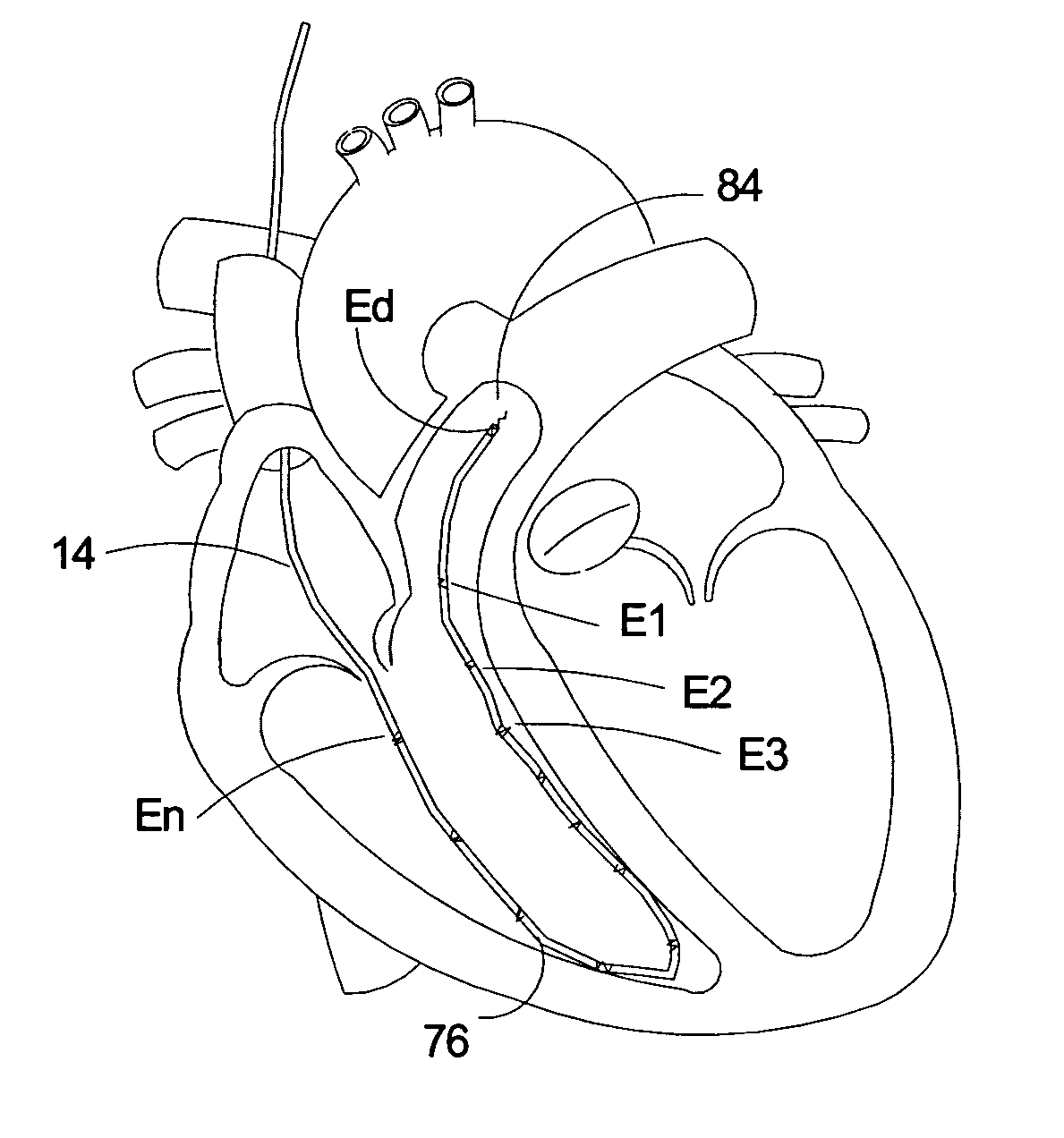
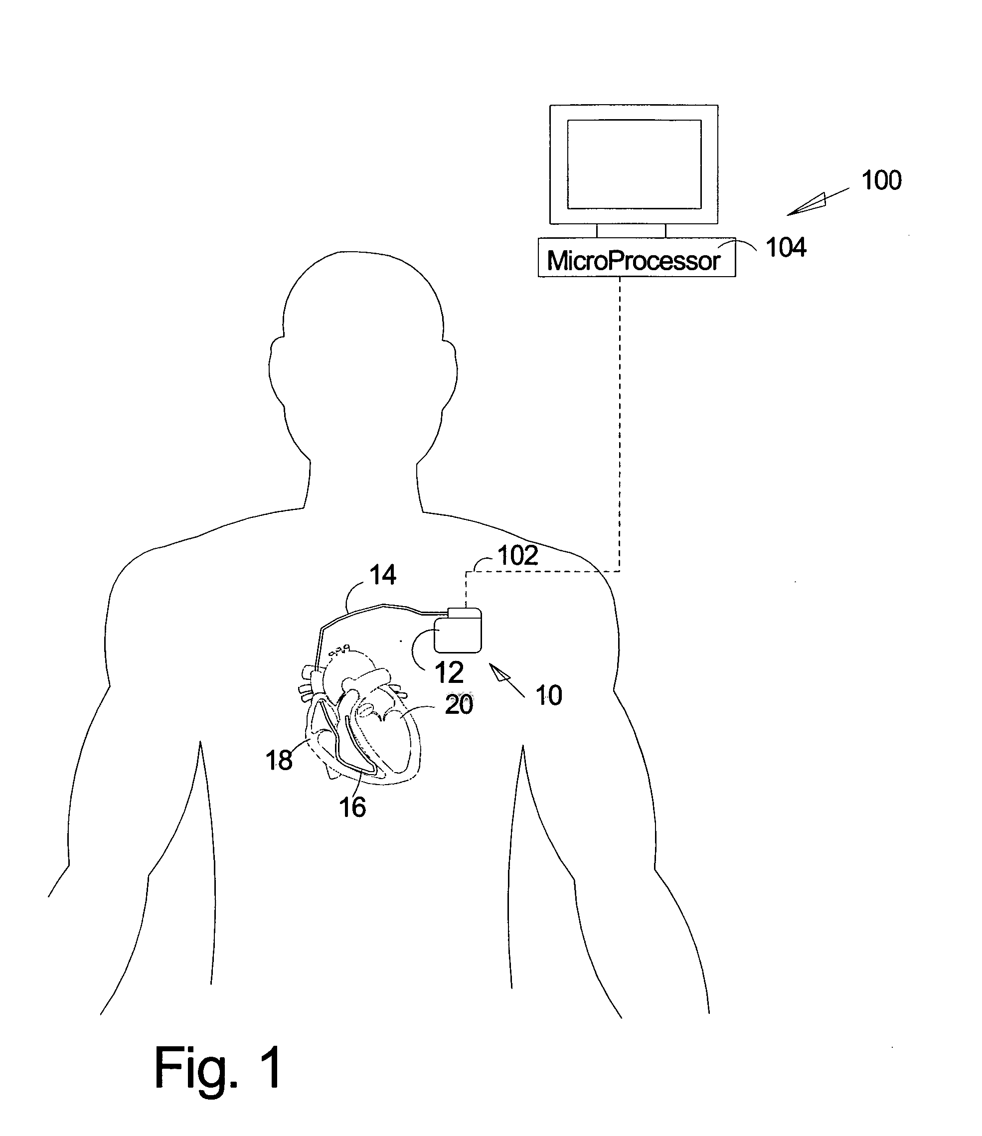
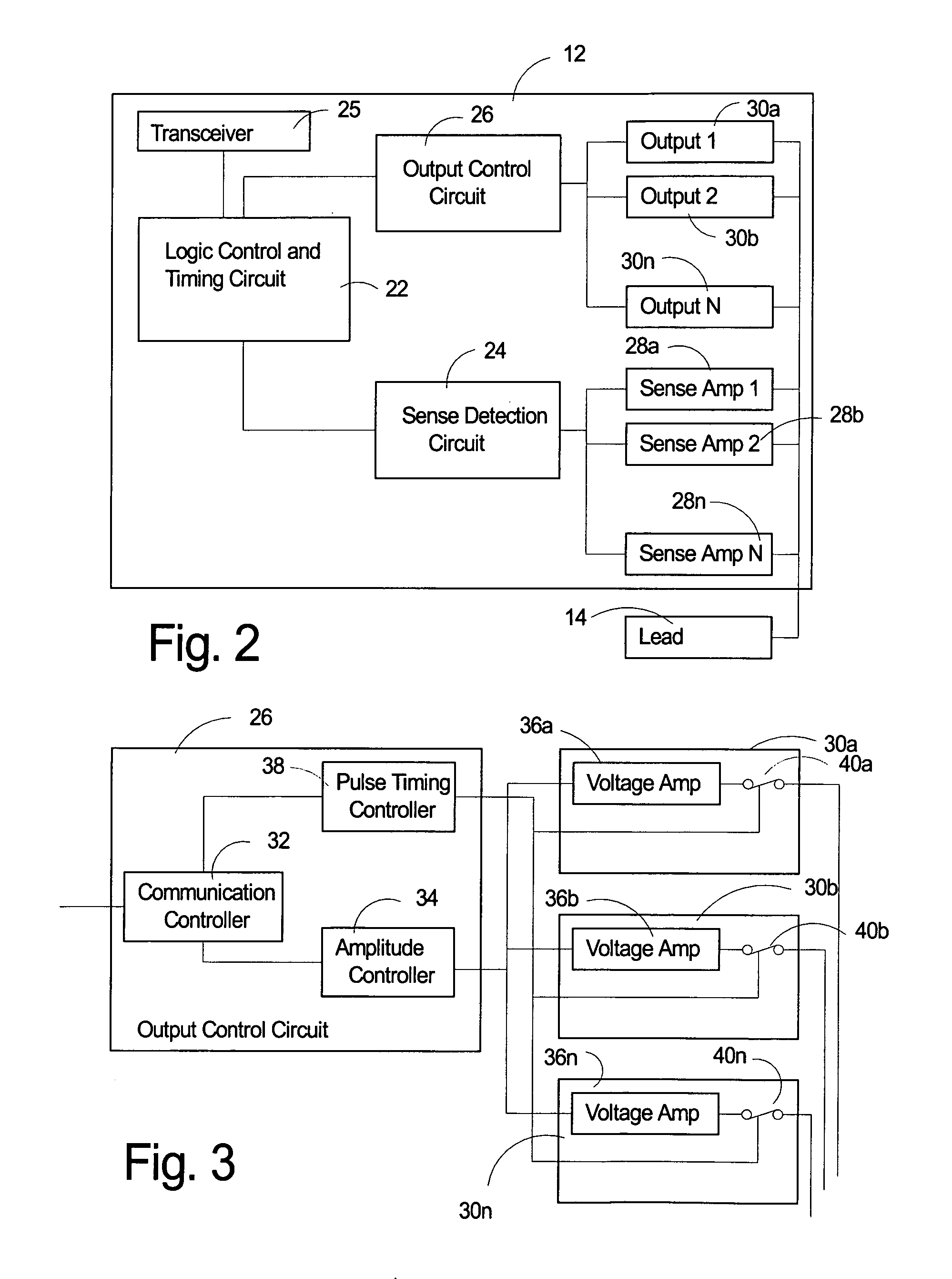
[0057] The subject invention pertains to an implantable cardiac stimulation system 10 including a cardiac stimulator 12 with various electronic circuits, and a multi-electrode lead 14 attached to the stimulator 12, as shown. The lead 14 has a distal end 16 disposed in the right ventricle 18 of heart 20. The system 10 is adapted to deliver therapy in the form of electrical pulses. The cardiac stimulator 12 contains electronic components common to current cardiac stimulators such as a battery, microprocessor control circuit, ROM, RAM, an oscillator, reed switch and antenna for communication, output circuits, and sense circuits. These components are well known to those of skill in the art. It is believed that a standard pacemaker, capable of unipolar or bipolar pacing and equipped with what is known as an “IS-1” connector could be used to deliver the therapy described herein, provided a sufficient number of electrodes are deployed. These electrodes may be of small size, to reduce power...


PUM


Abstract

A method for treatment of congestive heart failure from the right side of the heart, by stimulating at numerous points along the right-ventricular septum to produce a fused line of stimulation upon breakthrough of wave fronts into the left ventricular septum and an LV action potential that simultaneously propagates toward the apex, base, and left free wall. Five electrodes, all in contact with the septum and spaced approximately 1.5 cm apart, produce a fused action potential in an average adult human within 10 ms of delivering simultaneous pacing pulses. Breakthrough of this fused region of stimulation will occur within 20 ms of delivering the pacing pulses. The most proximal electrode may be located in or near the right-ventricular apex. The most distal electrode may be located somewhere near the right-ventricular outflow tract, generally somewhere near the moderator band.

Description

[0001] This application is a continuation-in-part of U.S. application Ser. No. 10 / 605,476, filed Oct. 1, 2003, which is a divisional application of U.S. application Ser. No. 10 / 075,808, filed Feb. 13, 2002, now U.S. Pat. No. 6,643,546 issued Nov. 4, 2003, which claimed the benefit of U.S. provisional application 60 / 298,449 filed Feb. 13, 2001.BACKGROUND OF THE INVENTION [0002] This invention pertains to a method and apparatus for applying cardiac stimulation using multiple electrodes, and more particularly, to a method and apparatus for improving left ventricular function by multiple site pacing from the right ventricle. [0003] The heart is a mechanical pump that is stimulated by electrical impulses. The mechanical action of the heart results in the flow of blood. During a normal heartbeat, the right atrium (RA) fills with blood from the returning veins. The RA then contracts and this blood is moved into the right ventricle (RV). When the RV contracts it pumps that blood to the lung...

Claims


Application Information

Patent Timeline
Patent Type & Authority Applications(United States)
IPC IPC(8): A61N1/362A61N1/368
CPCA61N1/3627A61N1/3686A61N1/3684A61N1/368A61N1/36842A61N1/36843
Inventor PRENTICE, JOHN K.FUKA, MARY Z.
Owner QUETZAL BIOMEDICAL
Why Eureka
Try Eureka