Chimeric adenoviruses for use in cancer treatment
a technology of chimeric adenoviruses and cancer cells, applied in the field of molecular biology, can solve the problems of often failing treatment, and achieve the effect of inhibiting the growth of colon cancer cells
- Summary
- Abstract
- Description
- Claims
- Application Information
AI Technical Summary
Benefits of technology
Problems solved by technology
Method used
Image
Examples
example 1
Viral Purification and Quantitation
[0108] Viral stocks were propagated on 293 cells and purified on CsCl gradients (Hawkins et al., 2001). The method used to quantitate viral particles is based on that of Shabram et al. (1997) Human Gene Therapy 8:453-465, with the exception that the anion-exchanger TMAE Fractogel was used instead of Resource Q. In brief, a 1.25 ml column was packed with Fractogel EMD TMAE-650 (S) (catalog # 116887-7 EM Science, Gibbstown, N.J. 08027). HPLC separation was performed on an Agilent HP 1100 HPLC using the following conditions: Buffer A=50 mM HEPES, pH 7.5; Buffer B=1.0 M NaCl in Buffer A; flow rate of 1 ml per minute. After column equilibration for not less than 30 minutes in Buffer A, approximately 109-1011 viral particles of sample were loaded onto the column in 10-100 ul volume, followed by 4 column volumes of Buffer A. A linear gradient extending over 16 column volumes and ending in 100% Buffer B was applied.
[0109] The column effluent was monitore...
example 2
Bioselection
[0111] Viral serotypes representing subgroups Ads B-F were pooled and passaged on sub-confluent cultures of the target tumor cell lines at a high particle-per-cell ratio for two rounds to invite recombination to occur between serotypes. Supernatant (1.0, 0.1 0.01, 0.001 ml) from the second round of the high viral particle-per-cell infection, subconfluent cultures, was then used to infect a series of over-confluent T-75 tissue culture flasks of target tumor cell lines PC-3, HT-29, Panc-1 and MDA-231. To achieve over-confluency, each cell line was seeded at split ratios that allowed that cell line to reach confluency between 24 and 40 hours post seeding, and the cells were allowed to grow a total of 72 hours post seeding prior to infection. This was done to maximize the confluency of the cells to mimic growth conditions in human solid tumors.
[0112] Cell culture supernatant was harvested from the first flask in the 10-fold dilution series that did not show any sign of CPE...
example 3
Serotype Characterization
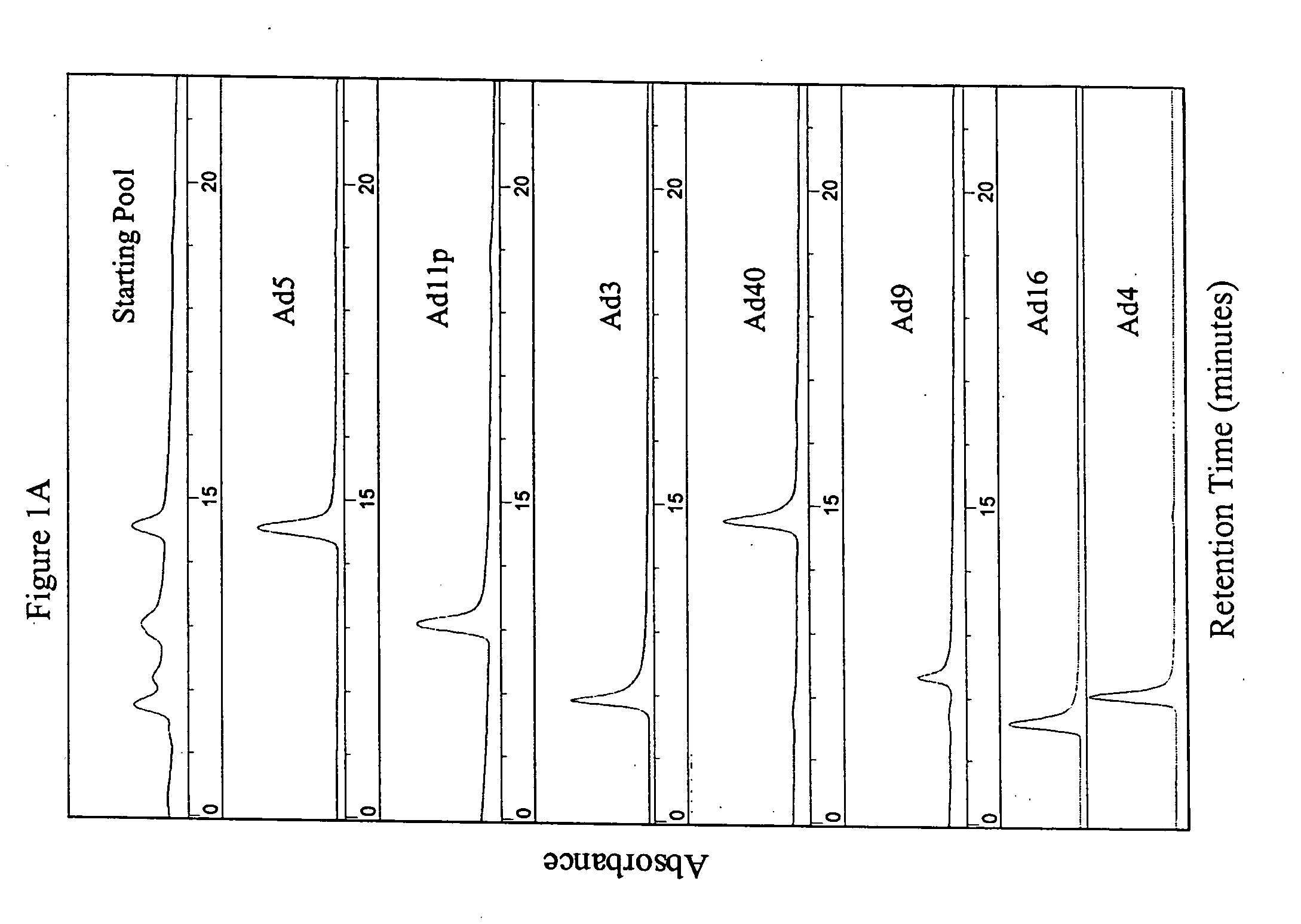
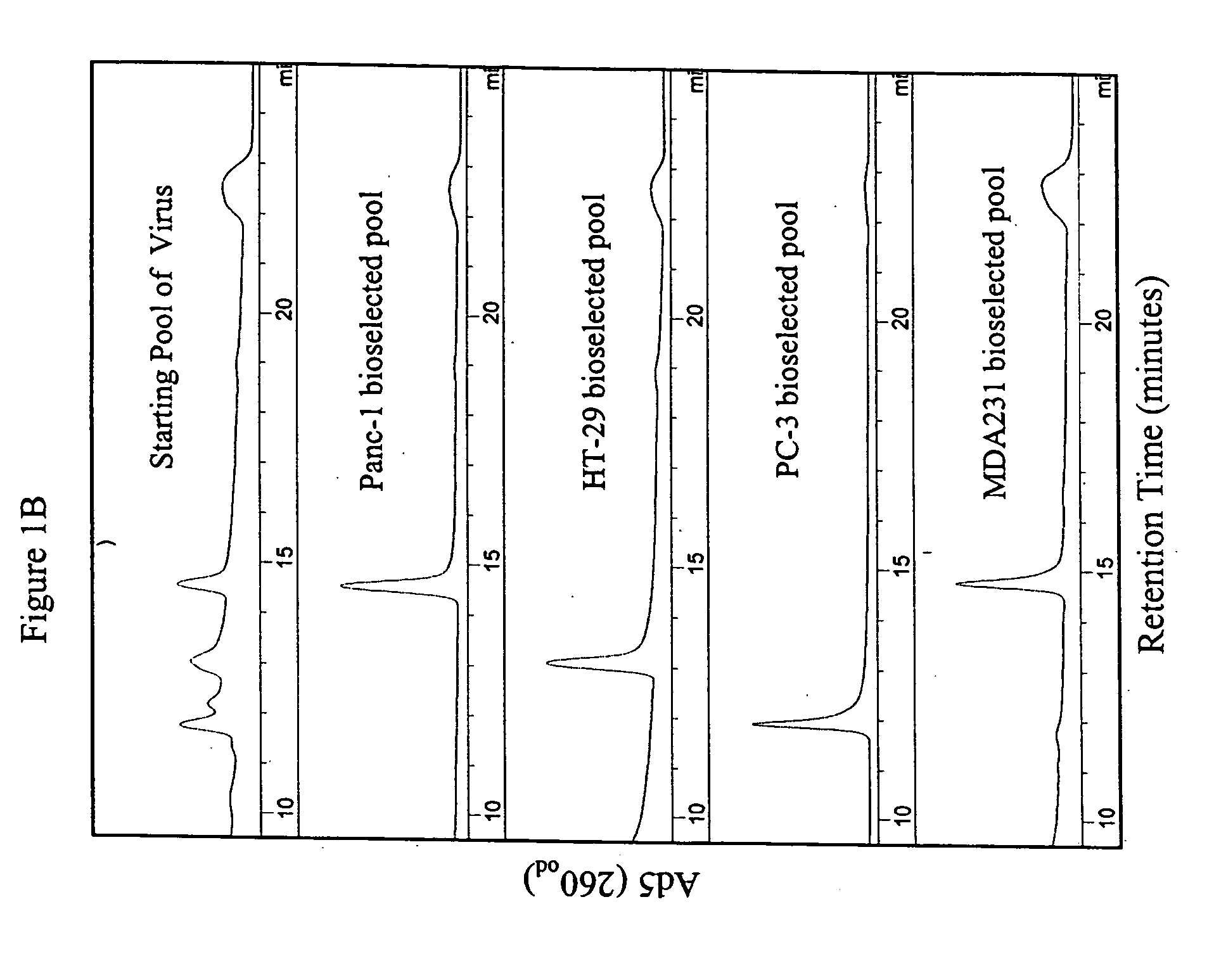
[0114] The parental adenoviral serotypes comprising the viral pools or the isolated ColoAd1 adenovirus were identified using anion-exchange chromatography similar to that described in Shabram et al. (1997) Human Gene Therapy 8:453:465, with the exception that the anion-exchanger TMAE Fractogel media (EM Industries, Gibbstown, N.J.) was used instead of Resource Q. as described in Example 1 (see FIG. 1).
[0115] Adenovirus type 5 eluted at approximately 60% Buffer B during the gradient. The other serotypes (3, 4, 9, 11 p, 16, 35, and 40) each eluted at a characteristic retention time consistent with the retention times on Q Sepharose XL published by Blanche et al. (2000) Gene Therapy 7:1055-1062.
PUM
| Property | Measurement | Unit |
|---|---|---|
| Therapeutic | aaaaa | aaaaa |
Abstract
Description
Claims
Application Information
 - R&D
- Intellectual Property
- Life Sciences
- Materials
- Tech Scout
- Unparalleled Data Quality
- Higher Quality Content
- 60% Fewer Hallucinations
Browse by: Latest US Patents, China's latest patents, Technical Efficacy Thesaurus, Application Domain, Technology Topic, Popular Technical Reports.
© 2025 PatSnap. All rights reserved.Legal|Privacy policy|Modern Slavery Act Transparency Statement|Sitemap|About US| Contact US: help@patsnap.com


