Method for detection of Mycobacterium tuberculosis antigens in biological fluids

a technology of biological fluids and antigens, applied in the field of method for detection of mycobacterium tuberculosis antigens in biological fluids, can solve the problems of poor sensitivity and specificity, and inability to detect tst with ppd, etc., and achieve the effect of reducing the cross-reaction of tst with ppd and immunization of persons with bcg

Inactive Publication Date: 2005-12-08
CHANG GUNG UNIVERSITY
View PDF2 Cites 26 Cited by
  • Summary
  • Abstract
  • Description
  • Claims
  • Application Information

AI Technical Summary

Benefits of technology

[0007] The main objective of the present invention is to provide an improved me

Problems solved by technology

However, this often takes 8 weeks, and the results sometime inaccurate.
However, its sensitivity and specificity

Method used


Image

  • Method for detection of Mycobacterium tuberculosis antigens in biological fluids
  • Method for detection of Mycobacterium tuberculosis antigens in biological fluids
  • Method for detection of Mycobacterium tuberculosis antigens in biological fluids

Examples


Embodiment Construction

Preparation of the M. tuberculosis Antigens

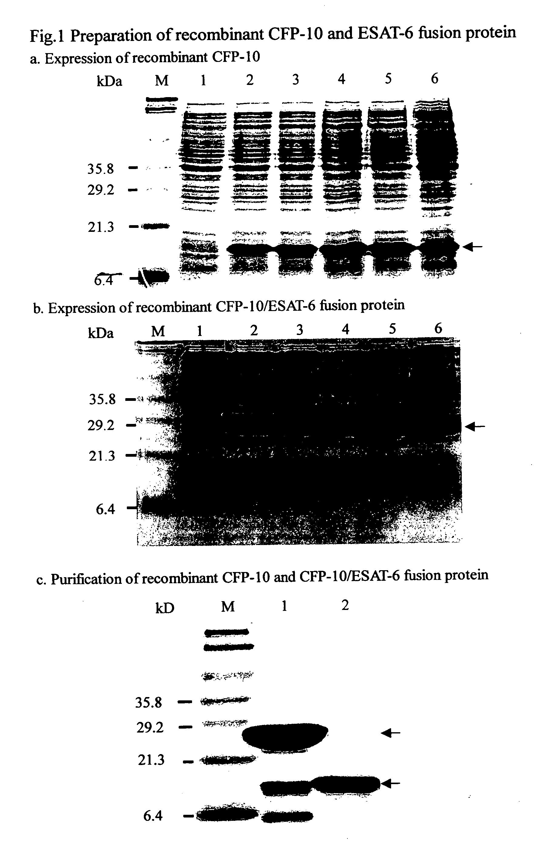
[0026] Antigens used for the immunoassays are prepared from recombinant proteins of ESAT-6, CFP-10 and CF-ES fusion proteins. Specific primers targeted to the lhp, esat-6 genes of M. tuberculosis are amplified by PCR technique. Amplified PCR products are than inserted into pGEM-T easy vector and transformed into E. coli (DH5a). Single colony of CFP-10 and ESAT-6 producing E. coli were selected from medium containing antibiotics. After sequencing to ensure the proper CFP-10 and ESAT-6 proteins were secreted, both CFP-10 and ESAT-6 proteins were extracted by restriction enzymes and fused together. Fused protein was reinserted into expressing vector of pET-29 system and transform into E. coli (BL21DE3).

Characterization of Antigens

[0027] Most of the studies identified antigens that are shared between M. tuberculosis, BCG and environmental mycobacteria. Although, potentially useful in vaccine design, these antigens may not be useful in spec...


PUM


Abstract

A method for detection of mycobacterium tuberculosis antigens in biological fluids provides immunoassay methods, diagnostic kits, and an immunochromatoraphic assay device for detection of Mycobacterium tuberculosis antigens in biological specimens, preferably body fluids and tissues. The preferred body fluids are blood, serum, plasma, urine, pulmonary fluid, sputum, cerebrospinal fluid, and the preferred tissue is the lung biopsy specimen. The immunoassays require two primary antibodies against RD1, RD2, or RD3 of Mycobacterium tuberculosis. At least one of the primary antibodies is attached to a solid carrier. Optional, a second antibody against an animal species producing one of the primary antibodies can be added. Either the other primary antibody or the secondary antibody is labeled with a detection agent, which can be an enzymatic marker, a fluorescent or luminescent agent, a radio active label or a color particle. The biological specimens may be used directly, concentrated or diluted for the immunoassays.

Description

BACKGROUND OF THE INVENTION [0001] 1. Field of the Invention [0002] The present invention relates to a method for detection of mycobacterium tuberculosis antigens, and more particularly to a method for detection of mycobacterium tuberculosis antigens in biological fluids. [0003] 2. Description of Related Art [0004] Tuberculosis (TB) is caused by repeated exposure to airborne droplets contaminated with a rod-shape bacterium, Mycobacterium tuberculosis. The TB bacterium is also known as the tubercle bacillus. A person with active pulmonary tuberculosis can spread the disease by coughing and sneezing. Once the person is infected with Mycobacterium tuberculosis, the infection will slowly progress to disease. More than 8 million new cases of Tuberculosis (TB) have been diagnosed each year and are responsible for more than three million deaths per year. Almost two and three quarter billion people (2.75 billion) or 33% of population are latently infected with TB. Give the large incidence o...

Claims


Application Information

Patent Timeline
IPC IPC(8): G01N33/53G01N33/554G01N33/569
CPCG01N33/554G01N33/5695
Inventor CHAN, ERR-CHENGYANG, MING-YING
Owner CHANG GUNG UNIVERSITY
Why Eureka
Try Eureka