Neonatal airway adaptor

a technology of airway adapters and neonatal tubes, which is applied in the field of airway adapters, can solve the problems of reducing the accuracy of capnographic measurement, affecting the insertion of other tubes and apparatuses, and difficulty in removing the et itself from the connector port,

Inactive Publication Date: 2005-12-22
ORIDION MEDICAL 1987
View PDF20 Cites 9 Cited by
  • Summary
  • Abstract
  • Description
  • Claims
  • Application Information

AI Technical Summary

Benefits of technology

[0011] The present invention seeks to provide a new airway adapter of novel construction which enables its use with neonatal patients without detracting significantly from the accuracy of capnographic measurements being performed on the patient, and without interfering unduly with the ventilation of the patient.
[0012] There is thus provided in accordance with a preferred embodiment of the present invention, a neonatal airway adapter which virtually eliminates void volumes (dead space) within the bore of the airways, such that mixing of the exhaled gases with void gases is reduced to minimal levels, and the waveform of the breath is maintained without unnecessary distortion. Furthermore, the virtual elimination of void volumes is clinically beneficial to the patient, since void volumes prevent the dispersion of the exhaled carbon dioxide, and cause a higher level of rebreathing. For this reason too, the neonatal airway adapter according to the present invention adds a minimal length to the airway circuit, in a situation where any excess length makes the dispersion of exhaled carbon dioxide more difficult.

Problems solved by technology

This is necessary since it may be difficult to remove the ET itself from its connector port, without disturbing the patient or his treatment.
Since, however, capnography measurements are generally not performed on a continuous basis, and the use of a sampling ET adapter would thus necessitate the sealing of the sampling port for most of the time, and the potential danger of bacterial growth in any debris lodged in the blind sampling tube, such sampling ET adapters are not generally used, a priori, for intubated neonatals.
Furthermore, the termination of the sampling ports in the wall of the ET adapter airway is likely to cause the ingestion of secretions and liquids into the gas analyzer through the sampling line, especially during pulmonary suction procedures, when liquids could be sucked straight from the lungs into the sampling port opening.
This, however, interferes with the insertion of other tubes and apparatus down the ET tube.
Internal mixing of the gases or alterations in the waveform reduce the accuracy of the capnographic measurement by slowing down the response time, and hence reduce the amount or accuracy of diagnostic information extractable from the capnograph.
As a consequence of this, the amount of void volume (also known as dead space) produced in such a connection is very variable, and for a tight fitting pair, could be very considerable.
Such liquids can block or partially block the sampling line, causing a reduction in the pressure of the gases passing through the airway adapter into the gas analyzer.
This pressure drop can cause alterations in the waveform, mixing of the gas, and alterations in the gas concentration , all of which reduce the accuracy of the gas analysis.
However, none of the airway adapters described therein are satisfactory for use with neonatals.
A major problem with neonatal patients is the very small flow involved.
This means that even a very small void volume can cause significant mixing of the neonatal's exhaled breaths, and inaccurate capnographic results.
Furthermore, the fast breathing rate of neonatals makes a fast response time even more critical than for other patients.
In addition, it is impractical to disconnect the ET tube of an intubated neonatal in order to change the ET adapter to fit the sampling device to be used.
Finally, closed circuit systems which include suction are widely used, and it is impractical to break such circuits in order to insert and remove capnographic adapters as needed.

Method used


Image

  • Neonatal airway adaptor
  • Neonatal airway adaptor
  • Neonatal airway adaptor

Examples


Embodiment Construction

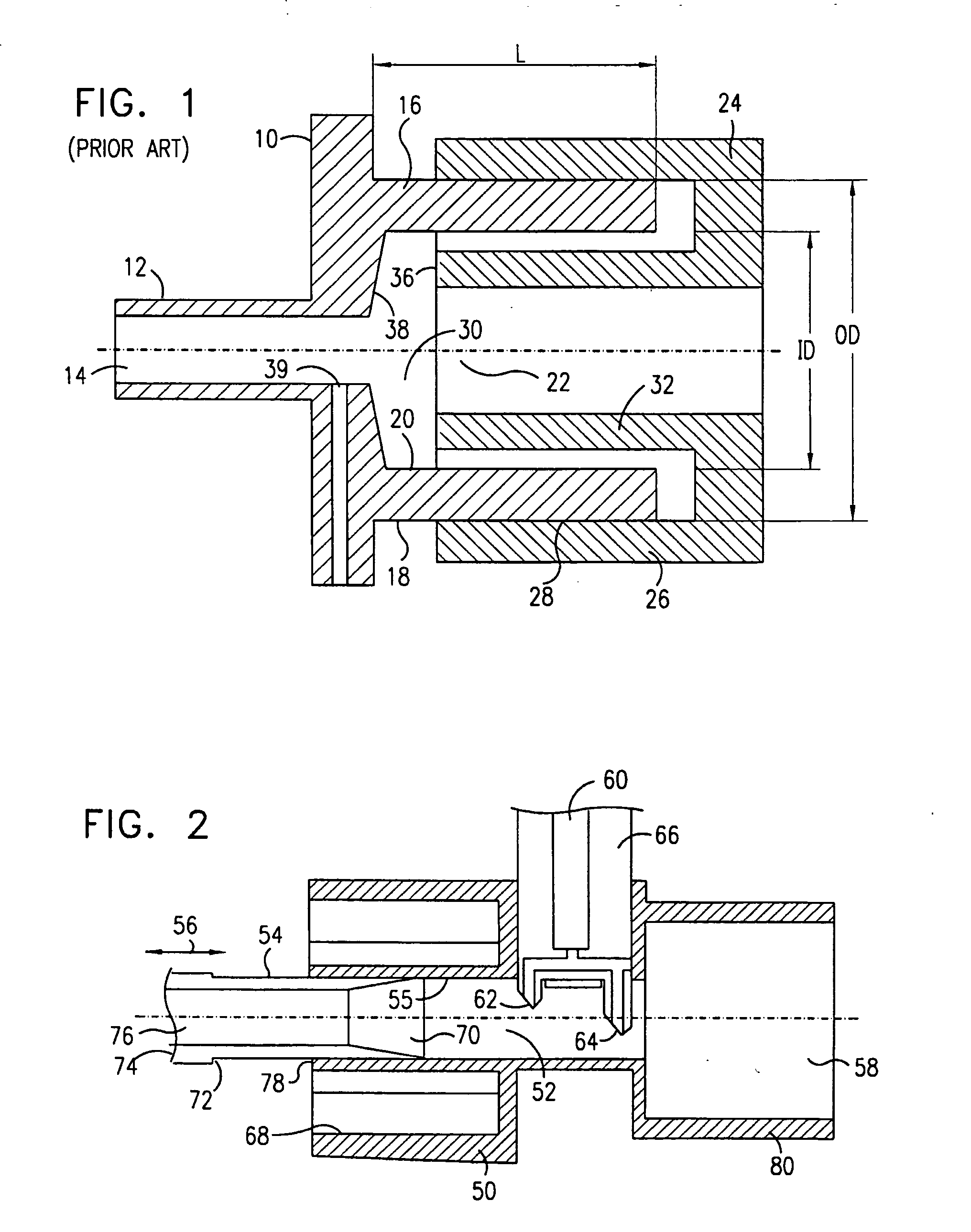
[0042] Reference is now made to FIG. 1, which is a cross-sectional view of a prior art airway connection made between an endotracheal tube (ET) adapter 10 and a ventilation tube connector 24, typically attached at its remote end to breathing apparatus. One end of the ET adapter 10 is formed into a narrow tubular section 12, having a wall and a central bore 14, over which the end of ET tube itself is fitted. Neonatal ET tubes are available in sizes ranging from 2.5 to 4 mm. The exhaled and breathing support gases are passed from and to the patient through this bore 14. The ET tube is attached over this tubular section which generally has an inner diameter (ID) of 3.5 mm, this being suitable for most neonatal ET tubes. This ET adapter can also be provided with a gas monitoring port, terminating at a hole 39 in the ET adapter wall.

[0043] At the other end of the ET adapter, there is a wide bore tubular opening having a wall 16 and a central bore 22 of similar size to that of the ventil...


PUM


Abstract

This invention is a neonatal airway adapter (10) with a sliding internal passage (54), which virtually eliminates void volumes (112) within the bore (76) of the airways, such that mixing of the exhales pages with void gases is reduced, and the waveform of the breath is maintained without undue distortion. Furthermore, the virtual elimination of void volumes reduces the level of re-breathing. The neonatal airway adapter connects to endotracheal tube adapters is such a way as to nullify the effect of the differing internal diameters (20) and internal lengths which are used in ET adapters currently available. The cross section of the internal bore of the airway through which the breath flows from the ET adapter to the connector of the ventilator is maintained almost constant, especially in the region of the gas sampling point (66), to ensure that reasonable conditions of laminar flow, and accurate sampling are maintained.

Description

FIELD OF THE INVENTION [0001] The present invention relates in general to the field of airway adapters for the connection of an endotracheal tube to ventilation apparatus, particularly for use with neonatal patients. BACKGROUND OF THE INVENTION [0002] Airway adapters are generally used with patients being given respiratory assistance, such as patients under anesthesia, or patients on life support systems, to connect between the endotracheal tube (ET tube) and the ventilating tube of the breathing apparatus. These tubes convey breathing gases to the patient and exhaled breath away from the patient. The airway adapter is in the form of a short connector of tubular shape, and is required to make a connection between the generally very different cross sections of these two tubes. The airway adapter connected directly to the endotracheal tube is known as the ET adapter. Airway adapters also have the function of providing a demountable joint in the ventilation path, for the insertion of o...

Claims


Application Information

Patent Timeline
Patent Type & Authority Applications(United States)
IPC IPC(8): A61M16/08A61M16/04
CPCA61M16/08A61M16/085Y10S128/911Y10S128/912A61M2230/43
Inventor COLMAN, LEWISLEVITSKY, GERSHON
Owner ORIDION MEDICAL 1987
Why Eureka
Try Eureka