

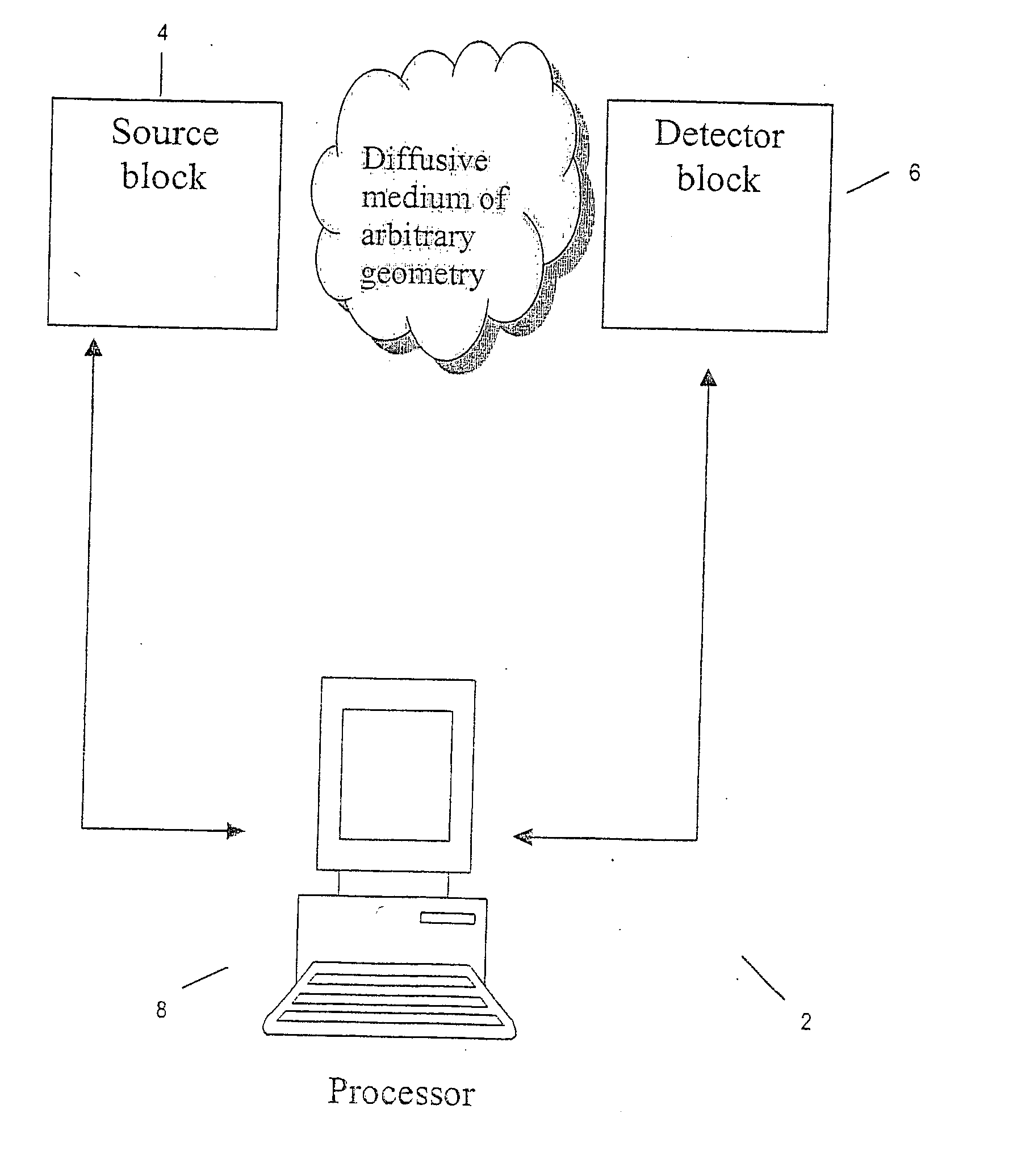
Imaging volumes with arbitrary geometries in contact and non-contact tomography
a technology of arbitrary geometries and images, applied in the field of clinical imaging modality, can solve the problems of inability to implement a limited number of detector channels, inapplicability, and significant disadvantages of fiber guides, and achieve the effect of retaining accuracy and capacity, achieving computational simplicity and efficiency of analytical methods
- Summary
- Abstract
- Description
- Claims
- Application Information
AI Technical Summary
Benefits of technology
Problems solved by technology
Method used
Image

Examples
example 1
KA is Several Orders of Magnitude Faster Than More Rigorous Numerical Methods Such as FD or ET
[0081] Referring to FIG. 4, the computing times achieved by using the KA as defined by equation (12) is compared to the extinction theorem (ET) and finite-differences solution (FD). While both ET and FD methods have a strong non-linear dependence as the area of the surface and the volume investigated increases, the KA method scales linearly with the size of the problem and more importantly it is faster than the ET and FD methods by almost three-orders of magnitude.
[0082] By way of a practical example, the number of discretization points for a sphere of radius 2 cm needed in order to maintain a one transport mean free path (ltr=D / 3) distance between points (N˜5000) was used to compare the speed of the KA and ET methods. In this case, it takes the KA methods 70 seconds and the ET method 50 minutes to solve the problem, indicating that the KA as approximately 40 times faster than the ET meth...
example 2
The Errors of the KA Due to Shadowing Effect
[0084] So-called shadowing effect appears when certain surface areas are blocked from the source by the geometry of the interface. Since the KA only considers first order reflections at the interface, errors appear in the proximities of the shadow regions of a source. Furthermore, since these shadow areas are not taken into account in the KA, it predicts higher values of the intensity for these areas.
[0085] Referring to FIGS. 5 (a)-(d), the shadow effect is demonstration of for the KA method and for the infinite case for a cylinder of R=2.5 cm with a sine profile in the boundary of amplitude 0.5 cm, and period π / 4. The error is plotted as a percentage of the absolute photon field strength at each point. Error committed for different source locations using the KA are shown in (a) and (c) and using the homogeneous Green's function in (b) and (d). The following source locations were considered: (ρ=2.3 cm, θ=0) for (a) and (b), and (ρ=1.5 cm...
example 3
The Errors of the KA When Imaging Small Volumes with Weak Absorption
[0087] A comparison of the intensity generated by a point source in a cylindrical geometry using the KA versus the exact solution, (ET) with different several radius and optical properties is shown in FIG. 6. Error in the KA were plotted against absorption coefficient for different radii and scattering coefficients for a case of a smooth cylinder (i.e., no shadowing effect).
[0088] The smaller the dimensions of the geometry and the smaller the absorption coefficient the larger the error committed by the KA method. As seen in this figure, the error increases as the volume of the medium or the absorption decreases. From FIG. 6(a), it can be concluded that in order to make practical use of the KA (assuming a minimum of 5% accuracy), either large volumes and therefore small curvatures need to be present (R>2.5 cm), or very large absorption coefficients are needed (μa>0.1 l / cm).
PUM
| Property | Measurement | Unit |
|---|---|---|
| diameters | aaaaa | aaaaa |
| distance | aaaaa | aaaaa |
| frequencies | aaaaa | aaaaa |
Abstract
Description
Claims
Application Information