Image analysis

a digital image and image technology, applied in the field of automatic analysis of digital images, can solve the problems of difficult to obtain the qualification to perform such examinations, requiring frequent review, and time-consuming, labor-intensive and expensive processes, and achieve the effects of reducing the complexity of some samples, reducing the difficulty of performing such examinations, and reducing the quality of images

Active Publication Date: 2006-01-05
QINETIQ LTD
View PDF5 Cites 34 Cited by
  • Summary
  • Abstract
  • Description
  • Claims
  • Application Information

AI Technical Summary

Benefits of technology

[0011] Property co-occurrence matrices (PCMs) are described e.g. in Electronics and Communication Engineering Journal, pp 713, Vol 5, No 2, 1993 (Co-occurrence Matrices for Image Analysis, J F Haddon and J F Boyce), and are an extension or generalisation to the standard grey level co-occurrence matrices described e.g. in IEEE Trans. Syst., Man, Cybern., Vol SMC-3, pp 610-621, 1973 (Texture Features for Image Classification, R M Haralick, K Shanmugan and I Dinstein). They are multidimensional histograms in which each element is the frequency with which selected properties co-occur. By generating a PCM using the properties of local mean and local standard deviation of intensity in neighbourhoods of image pixels, analysis of such a PCM can thus distinguish pixels contributing to regions of, say, relatively low local mean and relatively high local standard deviation (such as the dark, textured regions representing epithelial cells in the preferred implementation of this aspect of the invention) and pixels contributing to regions of, say, relatively high local mean and relatively low local standard deviation (such as the lighter, more uniform regions representing “background” tissue in the preferred implementation of this aspect of this invention), or to regions of other combinations of those properties in other applications of the invention.
[0012] These and other aspects of the invention will now be more particularly described, by way of example, with reference to the accompanying drawings and in the context of an automated system for grading cancer on the basis of tubule formations in digital images of histological slides of potential carcinomas of the breast.

Problems solved by technology

This is a time consuming, labour intensive and expensive process.
Qualification to perform such examination is not easy to obtain and requires frequent review.
This problem is exacerbated by the complexity of some samples, especially in marginal cases where there may not be a definitive conclusion.
If sufficient trained staff are not available this impacts upon pressures to complete the analysis, potentially leading to erroneous assessments and delays in diagnosis.
These problems mean that there are practical limitations on the extent and effectiveness of screening for breast cancer with the consequence that some women are not being correctly identified as having the disease and, on some occasions, this failure may result in premature death.
Conversely, others are being incorrectly diagnosed with breast cancer and are therefore undergoing potentially traumatic treatment unnecessarily.

Method used


Image

  • Image analysis
  • Image analysis
  • Image analysis

Examples


Embodiment Construction

General System Configuration

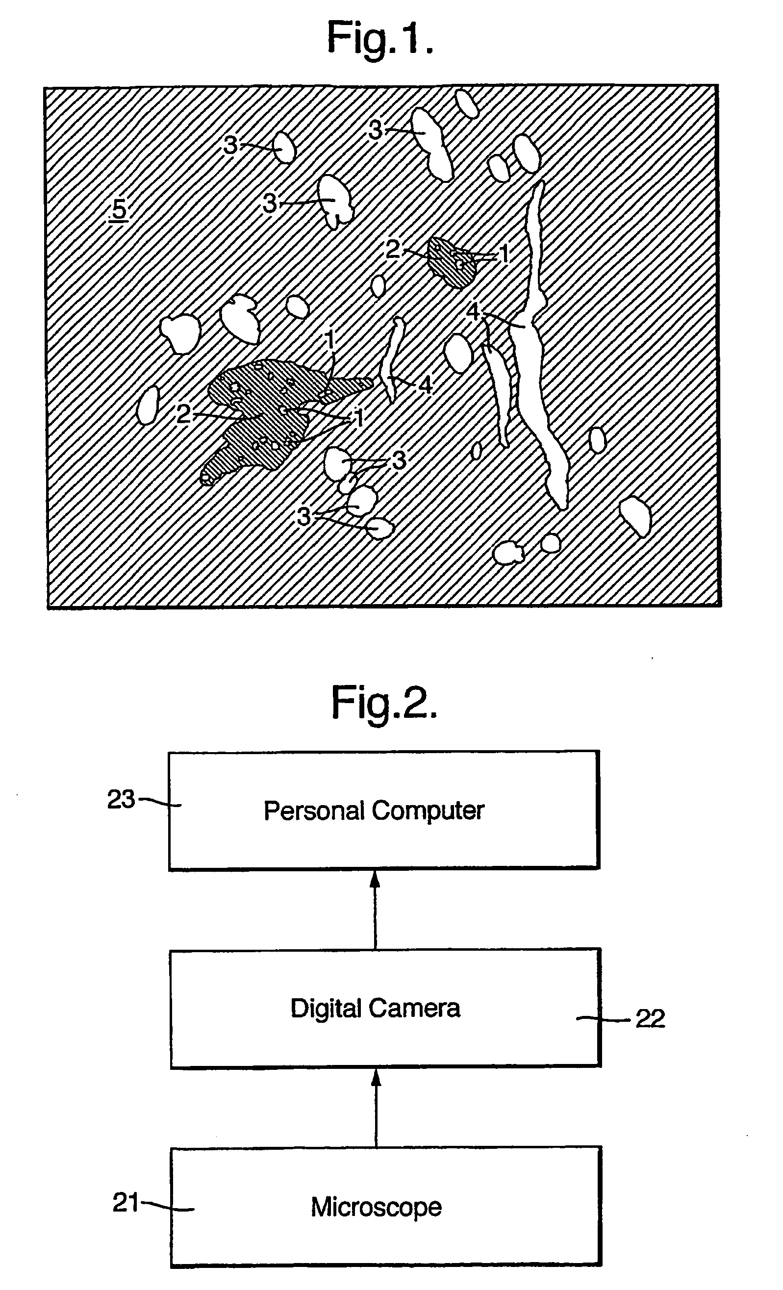
[0027]FIG. 2 shows a typical computer based system for image capture and processing for implementing the present invention. Sections are cut from breast tissue samples, placed on slides and stained in accordance with conventional techniques. A pathologist scans the slides in a microscope 21, selects regions which appear to be most promising in terms of the analysis to be performed, and they are photographed with a digital camera 22. Images from the camera 22 are downloaded to a personal computer (PC) 23 where they are stored and processed as described below. In a system utilised by the inventors, the microscope provided optical magnification of 10× and the digital images were 1476 pixels across by 1160 down. Other magnifications and digitised sizes can be used without compromising the algorithm more particularly described below provided that some system parameters such as cell size, the maximum bridged gap in dilation and shape criteria are adjusted acc...


PUM


Abstract

A method for the automated analysis of digital images, particularly for the purpose of assessing the presence and severity of cancer in breast tissue based on the relative proportions of tubule formations and epithelial cells identified in digital images of histological slides. The method includes the step of generating a property co-occurrence matrix (PCM) from some or all of the pixels in the image, using the properties of local mean and local standard deviation of intensity in neighbourhoods of the selected pixels, and segmenting the image by labelling the selected pixels as belonging to specified classes based upon analysis of the PCM. In this way relatively dark and substantially textured regions representing epithelial cells in the image can be distinguished from lighter and more uniform background regions Other steps include identifying groups of pixels representing duct cells in the image based on intensity, shape and size criteria, dilating those pixels into surrounding groups labelled as epithelial cells by a dimension to correspond to an overall tubule formation, and calculating a metric based on the ratio of the number of duct pixels after such dilation to the total number of duct and epithelial pixels. Other uses for the method could include the analysis of mineral samples containing certain types of crystal formations.

Description

FIELD OF THE INVENTION [0001] The present invention relates to the automated analysis of digital images. It is more particularly concerned with the automated identification of different cell groupings in digital images of histological or cytology specimens and most particularly for the purpose of assessing the presence and severity of cancer in breast tissue based on the relative proportions of tubule formations and epithelial cells identified in digital images of tissue sections, and it is in this context that the invention is principally described herein. The invention may, however, also find application in the analysis of various other kinds of structure presenting image components which are amenable to identification in a similar way, for example in the analysis of mineral samples containing certain types of crystal formations. BACKGROUND AND SUMMARY OF THE INVENTION [0002] Many thousands of women die needlessly each year from breast cancer, a cancer from which there is theoreti...

Claims


Application Information

Patent Timeline
Patent Type & Authority Applications(United States)
IPC IPC(8): G06F15/00G06T5/00G06T7/00G06T7/40
CPCG06T7/0012G06T7/0081G06T7/404G06T2207/10056G06T2207/30096G06T2207/20148G06T2207/30024G06T2207/30068G06T2207/10116G06T7/11G06T7/45G06T7/136G06V20/695G06V20/698
Inventor HADDON, JOHN FRANCISWATSON, SHARON KATRINA
Owner QINETIQ LTD
Why Eureka
Try Eureka