Image-type intubation-aiding device

a technology of intubation and aiding device, which is applied in the field of electromechanical surgical image examination instruments, can solve the problems of inability to the disadvantage of ccd size, and the difficulty of assembly and maintenance, so as to achieve quick intubation and quickly find the position of the trachea

Inactive Publication Date: 2006-01-05
MEDICAL INTUBATION TECH CORP
View PDF21 Cites 132 Cited by
  • Summary
  • Abstract
  • Description
  • Claims
  • Application Information

AI Technical Summary

Benefits of technology

[0008] The primary object of the present invention is to provide an image type intubation-aiding device comprising a small-size image sensor and a light source placed in an endotracheal tube to help doctors with quick intubation. Surgeon can rotate or move the probing device through the handle to quickly find the position of trachea. The image type intubation-aiding device of the present invention also applies to other hollow organs.
[0009] Another object of the present invention is to provide an image type intubation-aiding device, which makes use of the advantages of a CMOS image sensor like small size and power saving and new optical techniques to increase the spot ratio of nidus.
[0010] Another object of the present invention is to provide an image type intubation-aiding device, wherein a tiny CMOS image sensor and light emitting diodes (LED) or organic light emitting diodes (OLED) used as the illumination light source replace the conventional expensive and vulnerable fiber-type endoscope to effectively lower the cost of medical treatment.
[0011] Another object of the present invention is to provide an image type intubation-aiding device, whereby disposable endoscopes are available to avoid infection of the human body due to repetitive use of conventional endoscopes.

Problems solved by technology

Moreover, the size of CCD is generally large.
The above fiber-type endoscope has the disadvantages of high price and complexity and difficult assembly and maintenance.
Because, the above fiber-type endoscope has a high price, it is usually used repetitively for many times so that infection may occur due to difficult sterilization.

Method used


Image

  • Image-type intubation-aiding device
  • Image-type intubation-aiding device
  • Image-type intubation-aiding device

Examples


Embodiment Construction

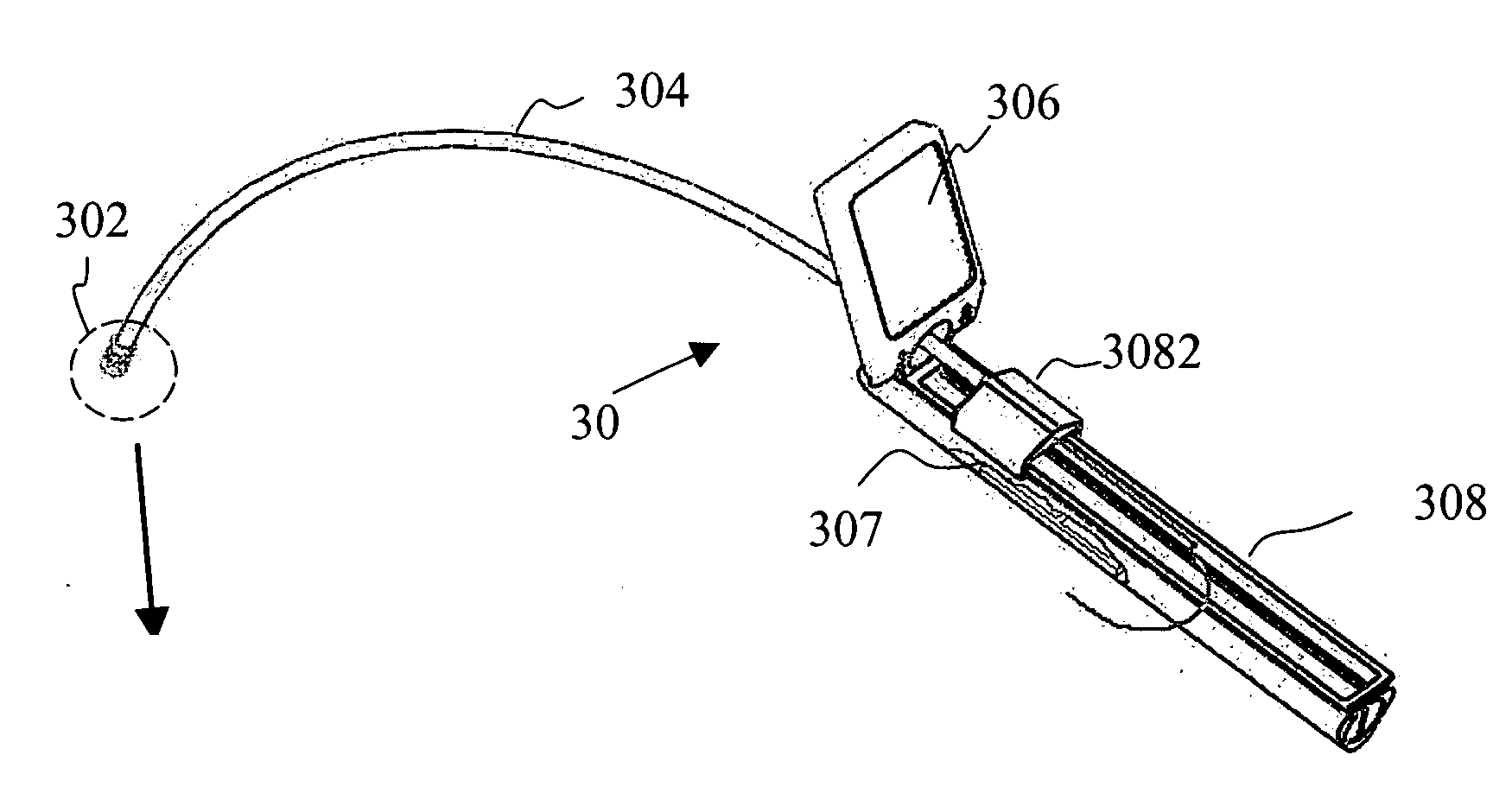
[0024] The present invention proposes an image type intubation-aiding device. As shown in FIG. 3a, an image type intubation-aiding device 30 comprises a probing device 302 made of material compatible with the human body. As shown in FIG. 3b, the probing device 302 comprises a housing 3022 with a diameter smaller than 15 mm. The housing 3022 is pervious to light or has several holes for light penetration. A light-collecting lens 3024 is disposed in the housing 3022. The light-collecting lens 3024 can be integrally formed with the housing 3022. The light-collecting lens 3024 is used for light collection to produce an optical signal. A light source module 3026 is disposed behind the housing 3022 for illuminating the front through the light-collecting lens 3024. An optical and imaging device 3028 is disposed behind the light source module 3026 for converting the optical signal into an electric signal like a digital signal or an analog signal. The image type intubation-aiding device 30 a...


PUM


Abstract

An image-type intubation-aiding device comprises a small-size image sensor and a light source module both placed into an endotracheal tube to help doctors with quick intubation. Light from light emission devices in the light source module passes through a transparent housing and is reflected by a target and then focused. The optical signal is converted into a digital or analog electric signal by the image sensor for displaying on a display device after processing. Doctors can thus be helped to quickly find the position of trachea, keep an appropriate distance from a patient for reducing the possibility of infection, and lower the medical treatment cost. Disposable products are available to avoid the problem of infection. The intubation-aiding device can be used as an electronic surgical image examination instrument for penetration into a body. Moreover, a light source with tunable wavelengths can be used to increase the spot ratio of nidus.

Description

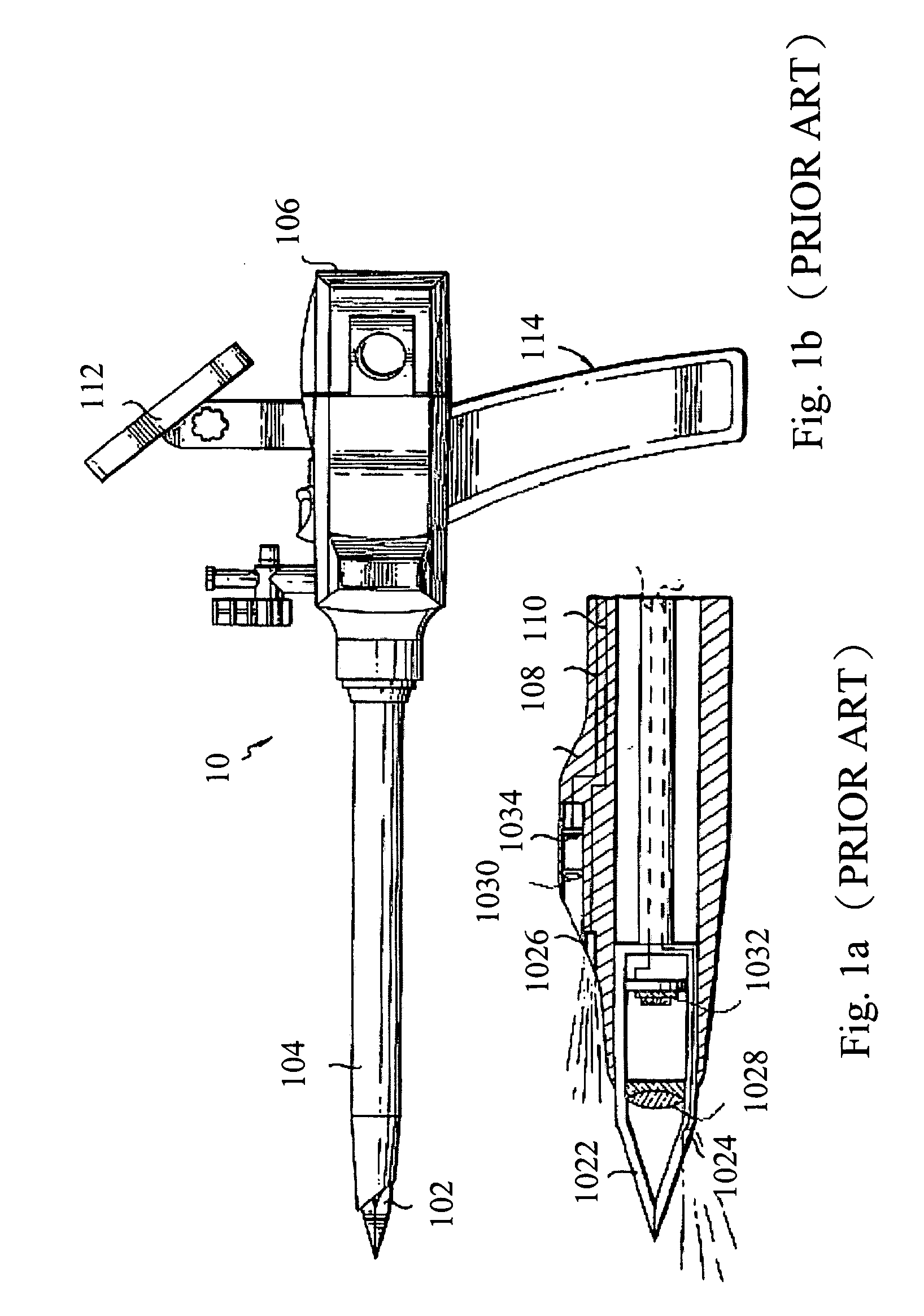
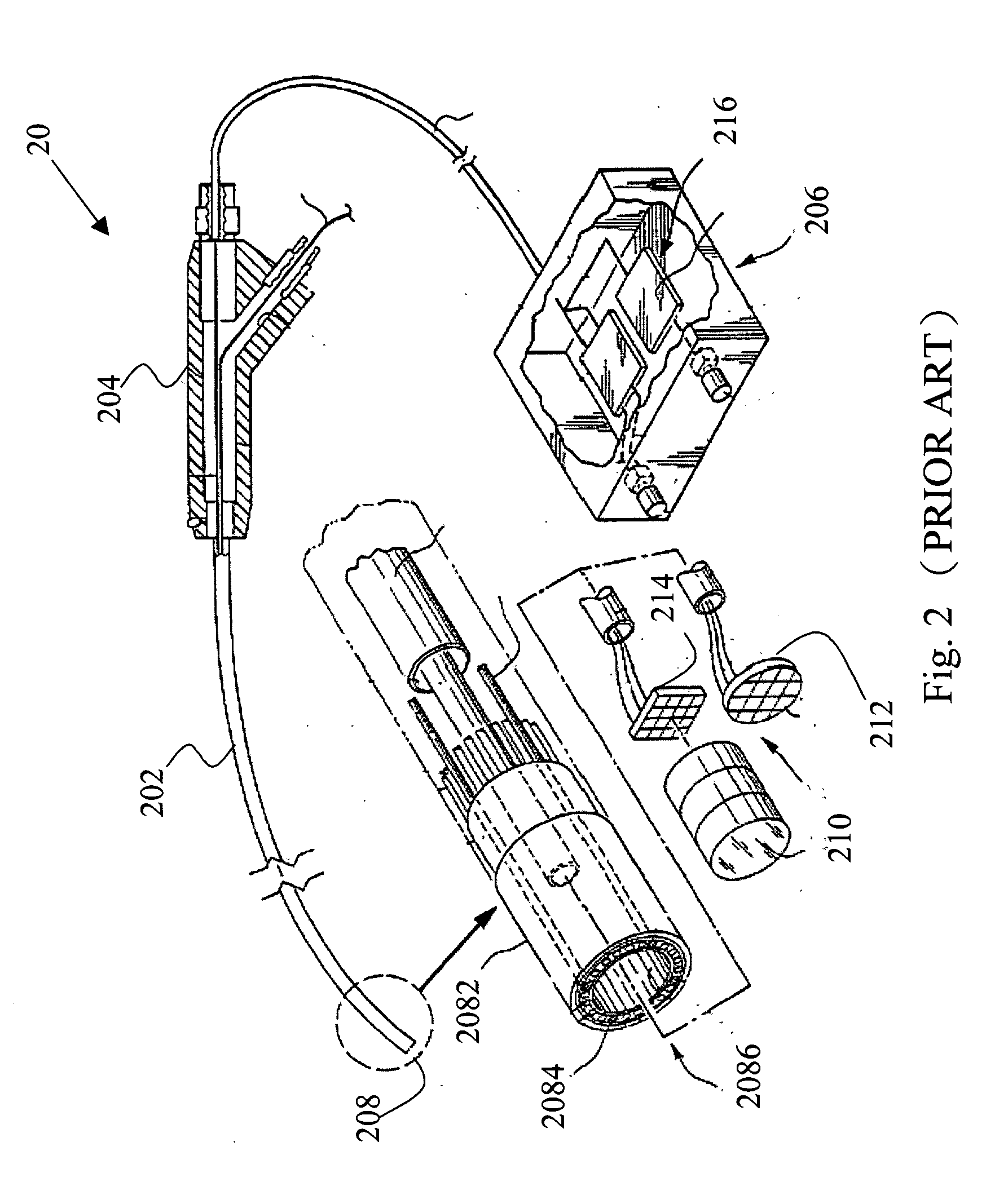
FIELD OF THE INVENTION [0001] The present invention relates to an electronic surgical image examination instrument for penetration into a body and, more particularly, to an image-type intubation-aiding device for helping a doctor with the intubation of tracheal tube. BACKGROUND OF THE INVENTION [0002] An endoscope is an instruments widely used in medicine. It is generally used to examine hollow internal organs or cavities. An endoscope can increase the brightness within the range of a wound and can also enlarge the field of vision for a doctor. A doctor can make use of an endoscope to perform an operation for many wounds without resulting in a larger wound. [0003] Conventionally, many fibers bundled together with a charge couple device (CCD) used to take pictures to form an endoscope, which is used to penetrate hollow organs (e.g., stomach, large intestine and trachea) to get tissue images for determining the type and development degree of diseases. Light from a light source is tran...

Claims


Application Information

Patent Timeline
Patent Type & Authority Applications(United States)
IPC IPC(8): A61B1/06A61B1/04
CPCA61B1/00052A61B1/0607A61B1/0638A61B1/0676A61B1/0684A61B1/267A61B17/34A61B19/5202A61B19/5225A61B2010/045A61B2019/521A61B2019/5229A61M16/04A61M16/0434A61M16/0488A61M2205/273A61B1/05A61B90/30A61B90/37A61B2090/309A61B2090/372A61M16/0418
Inventor SUN, WEI-ZENHSU, JUNG-HSIANGWENG, PING-KUOHUANG, KER-JERZHAN, ZHI-JUN
Owner MEDICAL INTUBATION TECH CORP
Why Eureka
Try Eureka