Systems and methods for localized image registration and fusion

Active Publication Date: 2006-01-05
SIEMENS MEDICAL SOLUTIONS USA INC
View PDF16 Cites 175 Cited by
  • Summary
  • Abstract
  • Description
  • Claims
  • Application Information

AI Technical Summary

Benefits of technology

[0012] The present invention provides new and improved systems and methods for co-registering, displaying and quantifying images from different medical devices or imaging modalities. By blending or replacing planar Regions-of-Interest (ROI) and/or Volumes-of-Interest (VOI) from different imaging devices, a novel display with improved utility can be generated. The size and location of the ROI or V

Problems solved by technology

However, if the resolution and noise characteristics are quite different, as is the case, for example, with functional and anatomical imaging, such as SPECT and CT images or MRI images, then the information delivered to an observer can be less than optimal, and more difficult to interpret with a simple IF technique.
Current methods for combining images also fail to address the need of multi-modality i

Method used


Image

  • Systems and methods for localized image registration and fusion
  • Systems and methods for localized image registration and fusion
  • Systems and methods for localized image registration and fusion

Examples


Example

[0020] Throughout this document, a Region-of-Interest (ROI) is meant to refer to a contractible, and thus a simply connected subset of image pixels within one slice (i.e. a two-dimensional plane)of a total image volume. The smallest ROI is one pixel, and the largest is the entire slice. A Volume-of-Interest (VOI) extends the notion of a ROI to three dimensions, with the smallest unit being a voxel, i.e. a three-dimensional pixel. That is, a VOI is a contractible, and thus simply connected subset of image voxels from the entire image volume in three dimensional space.

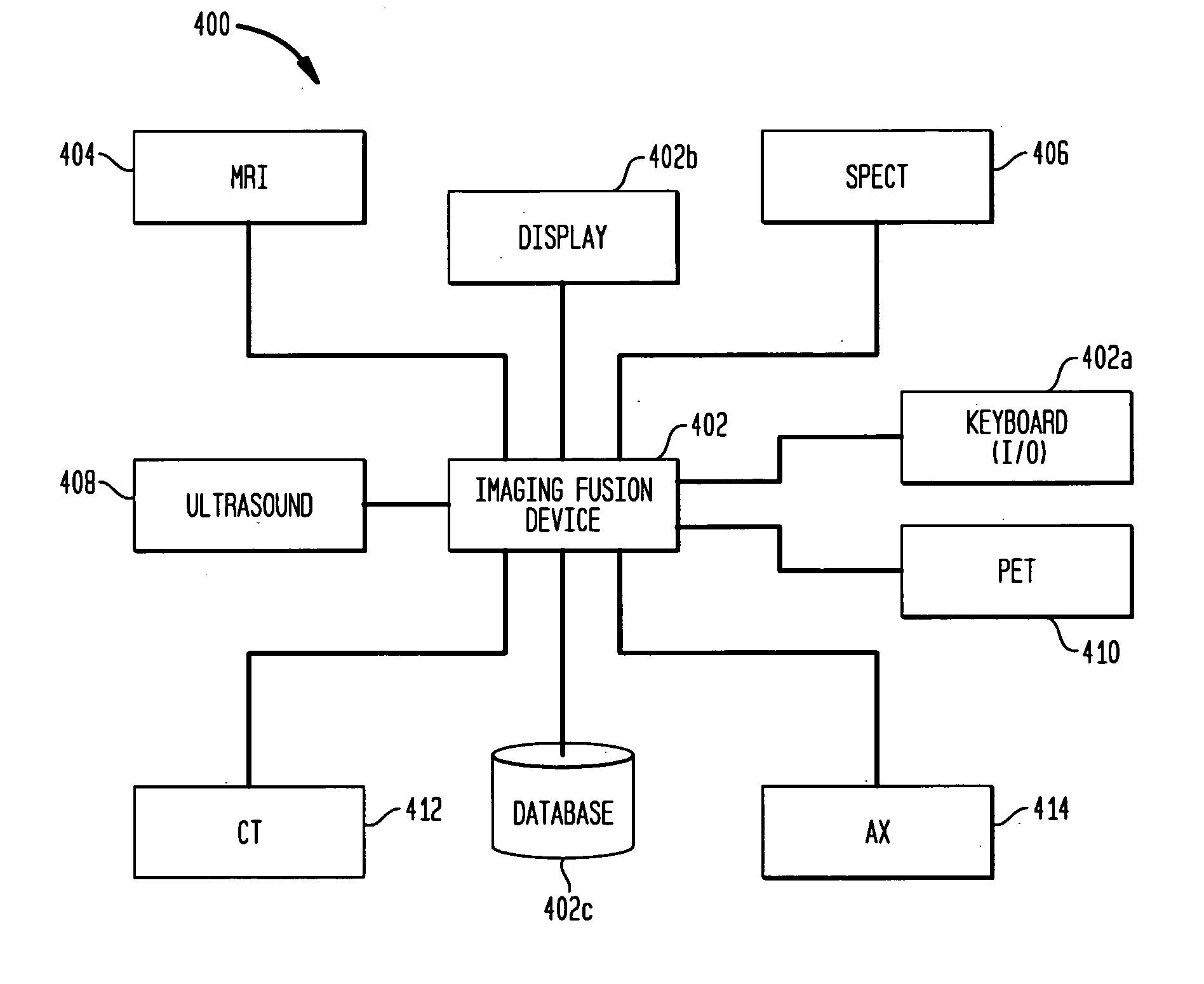
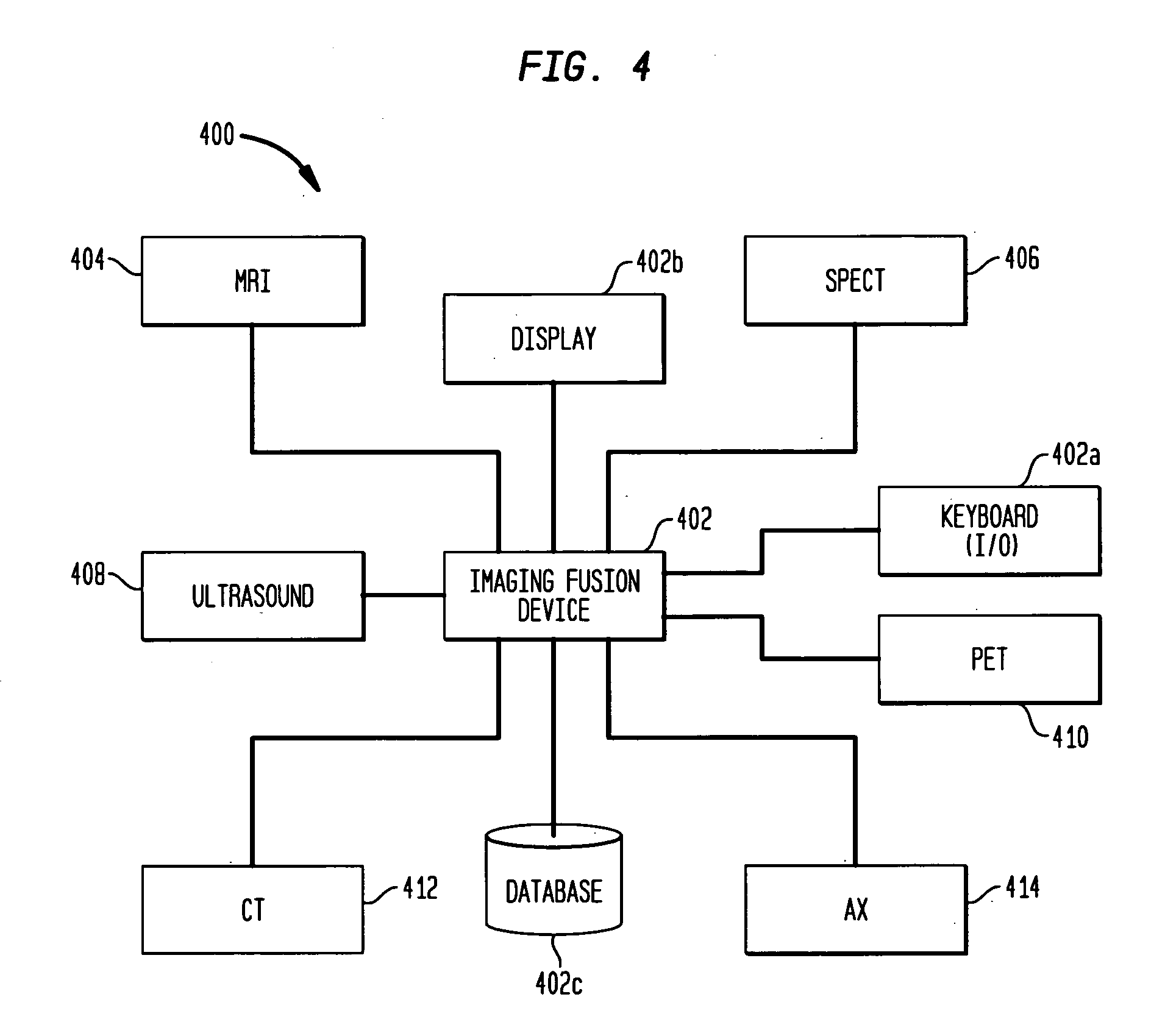
[0021] The present invention is able to produce blended images from disparate imaging devices, which produce data in different modalities. One advantage of the present invention is the ability to register and / or fuse a portion of a first image volume with a second image volume, without registering and / or fusing the entire image volumes. This is accomplished by allowing ROIs or VOIs to be selected (manually or automatica...


PUM


Abstract

Systems and methods are described for co-registering, displaying and quantifying images from numerous different medical modalities, such as CT, MRI and SPECT. In this novel approach co-registration and image fusion is based on multiple user-defined Regions-of-Interest (ROI), which may be subsets of entire image volumes, from multiple modalities, where the each ROI may depict data from different image modalities. The user-selected ROI of a first image modality may be superposed over or blended with the corresponding ROI of a second image modality, and the entire second image may be displayed with either the superposed or blended ROI.

Description

BACKGROUND OF THE INVENTION [0001] 1. Field of the Invention [0002] The present invention is generally directed to medical imaging. More particularly, the present invention is directed to advanced image fusion systems and methods for use in image-assisted biopsy, image-assisted treatment planning, image-assisted treatment and image-assisted surgery, and molecular imaging. [0003] 2. Description of the Related Art [0004] Nuclear medicine imaging tools, such as single-photon-emission-computed-tomography (SPECT) and positron-emission-tomography (PET) are known. Other types of medical imaging tools, such as magnetic resonance imaging (MRI), computed tomography (CT) or ultrasound (US), are also well known. Each of these known imaging techniques (modalities) provides a clinician with a different type of data and is useful for a different purpose, such as mainly functional (SPECT, PET) or mainly anatomical imaging (MR, CT, US). Until recently, images from only two modalities were combined i...

Claims


Application Information

Patent Timeline
IPC IPC(8): A61B5/05
CPCA61B6/00A61B2019/5289G01T1/1644A61B6/037G06T2210/41G06T15/503G06T11/00A61B2090/364
Inventor VIJA, A. HANSREMPEL, TRUDY DAYLE
Owner SIEMENS MEDICAL SOLUTIONS USA INC
Why Eureka
Try Eureka