
Method and device to recover diagnostic and therapeutic agents

a diagnostic agent and therapeutic technology, applied in the direction of biocide, ultrasonic/sonic/infrasonic diagnostics, biocide, etc., can solve the problems of diabetes mellitus patients, increased risk of contrast-induced acute renal failure, excess morbidity and mortality, etc., and achieve the effect of reducing the amount of the agen

Inactive Publication Date: 2006-01-19
UNIVERSITY OF VERMONT
View PDF3 Cites 36 Cited by
  • Summary
  • Abstract
  • Description
  • Claims
  • Application Information

AI Technical Summary

Benefits of technology

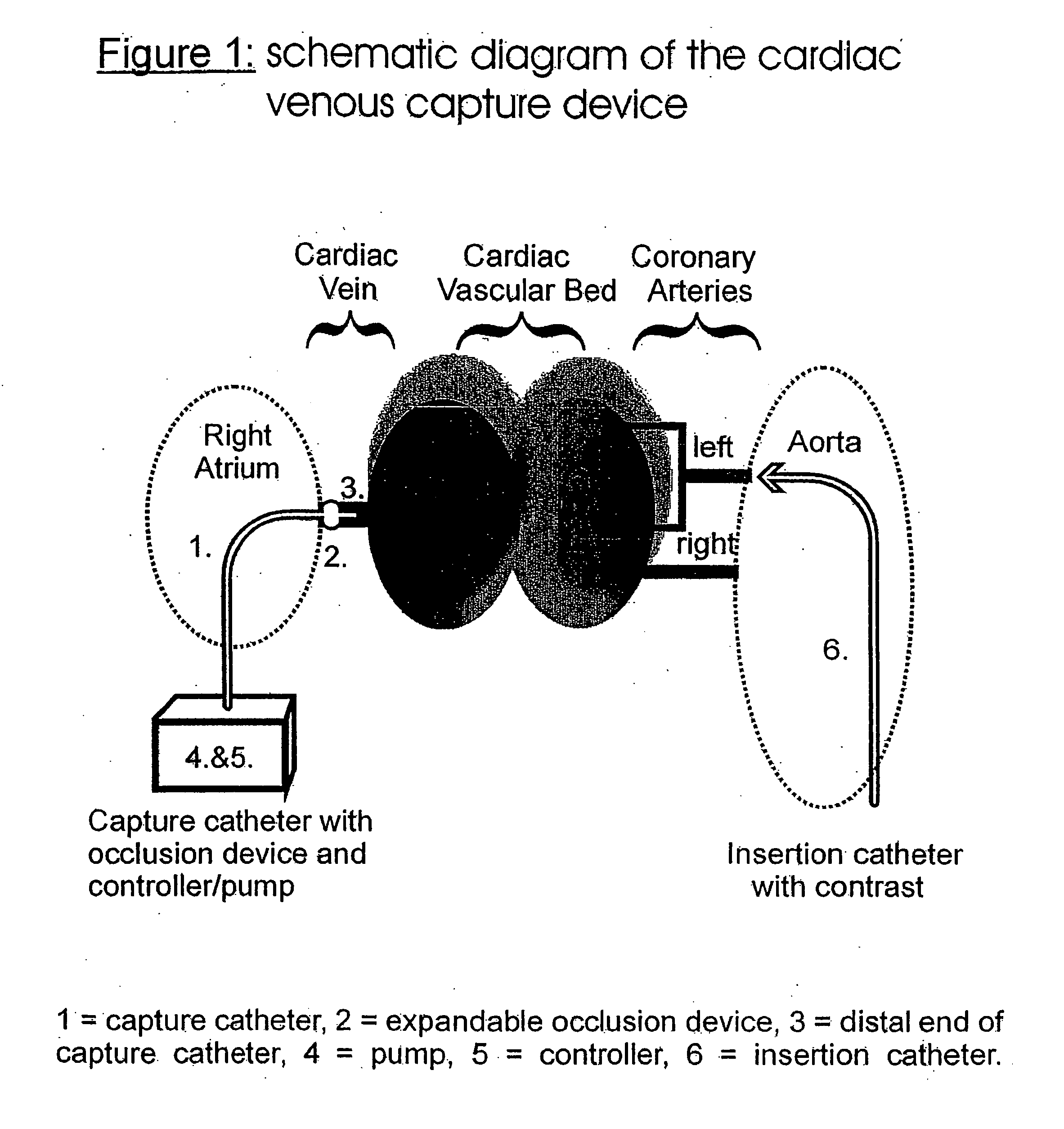
[0008] The invention provides methods and devices useful for recovering at least a portion of an agent that has been injected into a blood vessel serving an organ or tissue in the body. In this manner, the devices and methods of the present invention decrease the amount of the agent that leaves the organ or tissue and enters into the rest of the circulatory system.

Problems solved by technology

However, it has become common knowledge to those of skill in the art that the use of intravascular contrast agents is associated with an increased risk of contrast-induced acute renal failure (CIARF), which may lead to excess rates of morbidity and mortality.
Additionally, patients with diabetes mellitus and conditions that predispose to development of ARF, e.g., sepsis, are also at increased risk of CIARF.
However, these strategies have significant shortcomings with respect to efficacy, they require increased drug usage and / or prolonged hospitalization (both of which increase treatment costs), and, in the case of decreasing the amount of contrast agent used during the procedure, a lessened ability to adequately visualize the vasculature.

Method used


Image

  • Method and device to recover diagnostic and therapeutic agents
  • Method and device to recover diagnostic and therapeutic agents
  • Method and device to recover diagnostic and therapeutic agents

Examples


example 1

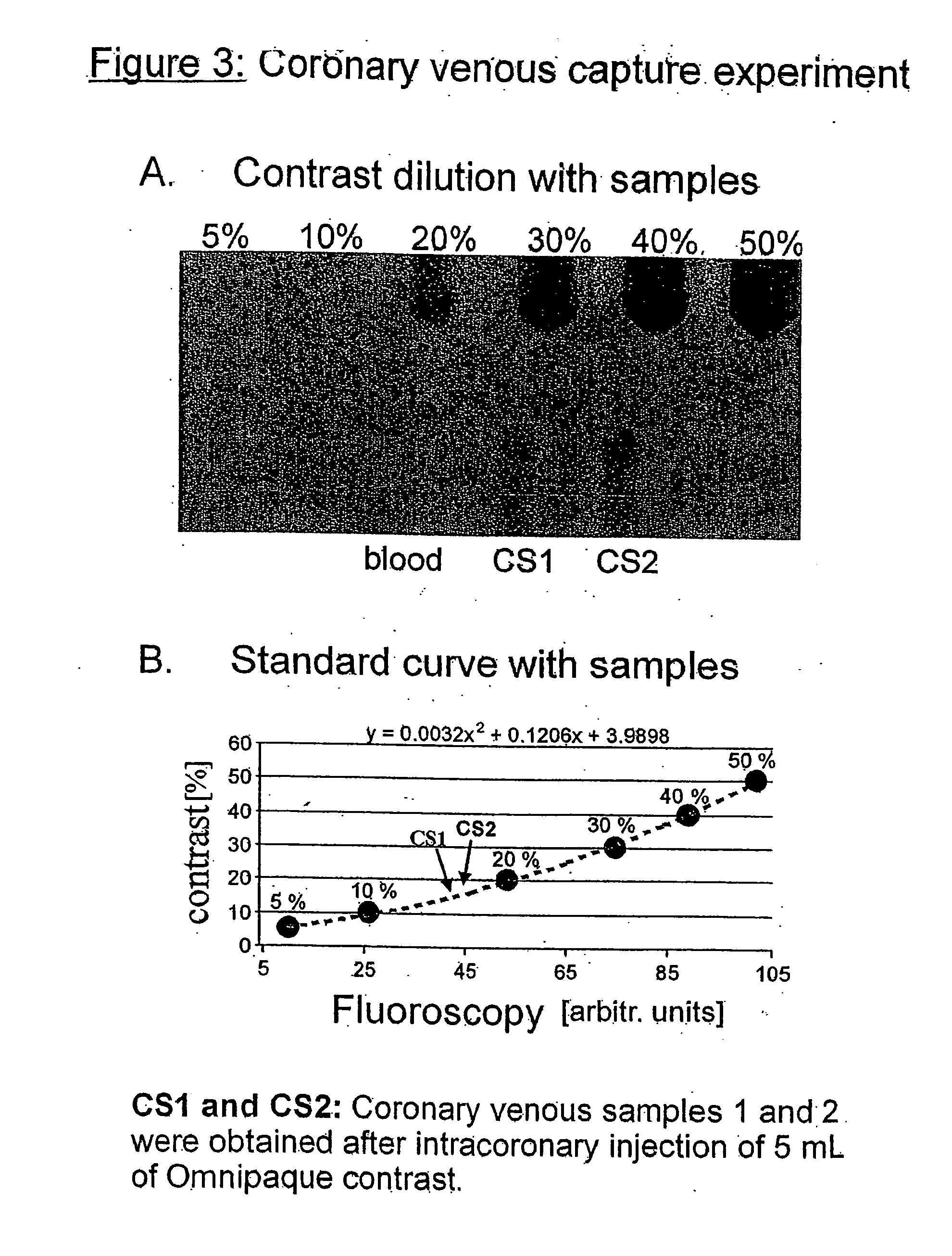
[0056] To demonstrate a process of the present invention, coronary angiography was performed in an anesthetized dog in which contrast medium was injected into the left main coronary artery and blood and contrast medium were withdrawn from the coronary sinus. In this example, the catheter balloon was inflated as soon as the contrast injection started. Furthermore, the withdrawal of blood and contrast medium from the coronary sinus was manually controlled and performed under fluoroscopic guidance following injection of the contrast into the left main coronary artery. The collected fluid was analyzed by quantitative fluoroscopy, as exemplified in FIG. 3.

[0057]FIG. 3A is a fluoroscopic image showing an array of blood samples containing known concentrations of contrast agent ranging from 0 to 50%. A sample that is 5% contrast agent and 95% blood is denoted 5%. A sample that is 50% contrast agent and 50% blood is denoted 50%. As can be appreciated from this figure, the fluoroscopic densi...

example 2

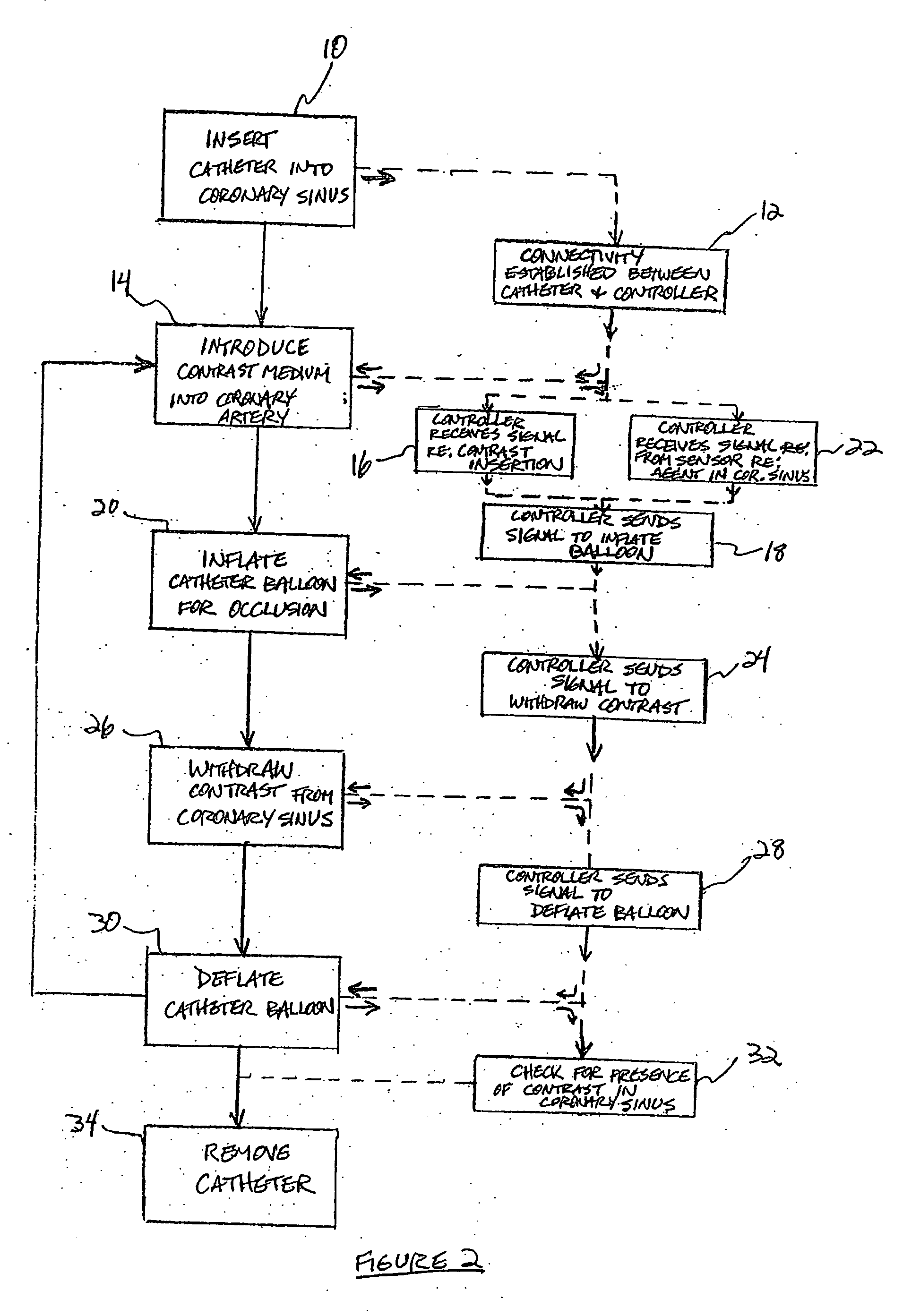
[0061] In a follow-on demonstration, it was shown that the electrical conductivity of contrast medium is markedly higher than the electrical conductivity of blood. The electrical resistance of 10 ml of blood was measured (in Ohms), and compared with the separately measured electrical resistance of 10 ml of a contrast medium, Iohexol. With Conductivity=1 / Resistance, the difference in conductivity between blood and Iohexol was found to be approximately four-fold. Thus, it was concluded that changes of blood conductivity can potentially be used to detect the presence of contrast in the coronary sinus and trigger a withdrawal pump that is controlled by an automated controller.


PUM

PropertyMeasurementUnit
Fractionaaaaaaaaaa
Diameteraaaaaaaaaa
Diameteraaaaaaaaaa

Abstract

The invention relates to a method and device for recovering therapeutic and diagnostic agents from the venous outflow of a tissue or organ. A method for recovering an agent includes inserting a catheter into a vascular outflow of a tissue or organ, occluding the flow of blood / fluid through the vascular outflow, and withdrawing blood / fluid in the vascular outflow into the catheter to recover at least a portion of the agent administered to the vascular inflow of the organ or tissue. Also provided is a device to capture an agent administered to a vascular inflow, the device having a catheter having an expandable occlusion device to occlude a flow of fluid through the vascular outflow, a pump operably coupled to the catheter for withdrawing from the vascular outflow at least a portion of the agent administered to a vascular inflow of the organ or tissue, and an automated controller in signal communication with the catheter to control the expandable occlusion device and the pump to withdraw at least a portion of the agent from the vascular outflow.

Description

RELATED APPLICATIONS [0001] This application claims benefit under 35 U.S.C. 119(e) of U.S. Provisional Patent Application Ser. Nos. 60 / 584,238, filed Jun. 30, 2004, and 60 / 668,402, filed Apr. 5, 2005, the entire contents of which are incorporated herein by reference.BACKGROUND OF INVENTION [0002] 1. Field of Invention [0003] This invention relates to a method for recovering therapeutic and diagnostic agents during medical procedures, and a device for recovering such agents. [0004] 2. Description of Related Art [0005] The use of diagnostic or therapeutic agents to facilitate the examination or treatment of internal organs and tissues is common medical practice. A diagnostic agent or dye may be injected into the blood flow serving an organ or tissue as part of a radiographic procedure, such as an x-ray, performed to help diagnose any problems or abnormalities with the organ or tissue. A therapeutic agent may be introduced into the blood flow serving a tissue or an organ to improve the...

Claims


Application Information

Patent Timeline
IPC IPC(8): A61K48/00A61M31/00
CPCA61M1/0058A61M1/3621A61M1/3615A61M1/3613A61M2025/1052A61M1/77
Inventor LEWINTER, MARTIN M.DAUERMAN, HAROLD L.LUSTGARTEN, DANIEL L.MEYER, MARKUS
Owner UNIVERSITY OF VERMONT
Learn More