
Medical image processing system

a technology of image processing and image, applied in the field of medical image processing system, can solve the problems of system not being designed to be incorporated into, and the use of the system being limited to the same modality for re-imaging, and achieve the effect of improving the efficiency of image interpretation and diagnosis

Inactive Publication Date: 2006-02-16
FUJIFILM HLDG CORP +1
View PDF8 Cites 21 Cited by
  • Summary
  • Abstract
  • Description
  • Claims
  • Application Information

AI Technical Summary

Benefits of technology

[0016] The present invention has been developed in view of the circumstances described above, and it is an object of the present invention to provide a medical image processing system capable of determining objectively and rapidly whether additional imaging or a medical examination is required, and further determining the type of the required additional imaging or medical examination if it is determined necessary prior to image interpretation to be performed by a doctor, thereby further improving the efficiency of the image interpretation and diagnosis.
[0035] The referent of “magnified imaging” as used herein means imaging a partial region of a subject by magnifying it such that the resolution or sharpness of the image is enhanced.
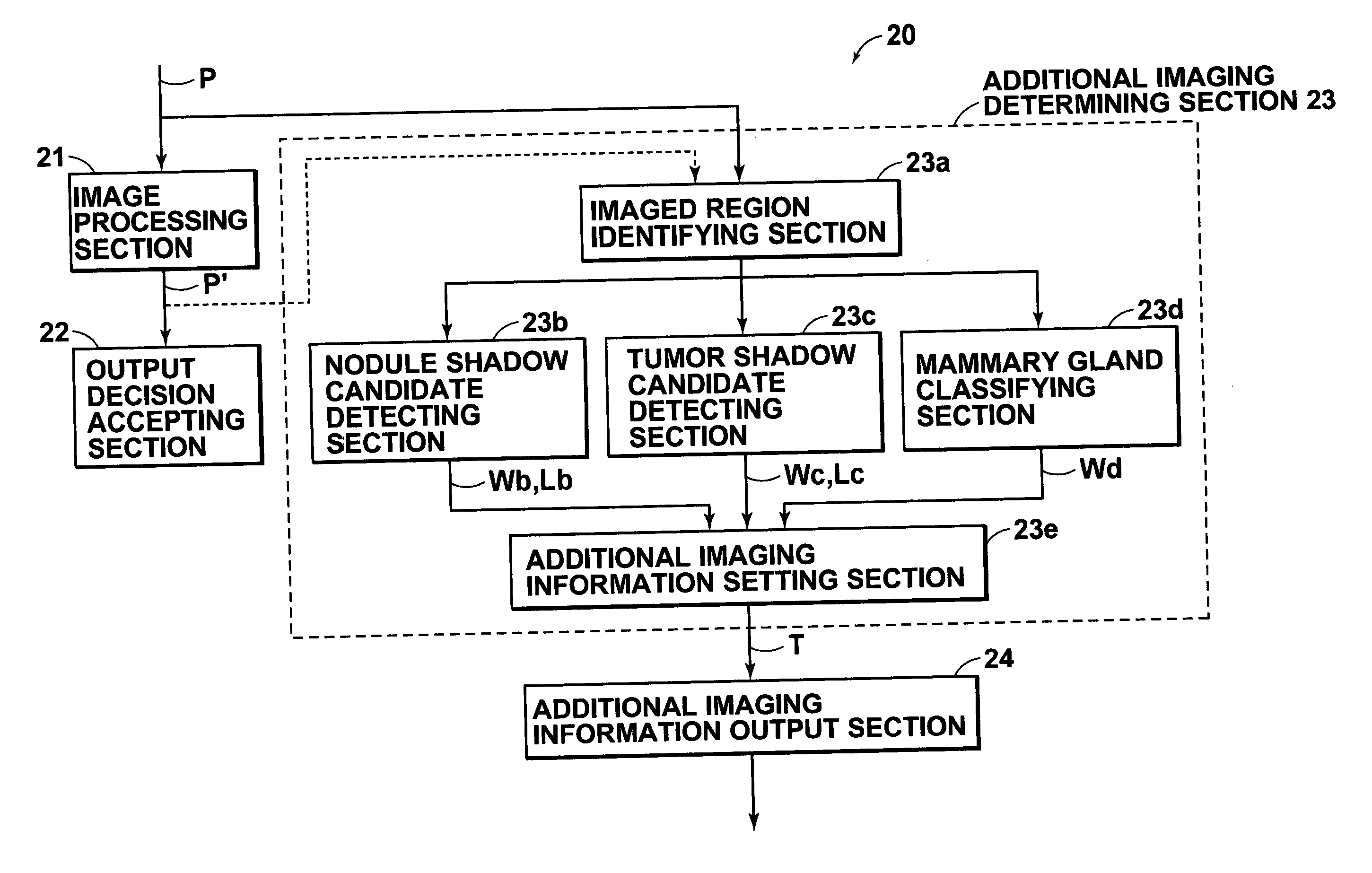
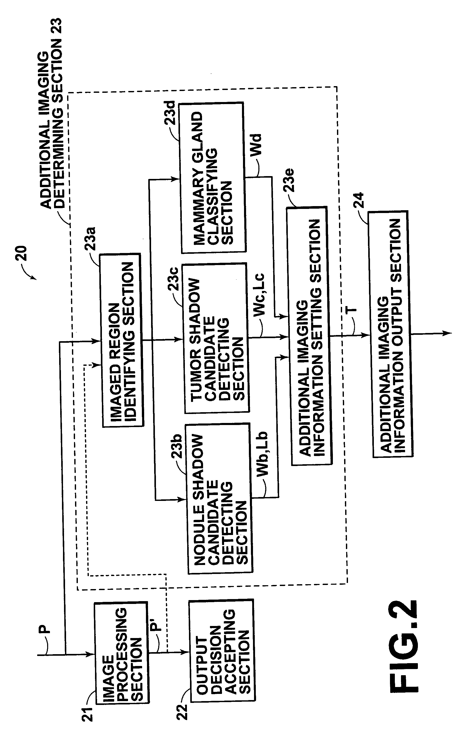
[0036] According to the medical image processing system of the present invention, the image quality inspection terminal has a determining section configured to perform a first determination process for determining whether additional imaging or an additional medical examination is required based on the medical image inputted thereto, and a second determination process for determining the type of the required additional imaging or medical examination, if the additional imaging or medical examination is determined necessary by the first determination; and the output section configured to output information which indicates that the additional imaging or medical examination is required and the type thereof, if the additional imaging or medical examination is determined necessary by the determining section. This allows the inspection engineer to direct additional imaging or an additional medical examination to be performed in advance by referring to the output from the output section. Consequently, it is possible to determine objectively and rapidly whether additional imaging or an additional medical examination is required, and to further determine the type of the additional imaging or medical examination, if the additional imaging or medical examination is determined necessary prior to the image interpretation to be performed by a doctor, thereby the efficiency of the image interpretation and diagnosis may be further improved.

Problems solved by technology

But the system is not designed to be incorporated into the image quality inspection terminal for performing the image quality inspection described above.
Still further, judging from the description contained in Japanese Unexamined Patent Publication No. 2001-187044, the use of the system is limited to the same modality for re-imaging.

Method used


Image

  • Medical image processing system
  • Medical image processing system
  • Medical image processing system

Examples


Embodiment Construction

[0044] Hereinafter, embodiments of the present invention will be described with reference to the accompanying drawings.

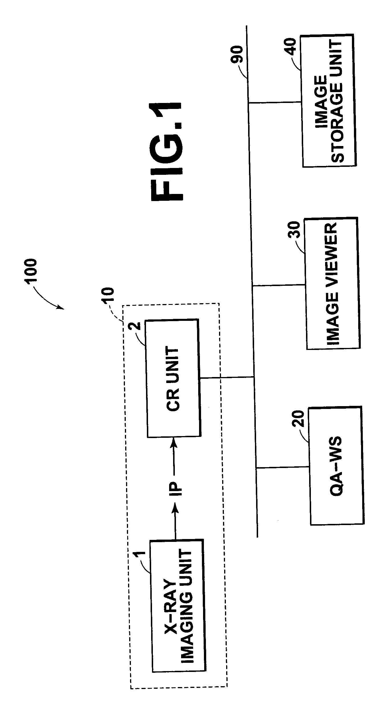
[0045]FIG. 1 is a block diagram of the medical image processing system 100 according to an embodiment of the present invention illustrating the overview thereof.

[0046] The medical image processing system 100 shown in FIG. 1 comprises: an X-ray image generation unit 10 (image generation unit) having an X-ray imaging unit 1 and a CR unit 2; a QA-WS 20 (image quality inspection terminal) for accepting a decision whether to output each of the medical images outputted from the X-ray image generation unit 10 and inputted thereto; an image viewer 30 (interpretation terminal) for displaying each of the medical images outputted from the QA-WS and inputted thereto; and an image storage unit 40 for storing various images, which are all connected to a network 90.

[0047] The X-ray imaging unit 1 emits X-rays to a test subject and receives X-rays transmitted through the test su...


PUM


Abstract

A medical image processing system having an image generation unit; an image quality inspection terminal; and an image interpretation terminal is provided. The system is capable of determining whether additional imaging or the like is required and the type objectively and rapidly prior to an image interpretation to be performed by a doctor to further improve the efficiency of the image interpretation and diagnosis. An additional imaging determining section is incorporated into a QA-WS (image quality inspection terminal). The determining section is configured to perform a predetermined image analysis on the image data generated by the image generation unit to determine whether additional imaging or the like is required and the type objectively. The inspection engineer performs image quality inspection, and directs additional imaging or the like, if necessary, by referring to the determination result at the QA-WS.

Description

BACKGROUND OF THE INVENTION [0001] 1. Field of the Invention [0002] The present invention relates to a medical image processing system. More specifically, the present invention is directed to a medical image processing system configured to generate digital medical images and inspect image qualities of the generated images to display qualified medical images for interpretation. [0003] 2. Description of the Related Art [0004] In the medical field, medical image processing systems are used to facilitate medical image interpretation works. The system has a medical image generation unit for generating a digital medical image; an image quality inspection terminal having an output decision accepting function for automatically or manually accepting a decision whether to output the medical image generated by the image generation unit and inputted to the terminal; and an interpretation terminal for displaying the medical image inputted from the image quality inspection terminal. [0005] The sy...

Claims


Application Information

Patent Timeline
IPC IPC(8): G06K9/00
CPCG06T7/0012G06T2207/30168G06T2207/30068G06T2207/30061
Inventor SHIMAYAMA, TOSHIHIDE
Owner FUJIFILM HLDG CORP
Learn More