Method for producing hypertrophic scarring animal model for identification of agents for prevention and treatment of human hypertrophic scarring

a hypertrophic scarring and animal model technology, applied in the field of producing a nonhuman animal model of hypertrophic scarring, can solve the problems of significant functional and aesthetic defects, achieve minimal scarring, reduce the risk of wound dehiscence (rupture), and improve the effect of volume and cellularity

Inactive Publication Date: 2006-02-16
GURTNER GEOFFREY C +1
View PDF0 Cites 86 Cited by
  • Summary
  • Abstract
  • Description
  • Claims
  • Application Information

AI Technical Summary

Benefits of technology

[0059] To directly examine the effect of human levels of strain on healing murine wounds, a simple strain device was developed that could be applied to incisional wounds, shown in FIG. 1A. Two separate full-thickness wounds were created on each mouse, shown in FIGS. 1B-C, and mechanical force was applied to one in a cyclical fashion beginning on day 4, which corresponds with the initiation of the proliferative phase of wound healing. The other wound was not strained and served as an internal control. Pilot studies had demonstrated that at day 4 re-epithelialization had occurred and the risk of wound dehiscence (rupture) was minimized. Prior experiments also demonstrated that this range of forces (6-10 N/mm2) would affect the tissues at the cellular level without exceeding the breaking limits (19 N/mm2) of the wound.
[0060] The timing of strain application was critical to the formation of hypertrophic scars. Strain during the earlier inflammatory phase (days 1-3) resulted in wound breakdown; strain during the proliferative phase of wound healing (day 3-10) resulted in exuberant scars, whereas strain during the remodeling phase after day 10 had little effect on subsequent scar formation. The unstrained wound healed with minimal scarring, shown in FIG. 1D, but the strained region developed into human-like hypertrophic scars with increased volume and cellularity, as shown in FIG. 1E (Linares et al., “The Histiotypic Organization of the Hypertrophic Scar in Humans,”J Invest Dermatol 59:323-331 (1972); White, C., Textbook of Dermatopathology. New York: McGraw Hill. 349-355 pp. (2004); Ehrlich et al., “Morphological and Immunochemical Differences Between Keloid and Hypertrophic Scar,”Am J Pathol 145:105-113 (1994), which are hereby incorporated by reference in their entirety). Histologically, the unstrained scar is small, as shown in FIG. 1F, whereas the strained scar is 10-20 fold larger, shown in FIG. 1G. There were no differences in scar formation when the strain device was activated over the cephalad or caudal wound, as might be expected to occur if Hox gene differences were responsible (Chauvet et al., “Distinct Hox Protein Sequences Determine Specificity in Different Tissues,”Proc Natl Acad Sci USA 97:4064-4069 (2000); Stelnicki et al., “Bone Morphogenetic Protein-2 Induces Scar Formation and Skin Maturation in the Second Trimester Fetus,”Plast Reconstr Surg 101:12-19 (1998); Stelnicki et al.

Problems solved by technology

Hypertrophic scarring commonly occurs following cutaneous wound

Method used


Image

  • Method for producing hypertrophic scarring animal model for identification of agents for prevention and treatment of human hypertrophic scarring
  • Method for producing hypertrophic scarring animal model for identification of agents for prevention and treatment of human hypertrophic scarring
  • Method for producing hypertrophic scarring animal model for identification of agents for prevention and treatment of human hypertrophic scarring

Examples


example 1

In Vivo Strain

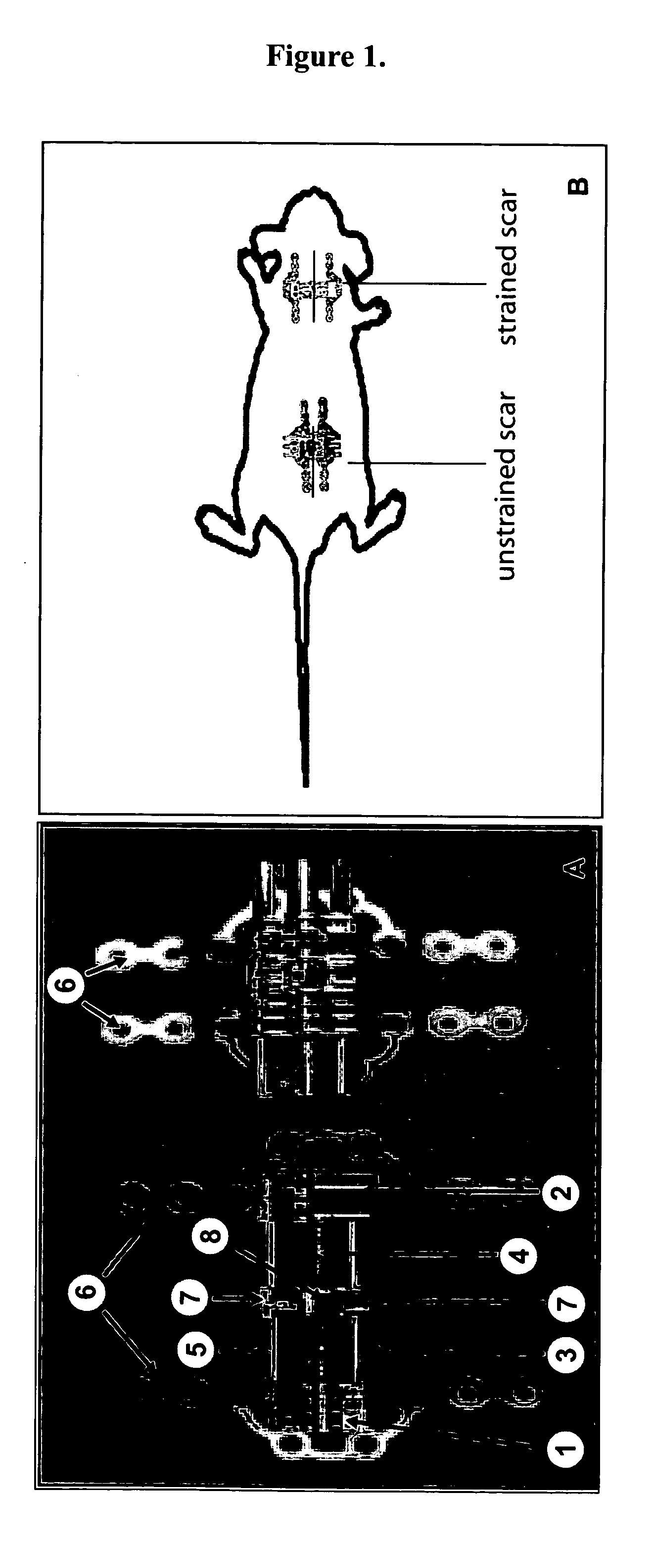
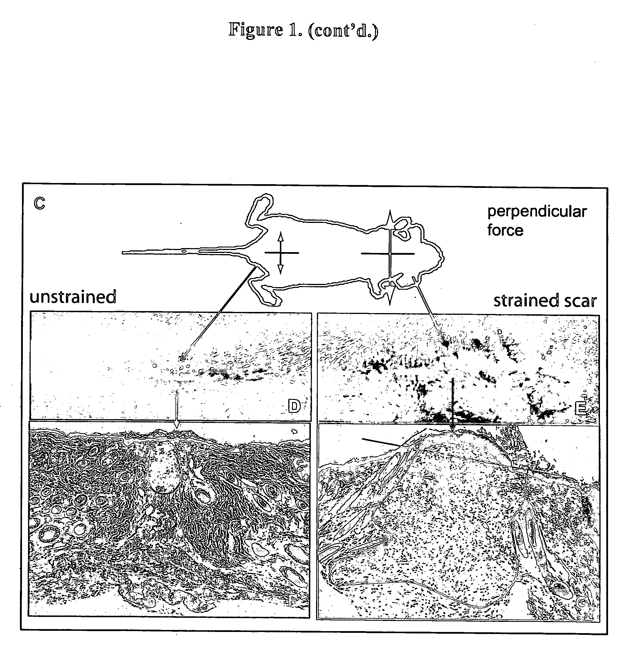
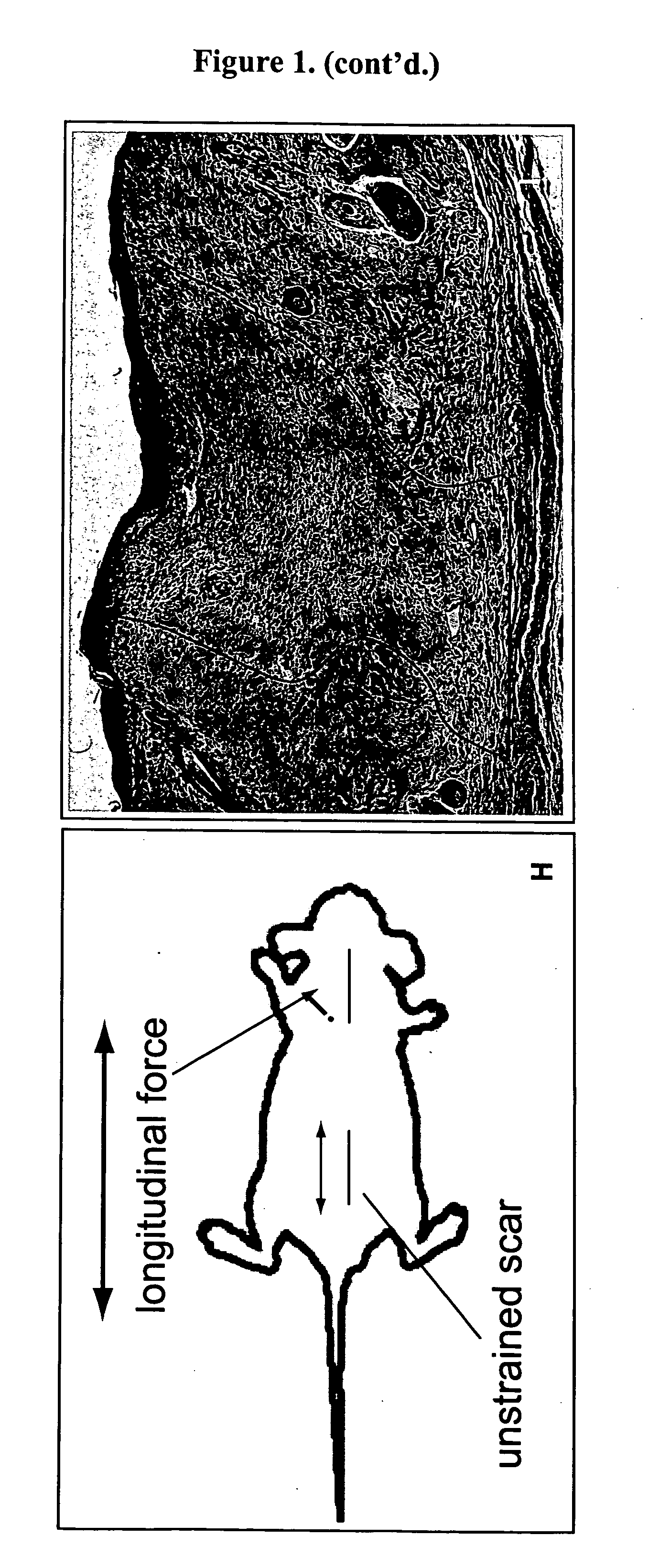
[0047] Four week old C57 / BL6 mice were first acclimated and housed under standard conditions, using protocols approved by the New York University Animal Care and Use Committee. Mouse strains B6.129S2-Trp53tm1Tyj / J (anti-apoptotic) and B6.129S2-Bcl2tm1Sjk / J (pro-apoptotic) (Jackson Laboratory, Bar Harbor, Me.) were used for the knockout studies. Two 2 cm linear full-thickness incisions (1.25 cm apart) were made on the dorsum of the mouse and then reapproximated with 6-0 nylon sutures. On post-incision day 4, the sutures were removed from the scars, and two biomechanical strain devices, shown in FIG. 1A, were carefully secured with 6-0 nylon sutures, as shown in FIG. 2B. The biomechanical strain devices were constructed from 22-mm expansion screw (Great Lakes Orthodontic Products, Tonawanda, N.Y., USA) and Luhr (Stryker-Leibinger Co, Freiburg, Germany) plate supports, as shown in FIG. 1A. One wound served as an internal control, with the device not activated, while mech...

example 2

In Vitro Strain

[0050] In order to study the molecular mechanisms of mechanical strain on a cellular level, human (HTERT-BJ1, Clonetech, Palo Alto, Calif.) and primary murine fibroblasts was examined in vitro. A novel in vitro model as designed and described by Holmes (Costa et al., “Creating Alignment and Anisotropy in Engineered Heart Tissue: Role of Boundary Conditions in a Model Three-Dimensional Culture System,”Tissue Eng 9:567-577 (2003); Knezevic et al., “Isotonic Biaxial Loading of Fibroblast-Populated Collagen Gels: A Versatile, Low-Cost System for the Study of Mechanobiology,”Biomech Model Mechanobiol 1:59-67 (2002); Zimmerman et al., “Structural and Mechanical Factors Influencing Infarct Scar Collagen Organization,”Am J Physiol Heart Circ Physiol 278:H194-200 (2000), which are hereby incorporated by reference in their entirety) was utilized. Briefly, this model maintains fibroblasts in a three-dimensional matrix (fibroblast plated collagen lattice, “FPCL”), thereby closel...

example 3

Cell Culture

[0051] Human HTERT-BJ1 cells were grown in DMEM (Invitrogen, Carlsbad, Calif.) supplemented with 10% fetal bovine serum (Invitrogen, Carlsbad, Calif.) and 1% antimycotic / antibiotic at 37° C. in a CO2 incubator. The cells were serum-starved for 18 h prior to conducting the in vitro experiments.


PUM

PropertyMeasurementUnit
Strain pointaaaaaaaaaa

Abstract

The present invention relates to a method of producing a non-human animal model of hypertrophic scarring. This involves producing an incision in a non-human animal and applying mechanical strain over the incision under conditions effective to produce hypertrophic scarring, thereby producing a non-human animal model of hypertrophic scarring. The present invention also relates to a method of determining the efficacy of an agent for prevention or treatment of a disease condition. This method involves providing a non-human animal having an incision over which mechanical strain is applied under conditions effective to produce hypertrophic scarring, administering an agent to the incision, and determining whether the agent is efficacious for prevention or treatment of a disease condition. Also provided is a non-human animal model of hypertrophic scarring. This involves a non-human animal having an incision over which mechanical strain has been applied under conditions effective to produce hypertrophic scarring.

Description

[0001] This application claims benefit of U.S. Provisional Patent Application Ser. No. 60 / 573,998, filed May 24, 2004, which is hereby incorporated by reference in its entirety.FIELD OF THE INVENTION [0002] The present invention relates to a method for producing a non-human animal model of hypertrophic scarring and the use of such a model for the development of agents for the prevention or treatment of hypertrophic scarring in mammals, including humans. BACKGROUND OF THE INVENTION [0003] The optimal result of human wound healing would be functional and scar-free healing (Martin, P., “Wound Healing—Aiming for Perfect Skin Regeneration,”Science 276:75-81 (1997)), but this is rarely the case. Each year more than 12 million traumatic and 1.25 million burn injuries result in disfiguring and dysfunctional hypertrophic scars (Singer et al., “Cutaneous Wound Healing,”N Engl J Med 341:738-746 (1999); Singer et al., “Evaluation and Management of Traumatic Lacerations,”N Engl J Med 337:1142-11...

Claims


Application Information

Patent Timeline
IPC IPC(8): A01K67/027A61K38/54
CPCA01K67/027A01K2267/03A01K2227/105
Inventor GURTNER, GEOFFREY C.BHATT, KIRIT A.
Owner GURTNER GEOFFREY C
Why Eureka
Try Eureka