

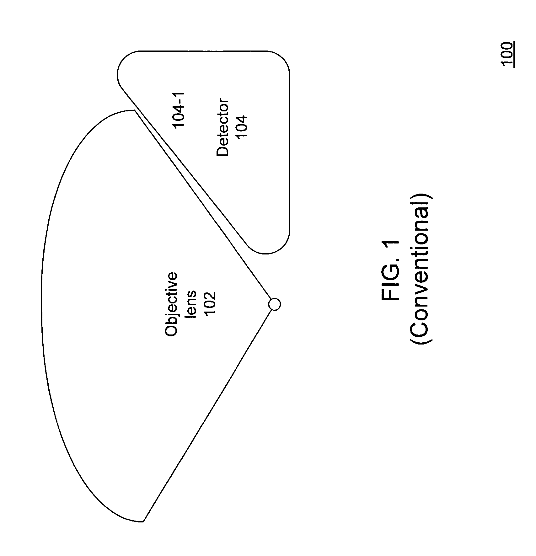
Apparatus and method for e-beam dark field imaging
a dark field and electron beam technology, applied in the direction of instruments, mass spectrometers, beam deviation/focusing by electric/magnetic means, etc., can solve the problems of higher lens aberration coefficient, inferior image resolution, interference with the collection efficiency of below-the-lens detectors
- Summary
- Abstract
- Description
- Claims
- Application Information
AI Technical Summary
Benefits of technology
Problems solved by technology
Method used
Image


Examples
Embodiment Construction
[0021] It is desirable to provide an SEM system with dark-field and bright-field imaging capabilities. It is further desirable for the system architecture to be designed to preserve the image resolution provided by an immersion lens without sacrificing dark-field image capability.
[0022]FIG. 4A is a three-dimensional depiction of an SEM dark field imaging system 400 with an improved behind-the-lens detection configuration in accordance with an embodiment of the invention. For purposes of explanation of the figure, certain components of the system are pointed to specifically. At the bottom of the figure, the immersion lens pole pieces 402 are shown. Next going up the column are the scan plates 404 and Wien filter 406. Towards the top, a scintillator detector 410 is shown, with an energy filter component 408 in front of the detector 410.
[0023]FIG. 4B is a cross-sectional schematic diagram of the SEM dark field imaging system 400. Depicted are various select components, including an i...
PUM
 Abstract
Description
Claims
Application Information