
System and method for acquisition and reconstruction of contrast-enhanced, artifact-reduced CT images

a technology of contrast enhancement and artifact reduction, applied in the field of radiographic imaging, can solve the problems of inability to provide independent data or feedback as to the energy and incident flux rate of photons, ct imaging would not be a viable diagnostic imaging tool, and inability to provide energy discriminatory data or otherwise count the number and/or measure the energy of photons actually received by a given detector elemen

Active Publication Date: 2006-05-25
GENERAL ELECTRIC CO
View PDF36 Cites 189 Cited by
  • Summary
  • Abstract
  • Description
  • Claims
  • Application Information

AI Technical Summary

Benefits of technology

[0020] In accordance with another aspect, the present invention includes a method of radiographic imaging is disclosed that includes the steps of acquiring energy discriminating CT data from an ROI and determining a material composition breakdown of the ROI from the energy discriminating CT data. The m

Problems solved by technology

A drawback of such detectors is their inability to provide independent data or feedback as to the energy and incident flux rate of photons detected.
While it is generally recognized that CT imaging would not be a viable diagnostic imaging tool without the advancements achieved with conventional CT detector design, a drawback of these integrating detectors is their inability to provide energy discriminatory data or otherwise count the number and / or measure the energy of photons actually received by a given detector element.
However, typically, images contain multiple materials and mixtures of materials that may yield similar contrasts in an x-ray projection or reconstructed CT image and make differentiation of the target objects difficult.
As a result, information available in the x-ray data and from the a priori knowledge of anatomy is not typically considered, which may lead to sub-optimal weighting.
However, such generalized x-ray energy weighting functions include limitations when actually implemented because they are globally implementated for all pixels in an image and do not specifically take into account the energy-dependent, x-ray attenuation properties of each tissue locally.
Furthermore, performance degradation is incurred due to the limited number of energy bins used during data acquisition and due to detector electronic noise.
Accordingly, when such generalized x-ray energy weighted acquisitions are performed instead of equally weighted acquisitions, increased noise is experienced in the reconstructed image.
Another drawback of generalized x-ray energy weighted approaches is that low energy photons are heavily weighted, which increases noise within a reconstructed image.
That is, typically, low energy photons can not penetrate the imaging subject effectively and, thus, the majority of low energy photons are attributable to noise.
Accordingly, when a generalized x-ray energy weighted approach is applied and low energy photons are heavily weighted, noise, which comprises a large percentage of the low energy photons, is increased within the reconstructed image.
However, it requires the viewing of multiple images during evaluation and diagnosis.
However, this additional information is not typically utilized during the display of medical images.
Beam hardening errors occur because the energy spectrum is different at different locations across the volume of the object.
As a result, conventional CT does not provide quantitative image values.
Beam-hardening errors are often present in conventional CT images because a given material may be located at different locations that are at varying distances from the x-ray source and, therefore, the given material provides differing contributions to the x-ray projection.
This non-linear relationship can cause beam-hardening artifacts such as non-uniformity, shading, and streaking.
These image artifacts can lead to misdiagnosis and limit the usefulness to perform quantitative analysis on CT images.
However, when the scanned object is highly heterogeneous and its attenuation characteristics deviate significantly from those of water, the water beam hardening correction become inadequate.
For example, in a head scan, where there is a large amount of bony structure present in addition to the soft tissues, water beam hardening correction is an insufficient compensation technique.
Accordingly, residual errors and image artifacts are present after water beam hardening correction is applied and dark banding between dense objects and degraded bone-brain interface may be found in the reconstructed image.
As such, should the actual bone structure deviate even slightly from the assumed or estimated the bone structure, over-correction or under-correction errors may cause less than optimal imaging results.

Method used


Image

  • System and method for acquisition and reconstruction of contrast-enhanced, artifact-reduced CT images
  • System and method for acquisition and reconstruction of contrast-enhanced, artifact-reduced CT images
  • System and method for acquisition and reconstruction of contrast-enhanced, artifact-reduced CT images

Examples


Embodiment Construction

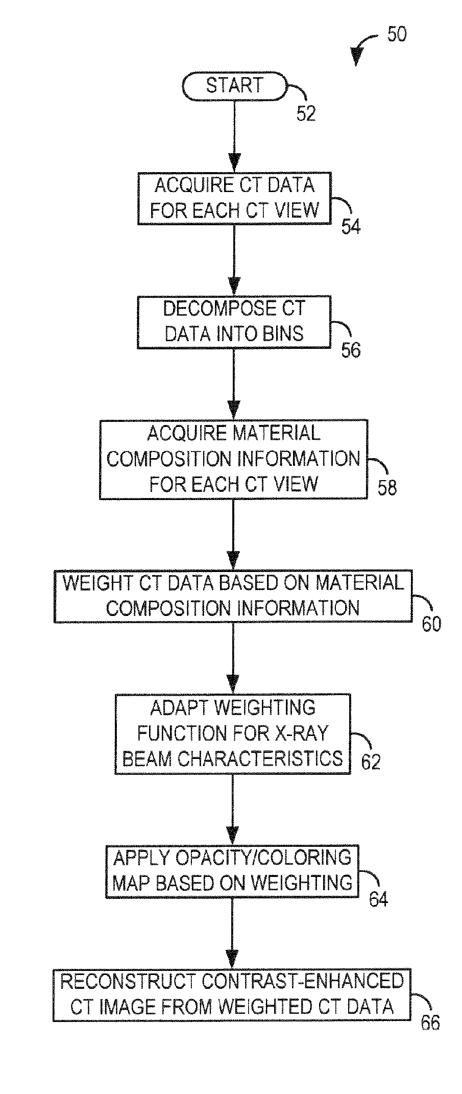
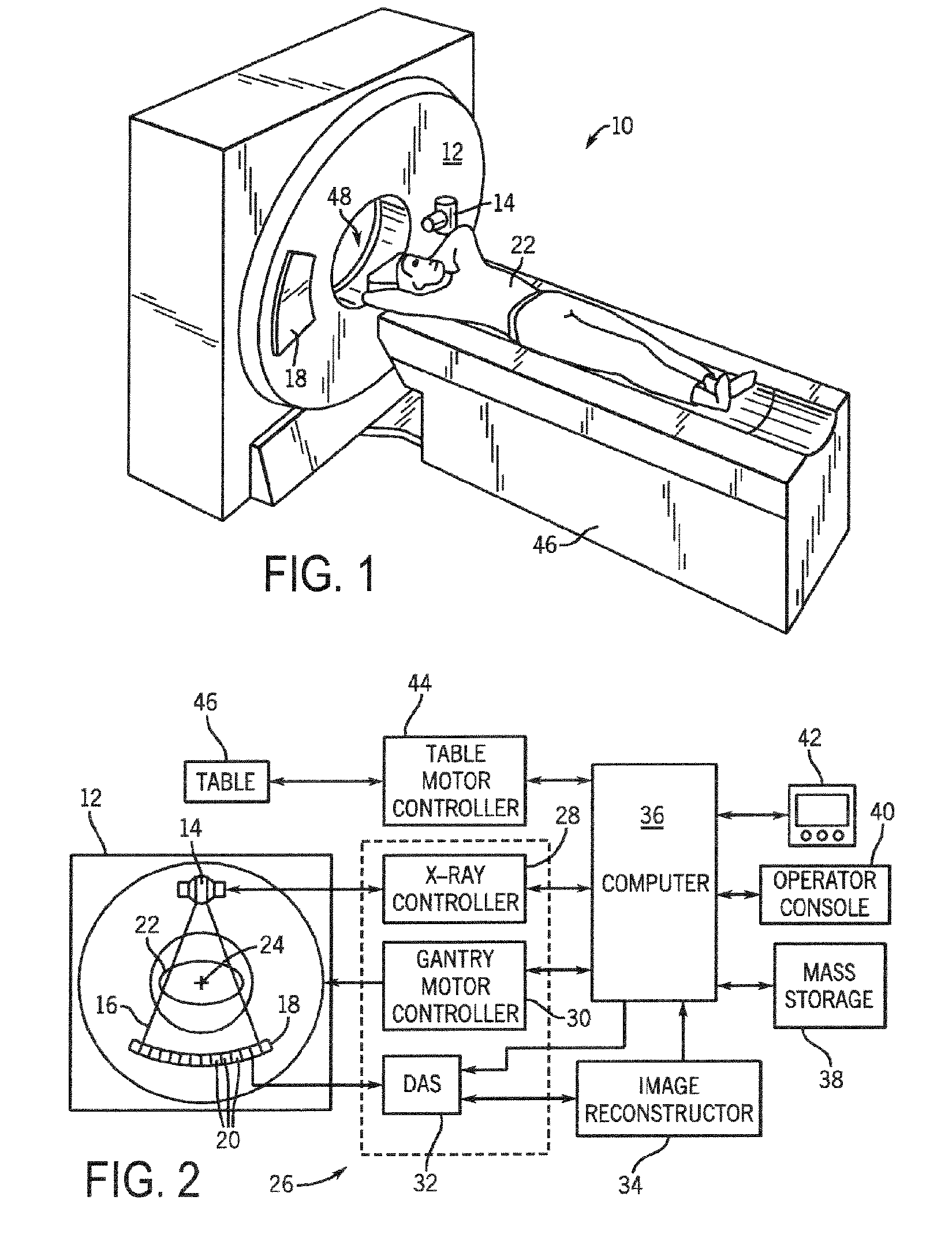
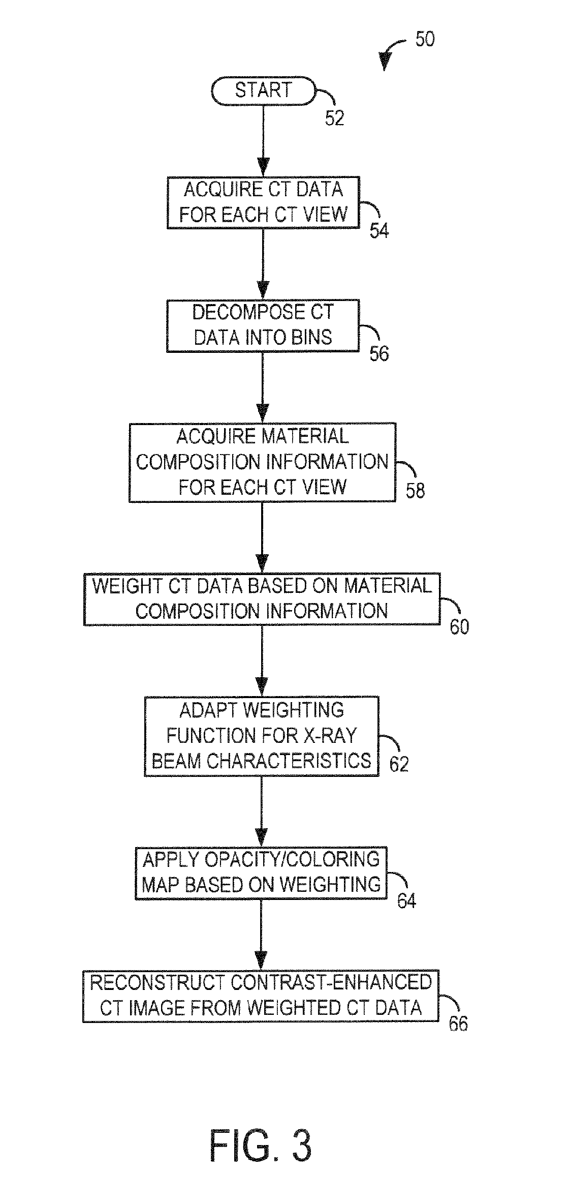
[0046] The present invention provides a system and method of reconstructing contrast-enhanced CT images that are substantially free of beam-hardening artifacts. The present invention is applicable with a photon counting (PC) radiographic system having a radiation energy detector configured to detect radiation energy at a given flux rate and output signals indicative of the detected radiation energy. The present invention is also applicable with an integrating energy selective detector, where the received radiation is registered in two or more energy ranges that may overlap through the use of either direct or indirect conversion detector materials using a layered design or depth of interaction to differentiate the energy bins. The present invention is also applicable with an energy integration detector and an x-ray source modulated to adjust the spectra for two or more different energy functions.

[0047] Referring now to FIGS. 1 and 2, a computed tomography (CT) imaging system 10 is s...


PUM


Abstract

A system and method are disclosed for reconstructing contrast-enhanced CT images that are substantially free of beam-hardening artifacts. An imaging system includes a radiation source configured to project radiation toward an object to be scanned and an energy discriminating detector assembly having a plurality of detector elements and configured to detect radiation emitted by the radiation source and attenuated by the object to be scanned. The imaging system also includes computer programmed to count a number of photons detected by each detector element and associate an energy value to each counted photon and determine a material composition of a CT view from the number of photons counted and the energy value associated with each counted photon. The computer is also programmed to apply a weighting to the CT view based on the material composition of the CT view and reconstruct an image with differential weighting based on the weighting of the CT view.

Description

BACKGROUND OF THE INVENTION [0001] The present invention relates generally to radiographic imaging and, more particularly, to a system and method for acquisition and reconstruction of contrast-enhanced and artifact-reduced CT images. The present invention further relates to system and method for processing CT data to increase contrast-to-noise ratio (CNR) and reduce artifacts in the reconstructed image. [0002] Typically, in radiographic systems, an x-ray source emits x-rays toward a subject or object, such as a patient or a piece of luggage. Hereinafter, the terms “subject” and “object” may be interchangeably used to describe anything capable of being imaged. The x-ray beam, after being attenuated by the subject, impinges upon an array of radiation detectors. The intensity of the radiation beam received at the detector array is typically dependent upon the attenuation of the x-rays through the scanned object. Each detector of the detector array produces a separate signal indicative ...

Claims


Application Information

Patent Timeline
IPC IPC(8): H05G1/60A61B6/00G01N23/00G21K1/12
CPCA61B6/032A61B6/4035A61B6/4241A61B6/482A61B6/542G01N2223/419G01N2223/423G06T11/006A61B6/5258
Inventor TKACZYK, J. ERICWALTER, DEBORAHDU, YANFENG
Owner GENERAL ELECTRIC CO
Learn More