Medical viewing system and method for detecting borders of an object of interest in noisy images

Inactive Publication Date: 2006-07-13
KONINKLIJKE PHILIPS ELECTRONICS NV
View PDF3 Cites 28 Cited by
  • Summary
  • Abstract
  • Description
  • Claims
  • Application Information

AI Technical Summary

Benefits of technology

[0023] The viewing system according to the invention further comprises enhancement means for enhancing the borders of the object of interest, using the location of said borders, and delivering an enhanced sequence of images. The knowledge of said location allows an outstanding enhancement of the contour of the object of interest. In the domain of angiography such an outstanding enhancement may help check the stent position and deployment.
[0024] The viewing system according to the invention also comprises measurement means for measuring characteristics of said object of interest using said location of borders. An advantage of said characteristics, which are for instance different widths of the

Problems solved by technology

As a matter of fact, clinical problems are associated with inadequate placement or expansion of the stent.
Inadequately expanded stents can locally disrupt blood flow and cause thrombosis.
During a PTCA it is possible to observe in real time the area of the stenosis in a sequence of angiographic images, but the precise stent placement is not easily visible for several reasons: the image sequence is rather noisy and its contrast is low due to the use of a low X-Ray dose, the stent lo

Method used


Image

  • Medical viewing system and method for detecting borders of an object of interest in noisy images
  • Medical viewing system and method for detecting borders of an object of interest in noisy images
  • Medical viewing system and method for detecting borders of an object of interest in noisy images

Examples


second embodiment

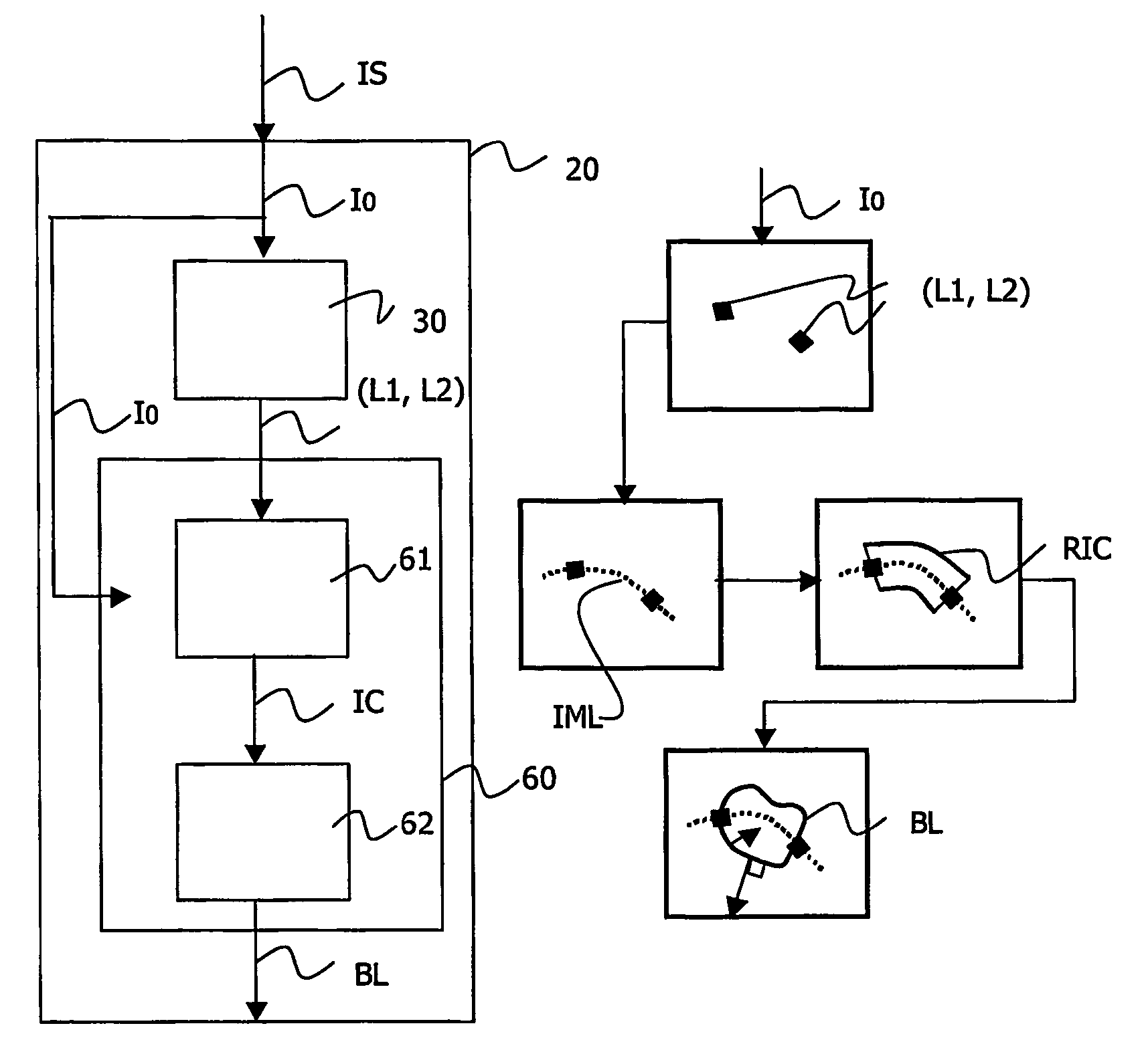
[0074] In the invention, the viewing system (as shown in FIG. 9) also comprises enhancement means 70 for enhancing said borders using said location of borders BL and for delivering an enhanced sequence of images EIS. Since said location of borders BL is known, the borders of the object of interest are enhanced very easily. Said borders are formed by points that have a gray level value. Said enhancement means 70 simply consist in increasing said gray level values so as to make the borders more visible. An outstanding enhancement is thus obtained and the main issue is to tune the enhancement so as to keep the enhanced sequence of images EIS acceptable for the practitioner.

third embodiment

[0075] In the invention, the viewing system (as shown FIG. 9) also comprises measurement means 71 for measuring characteristics CM of said object of interest using said location of borders BL. An interesting characteristic measure of a tubular object like a stent or an artery is a collection of widths of said object measured at several locations along a length of said object, for instance along said inter-marker line IML for a stent or along the tip for a stenosis. Variations of said widths may indicate whether the stenosis has been properly reduced or whether the stent has been properly expanded.

[0076] It is to be noted that the invention is not limited to width measures. The knowledge of the stent or of the artery borders also enables for instance an estimation of an agent contrast flow in the stenosis area to be derived, by measuring the mean contrast of contiguous sections of the artery at different times.

fourth embodiment

[0077] In the invention, the viewing system (as shown FIG. 9) also comprises 3D representation means 72 for delivering a three-dimensional (or 3D) representation 3DR of said object of interest using said location of borders BL. Such a 3D representation of a tubular object of interest, like an artery or a stent is easily obtained from two views (I0, I0′), which are preferably orthogonal views of said tubular object. It is not an issue in the domain of angiography, where an X-ray C-arm medical examination apparatus may provide two views in directions perpendicular to the axis of the tubular object and perpendicular to each other. It is also to be noted that very little distortion is introduced since the patient is placed at the center of the medical examination apparatus.

[0078] The localizers (L1, L2, L′1, L′2) detected in each sequence of images are matched and define a 3D referential in which all the points of the borders of the object of interest may be positioned in 3D. It is then...


PUM


Abstract

The invention relates to a viewing system and a method for detecting an object of interest in a sequence of images (IS). Said object of interest is detected by first locating localizers related to said object of interest and by locating borders (BL) related to said object of interest using the location (LI, LZ) of said localizers. The viewing system according to the invention is able to produce a sequence of enhanced images in which the object of interest is enhanced, to measure some characteristics and to build a three dimensional representation of said object of interest. The viewing system is also able to register and combine said sequence of enhanced images with a sequence of reference images.

Description

FIELD OF THE INVENTION [0001] The invention relates to a viewing system, comprising acquisition means for acquiring a sequence of images, detection means for detecting an object of interest in said sequence of images, and viewing means for displaying said sequence of images. The invention also relates to a method to be used in said system. The invention also relates to a medical examination apparatus coupled to such a system. [0002] The invention finds its application for example in medical imaging systems for the detection of objects of interest such as stents and artery walls in angiograms. BACKGROUND OF THE INVENTION [0003] A method for detecting stents in medical images is already known from the publication entitled “Deformable Boundary Detection of Stents in Angiographic Images”, by Ioannis Kompatsiaris et alii, in IEEE Transactions on Medical Imaging, Vol. 19, no 6, June 2000, pages 652-662. This document describes an image processing method for deformable boundary detection o...

Claims


Application Information

Patent Timeline
IPC IPC(8): A61B5/05G06T5/00
CPCG06T7/0012G06T7/0042G06T7/0083G06T7/0089G06T2207/10016G06T2207/10121G06T2207/20101G06T2207/30021G06T2207/30204G06T7/12G06T7/149G06T7/73
Inventor FLORENT, RAOULNOSJEAN, LUCILELELONG, PIERRERONGEN, PETER MARIA JOHANNES
Owner KONINKLIJKE PHILIPS ELECTRONICS NV
Why Eureka
Try Eureka