Methods for accelerating wound healing by administration of adipokines

a technology of adipokines and wound healing, applied in the field of wound healing acceleration by administration of adipokines, can solve the problems of impaired wound healing, inability to properly function, and inability to effectively promote so as to accelerate the healing process of damaged skin or skin wounds, accelerate the healing process of damaged skin, and promote the healing process.

Inactive Publication Date: 2006-08-10
HEALOR LTD
View PDF29 Cites 31 Cited by
  • Summary
  • Abstract
  • Description
  • Claims
  • Application Information

AI Technical Summary

Benefits of technology

[0012] It has further been found by the present inventors that the healing process of a damaged skin and skin wounds can be effectively promoted by administering to the skin cells colonizing the damaged skin or skin wound area an adipokine, an adipocyte modulator or a preadipocyte modulator.
[0013] Hence, the present invention provides, in one aspect, a method of inducing or accelerating a healing process of a damaged skin or skin wound, the method comprising administering to the skin cells colonizing the damaged skin or skin wound area a therapeutically effective amount of an agent selected from the group consisting of an adipokine, an adipocyte modulator, and a preadipocyte modulator, thereby inducing or accelerating the healing process of the damaged skin or skin wound.
[0014] According to another aspect of the present invention, there is provided a method of inducing or accelerating a healing process of a damaged skin or skin wound, the method comprising implanting onto the skin cells colonizing the damaged skin or skin wound area a therapeutically effective amount of adipocytes, preadipocytes or stem cells, thereby inducing or accelerating the healing process of the damaged skin or skin wound.
[0015] According to a further aspect of the present invention, there is provided a method of inducing or accelerating a healing process of a damaged skin or skin wound, the method comprising administering to the skin cells colonizing the damaged skin or skin wound area a therapeutically effective amount of a viral vector comprising a polynucleotide coding for an adipokine, thus transforming said skin cells to express and secrete said adipokine, thereby inducing or accelerating the healing process of the damaged skin or skin wound.

Problems solved by technology

Wound healing is impaired when these components, either individually or as a whole, do not function properly.
Yet skin ulceration in diabetic patients takes a staggering personal and financial cost (Knighton and Fiegel, 1993; Shaw and Boulton, 1997).
However, other mechanisms whereby the diabetic state associated with abnormal insulin signaling impairs wound healing and alters the physiology of skin has not been elucidated.
Another issue associated with impaired wound healing relates to infections of post surgical wounds occurring in 25% of patients hospitalized in surgical wards.
However, there is no teaching in the prior art for utilizing adipocytes, adipocyte or preadipocyte modulators, or molecules secreted by adipocytes, for inducing or accelerating the processes associated with wound healing.

Method used


Image

  • Methods for accelerating wound healing by administration of adipokines
  • Methods for accelerating wound healing by administration of adipokines
  • Methods for accelerating wound healing by administration of adipokines

Examples


example 1

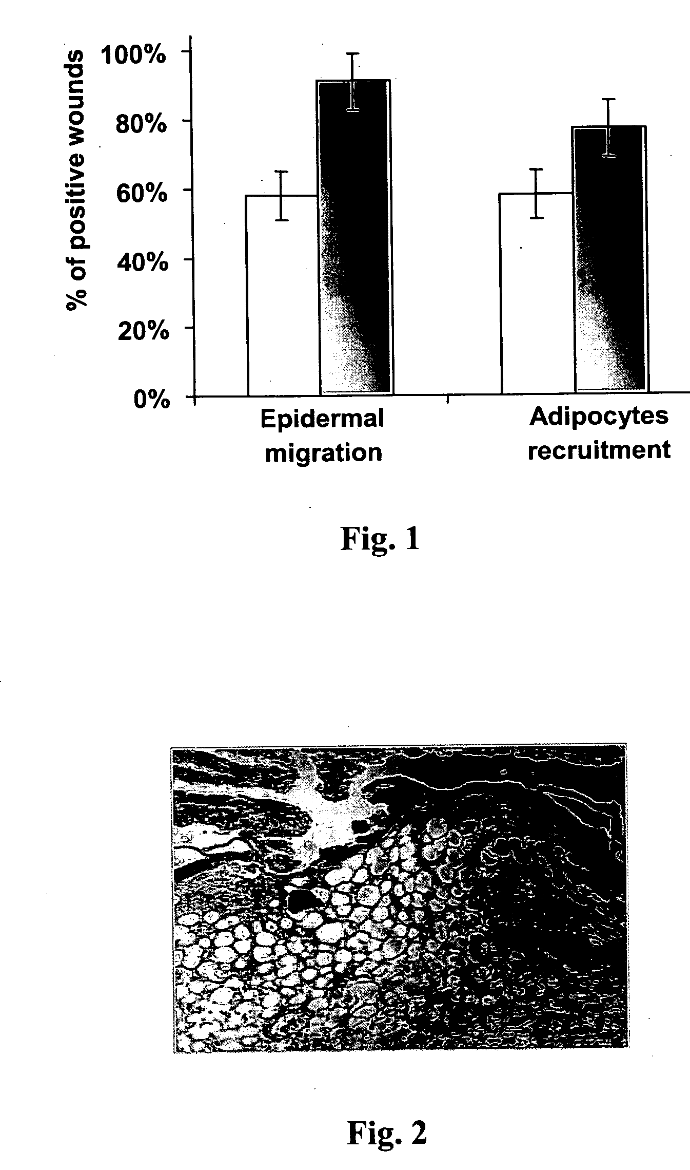
Association of Adipocytes with Migrating Keratinocytes During the Wound Healing Process

[0134] Migrating epidermal cells (keratinocytes) with recruited adipocytes were observed in 7-day-old wound tissue (FIG. 2). As can be seen in FIG. 1, migrating keratinocytes were observed across the wound gap in about 60% of the untreated wounds, and recruited adipocytes were also present in about 60% of the untreated wounds. FIG. 1 also shows that migrating keratinocytes and recruited adipocytes were observed in about 90% and 80% of insulin-treated wounds, respectively.

[0135] These results reveal that migration of keratinocytes to the wound gap area is closely associated with recruitment of adipocytes during an early stage of the wound healing process. The results thus indicate that migration of adipocytes to the wound area is involved in the wound healing process.

example 2

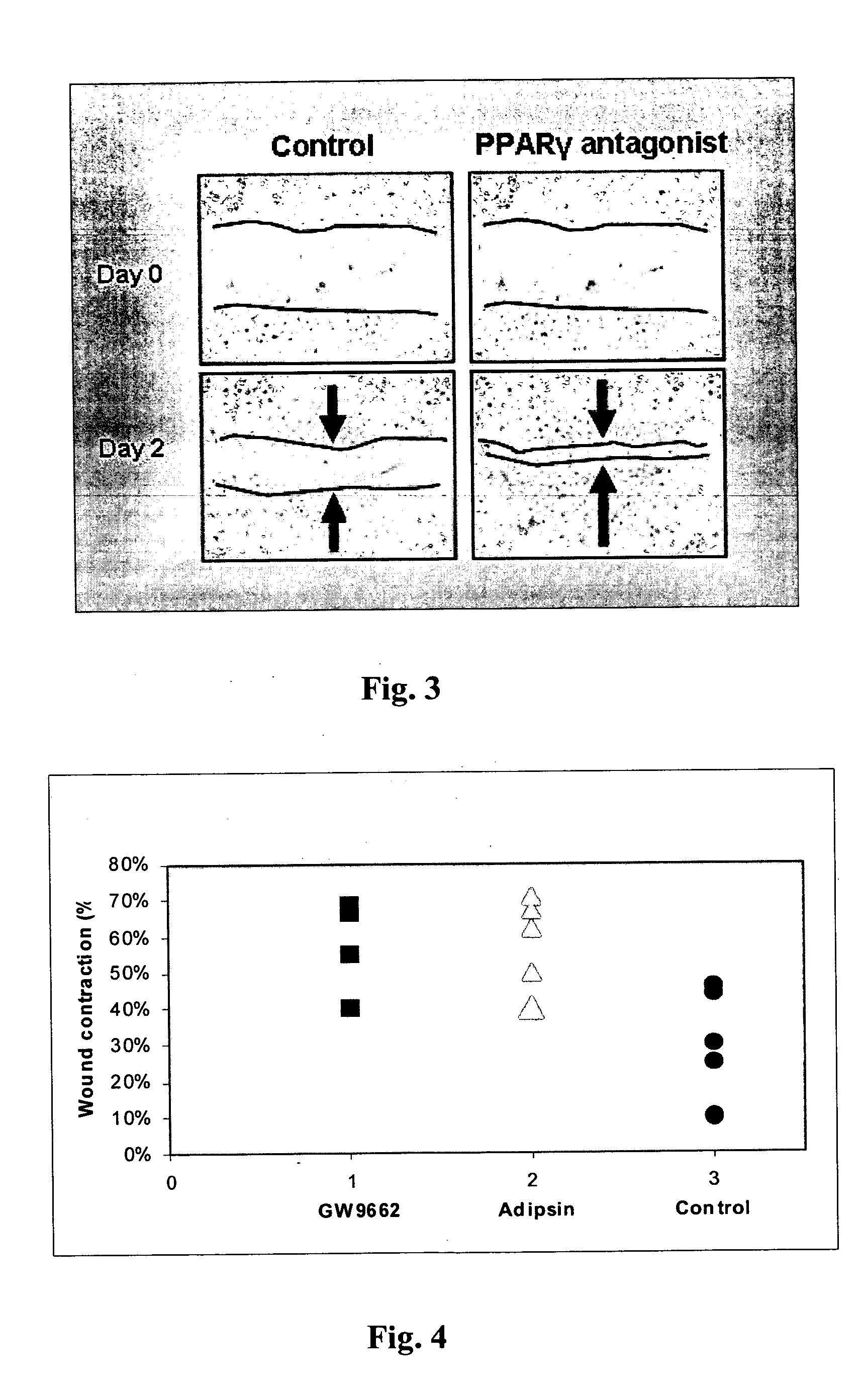
The Effect of PPARγ Modulators on Keratinocytes Migration and Wound Contraction

[0136] The effect of inhibiting or enhancing the activity of peroxisome proliferators-activated receptor gamma (PPARγ) on keratinocytes migration was evaluated utilizing the in vitro migration assay. Thus, primary keratinocytes were isolated and plated on 5 ml non-coated dishes until confluence in low Ca2+ medium, and an artificial crossover scratch was formed in each dish with a 200 μm tip. Cells were either non-treated (control) or treated daily with PPARγ modulators. As can be seen in FIG. 3, the treatment of cultured primary murine keratinocytes with the PPARγ antagonist GW9662 promoted keratinocytes migration. On the other hand, the treatment of cultured keratinocytes with the PPARγ agonist troglitazone inhibited keratinocytes migration (FIG. 8).

[0137] The effect of inhibiting or enhancing the activity of PPARγ using PPARγ antogonist and agonist, respectively, on wound healing was also evaluated in...

example 3

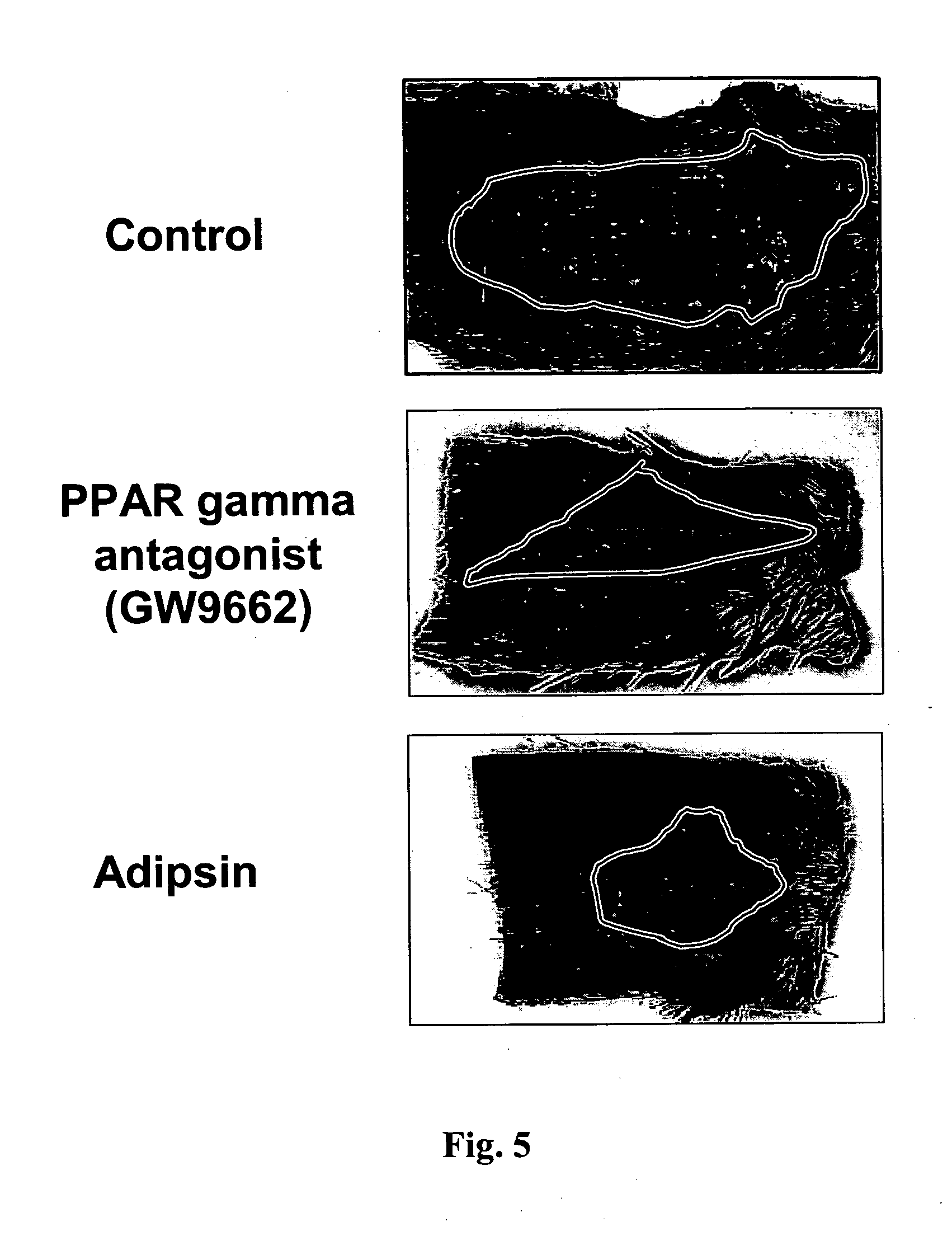
The Effect of Adipsin on Keratinocytes Migration and Wound Contraction

[0139] The effect of adipsin (complementing Factor D, which is secreted from adipocytes) on wound healing was evaluated in vivo. Accordingly, full thickness incision wounds were effected on the back of C57BL mice and were treated daily, for 6 days, with PBS buffer (control), or with 1 μM adipsin. The mice were sacrificed 6 days after wounding and the wounds were then analyzed. As illustrated in FIGS. 4-5 and 7, adipsin substantially promoted wound contraction. In addition, adipsin increased epidermal closure from about 15% to about 30%, and increased keratinocytes migration from about 30% to about 65%, as compared with the buffer control (FIG. 6).

[0140] These results demonstrate that an adipokine, such as adipsin, can effectively induce or accelerate wound healing.


PUM

PropertyMeasurementUnit
diameter×1aaaaaaaaaa
timeaaaaaaaaaa
thicknessaaaaaaaaaa

Abstract

Methods for inducing or accelerating a healing process of a damaged skin or skin wounds are described. The methods include administering to the skin cells colonizing the damaged skin or skin wound a therapeutically effective amount of an adipokine, an adipocyte or preadipocyte modulator, adipocytes, preadipocytes, or stem cells, or transforming the skin cells colonizing the damaged skin or skin wound such as to express and secrete an adipokine, thereby inducing or accelerating the healing process of the damaged skin or skin wound.

Description

CROSS-REFERENCE TO RELATED APPLICATIONS [0001] The present application is a continuation-in-part application of PCT application No. PCT / IL2004 / 000727, filed Aug. 05, 2004, in which the US is designated, and claims the benefit of US Provisional Patent Application No. 60 / 493,000, filed Aug. 7, 2003, now expired, the entire contents of each and both these applications being hereby incorporated by reference herein in their entirety as if fully disclosed herein.FIELD AND BACKGROUND OF THE INVENTION [0002] The present invention relates to methods and pharmaceutical compositions for inducing and / or accelerating cell proliferation and / or cell migration and / or cell differentiation and thereby accelerating the healing process of wounds. More particularly, the present invention relates to the use of bioactive molecules which are secreted by adipocytes (adipokines), and bioactive molecules which modulate adipocytes and preadipocytes differentiation, and / or adipocytes proliferation and / or activi...

Claims


Application Information

Patent Timeline
Patent Type & Authority Applications(United States)
IPC IPC(8): A61K38/19A61K38/18A61K38/48A61K
CPCA61K31/22A61K38/4826A61K35/28A61K35/35A61K38/22A61P17/02A61P43/00
Inventor BRAIMAN-WIKSMAN, LIORASOLOMONIK, INESSA
Owner HEALOR LTD
Why Eureka
Try Eureka