Methods of identifying genes which modulate myelination

Inactive Publication Date: 2006-08-17
ELAN PHARM INC
View PDF6 Cites 6 Cited by
  • Summary
  • Abstract
  • Description
  • Claims
  • Application Information

AI Technical Summary

Benefits of technology

[0019] In an embodiment, the expression of a molecule is increased

Problems solved by technology

Even though there is considerable remyelination and repair following the first immune attacks in multiple sclerosis (MS), there is a loss of ability to remyelinate over time, causing progressive neurodegeneration in patients in the secondary progressive phase of the disease.
These oligodendrocytes adhere to axons and form a few loose loops of membrane around them, but fail to produce internodes of compact myelin.
Failing to myelinate, these oligodendrocytes will undergo cell death.
There is not much known about why

Method used


Image

  • Methods of identifying genes which modulate myelination
  • Methods of identifying genes which modulate myelination
  • Methods of identifying genes which modulate myelination

Examples


example 1

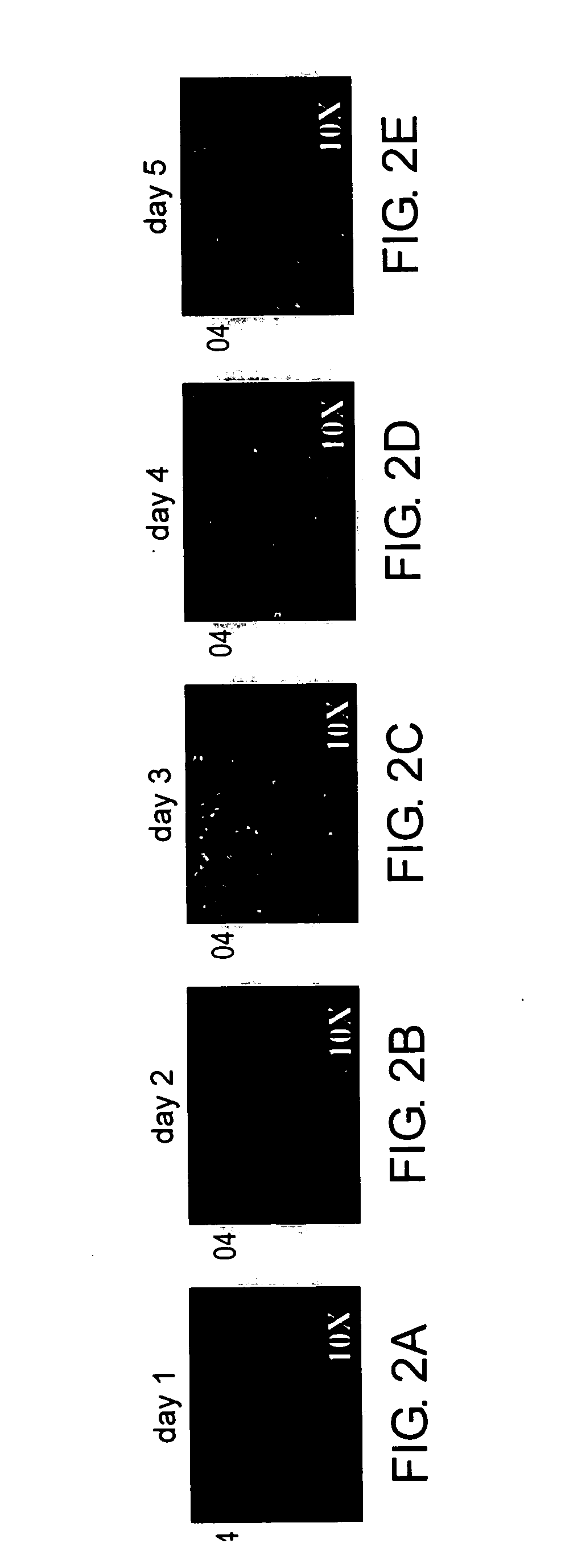
Development of Cell Culture Systems to Study Myelination

[0162] In order to address the myelinating and demyelinating aspects of the secondary progressive stage of MS, a cellular system was established to represent such MS lesions. A co-culture system of purified rat retinal ganglion cells (RGC) and optic nerve oligodendrocytes was used. This system recapitulates some significant aspects of MS lesions, namely, the oligodendrocytes loosely wrap membrane around axons, but fail to form internodes of compact myelin. The RGC neurons in these cell cultures are, for the most part, electrically inactive and may resemble dystrophic neurons observed in MS lesions. Furthermore, cell isolation, purification, and culture methods, conditions and compositions were developed, as described herein.

[0163] Purification of optic nerve oligodendrocytes was conducted as follows. Optic nerve oligodendrocytes are purified from postnatal day 5 (p5) Sprague-Dawley (“SD”) rat pups according to the methods des...

example 2

Use of siRNA as a Control for Oligodendrocyte Cell Culture Screening and Modulation of Myelination

[0184] It was also demonstrated herein that oligodendrocytes are competent for RNAi techniques. The criterion for choosing an endogenous target for RNAi requires that the target is relatively non-essential for oligodendrocyte survival and functioning. To accomplish this, the endogenously expressed cell adhesion molecule NCAM was selected for preliminary validation. Four RNAi oligonucleotides for NCAM were prepared (Qiagen). Oligodendrocytes were grown in mitogenic media for 3 days and differentiated for two days before transfecting with siNCAM (“siNCAMs 1-4,” which represent a set of four different siRNAs that were evaluated for use in RNAi experimentation as described herein).

[0185] The protocol for siRNA transfection into oligodendrocytes is as follows. All transfections were conducted using 24-well plates, The first solution included 50 μL OptiMEM medium, 0.8 μg plasmid DNA and / or ...

example 3

In Vitro Myelination Assay

[0188] Optic nerve or cortical oligodendrocytes were purified from postnatal day 2-day 5 old rat pups by immuno-panning with the A2B5 antibody (ATCC). Retinal ganglion cells were purified from 5 day old rats by immunopanning with the Thy1 antibody T11D7.

[0189] Purified oligodendrocytes were plated on PDL / merosin substrate-coated tissue culture dishes in defined DMEM Sato medium, promoting proliferation for two days. After 2 days, the medium was switched to defined DMEM Sato medium, containing factors promoting differentiation. Twenty-four hours after the switch to differentiation medium, ADORA1 agonists were added. Retinal ganglion cells were plated 24 hours thereafter, and medium consisted of astrocyte-conditioned medium with DMEM Sato medium mixed with defined Neurobasal™ Sato medium, containing growth factors promoting retinal ganglion cell survival and 0.5% FBS.

[0190] Cultures were grown for two weeks, with medium changes occurring every three days. ...


PUM

PropertyMeasurementUnit
Fractionaaaaaaaaaa

Abstract

The present invention relates to methods of identifying and/or characterizing genes that are modulated during myelination or remyelination, as well as in demyelinating diseases, such as in multiple sclerosis. The invention further provides methods of treating diseases related to myelination in a mammal, such as multiple sclerosis, by administering agents that promote remyelination of oligodendrocytes.

Description

CROSS-REFERENCE TO RELATED APPLICATION [0001] The present application is entitled to priority under 35 U.S.C. § 119(e), to U.S. Provisional Application No. 60 / 652,477, filed on Feb. 11, 2005, which application is hereby incorporated herein by reference in its entirety.REFERENCE TO A SEQUENCE LISTING [0002] This application includes a sequence listing in accordance with 37 C.F.R. 1.821 et seq., which sequence listing is hereby incorporated by reference in its entirety. FIELD OF THE INVENTION [0003] The application relates to methods of identifying and / or characterizing genes that are modulated in myelination, remyelination, and in demyelinating diseases, such as in multiple sclerosis. The invention further provides methods of treating multiple sclerosis in a mammal by administering agents that promote remyelination of oligodendrocytes. BACKGROUND [0004] Even though there is considerable remyelination and repair following the first immune attacks in multiple sclerosis (MS), there is a...

Claims


Application Information

Patent Timeline
IPC IPC(8): C12Q1/68
CPCC12Q1/6809C12Q1/6883C12Q2600/158
Inventor MEYER-FRANKE, ANKE
Owner ELAN PHARM INC
Why Eureka
Try Eureka