Apparatus and method for intravascular imaging

a technology of intravascular imaging and guidewires, which is applied in the field of imaging guidewires and catheters, can solve the problems of failure of drive cables operated by motors located outside the patient's body and connected to transducers, failure of drive cables, and failure of cables

Inactive Publication Date: 2006-10-19
MARTINELLI MICHAEL A
View PDF12 Cites 108 Cited by
  • Summary
  • Abstract
  • Description
  • Claims
  • Application Information

AI Technical Summary

Problems solved by technology

The path in the vascular cavity along which the imaging guidewire or catheter travels can often be tortuous causing difficulties for the various mechanical or electrical devices used for causing ultrasonic signals to scan surrounding tissue.
One problem encountered by known ultrasonic probes is failure of a drive cable operated by a motor located outside of the patient's body and connected to a transducer or reflector disposed within the probe.
Oftentimes the drive cable is unable to provide uniform rotation of the transducer or reflector, causing artifacts in the ultrasound image of tissue surrounding the probe.
Sometimes rapid and repetitive rotations of the drive cable will result in cable failure.

Method used


Image

  • Apparatus and method for intravascular imaging
  • Apparatus and method for intravascular imaging
  • Apparatus and method for intravascular imaging

Examples


Embodiment Construction

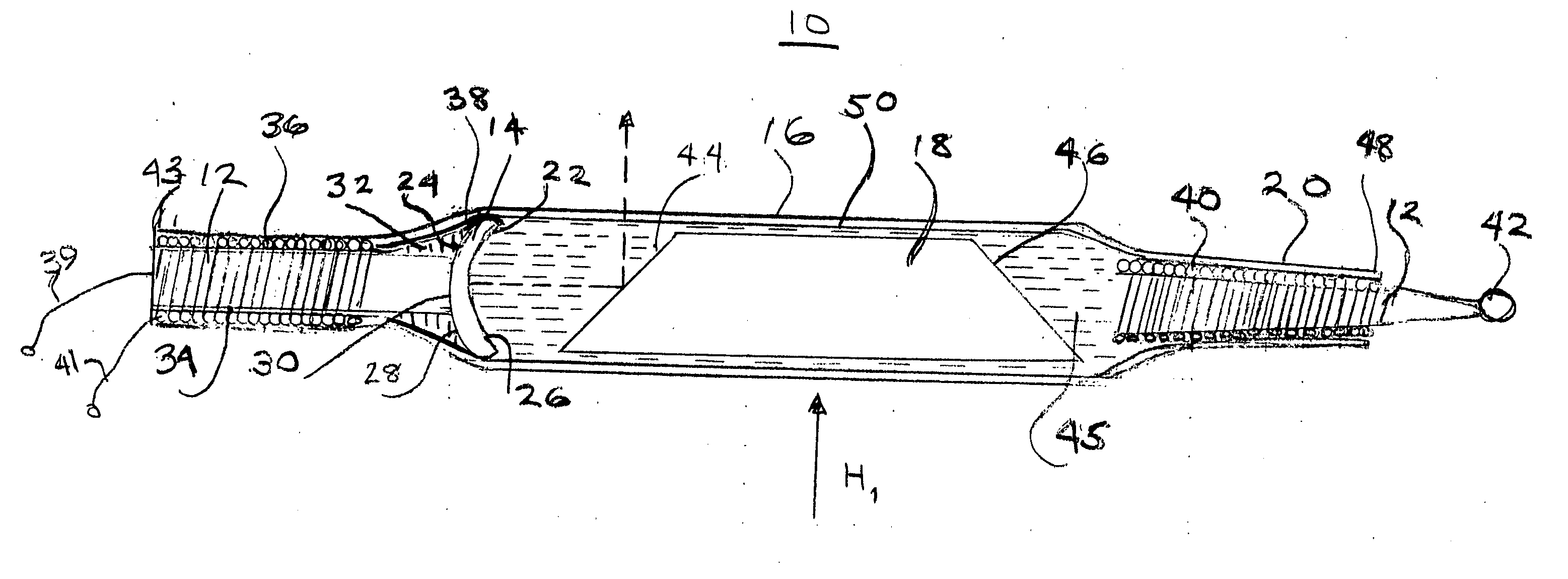
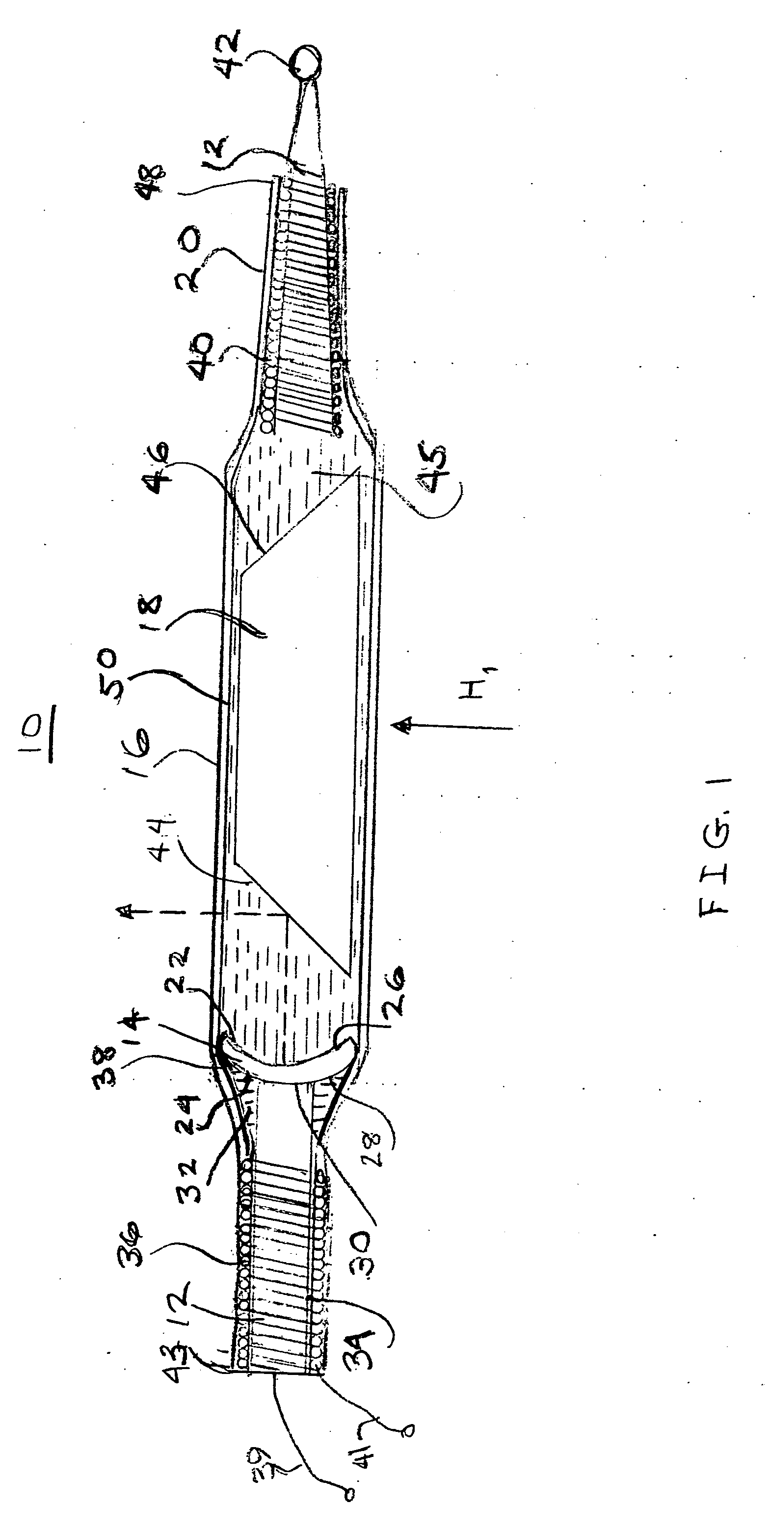
[0026] This invention provides an apparatus and method for imaging tissues from inside a patient's body by utilizing a rotating magnetic field generated outside of the patient's body to cause a substantially synchronous rotation of an ultrasonic signal inside the patient's body.

[0027] Referring to FIG. 1, there is shown a side sectional view of a catheter or distal end of an imaging guidewire 10 including a central core wire 12, a piezoelectric transducer 14, a tubular housing or sleeve 16, a permanently magnetized cylindrical slug 18, and a flexible tip guide 20. The slug 18 may be formed from a rare earth magnetic material such as neodymium iron boron or samarium cobalt or from a suitable ferrite material. The magnetization of slug 18, represented by the magnetic field vector H1, is substantially orthogonal to the longitudinal axis of slug 18. A metal cladding 17 over slug 18 is intended to enhance acoustic reflection and minimize corrosion of the rare earth material. The piezoel...


PUM


Abstract

A method and apparatus for intravascular imaging utilizes a rotating magnetic field generated outside of the patient's body to cause a substantially synchronous rotation of an ultrasonic signal inside the patient's body.

Description

CROSS-REFERENCE TO RELATED APPLICATIONS [0001] THIS APPLICATION CLAIMS THE BENEFIT OF U.S. PROVISIONAL APPLICATION NO. 60 / 671,008, FILED Apr. 13, 2005FEDERALLY SPONSORED RESEARCH [0002] Not Applicable BACKGROUND OF THE INVENTION [0003] 1. Field of the Invention [0004] The present invention relates to imaging guidewires and catheters, more particularly, to intravascular imaging guidewires and catheters that can scan an ultrasonic signal against tissues surrounding the guidewire or catheter by utilizing a rotating magnetic field applied from outside a patient's body to rotate a permanent magnet disposed within the guidewire or catheter. [0005] 2. Description of Related Art [0006] Ultrasonic imaging of tissue surrounding a vascular cavity has long been a tool for determining the condition of such tissue. Apparatus for introducing ultrasonic signals into a desired location in a vascular cavity have included imaging guidewires and catheters adapted to slide along a guidewire. The path in...

Claims


Application Information

Patent Timeline
Patent Type & Authority Applications(United States)
IPC IPC(8): A61M25/00
CPCA61B8/12A61B8/445G10K11/357A61B8/4461
Inventor MARTINELLI, MICHAEL A.
Owner MARTINELLI MICHAEL A
Why Eureka
Try Eureka