Magnetic resonance imaging apparatus and magnetic resonance imaging method

a magnetic resonance imaging and magnetic resonance imaging technology, applied in the direction of reradiation, measurement using nmr, instruments, etc., can solve the problems of inconvenience, difficulty in efficiently performing the imaging of a subject, artifacts, etc., and achieve the effect of improving imaging efficiency

Active Publication Date: 2006-11-16
GE MEDICAL SYST GLOBAL TECH CO LLC
View PDF6 Cites 59 Cited by
  • Summary
  • Abstract
  • Description
  • Claims
  • Application Information

AI Technical Summary

Benefits of technology

[0008] Therefore, an object of the present invention is to provide a magnetic resonance imaging apparatus and magnetic resonance imaging method, which may further improve the efficiency of imaging.
[0011] In accordance with the present invention, a magnetic resonance imaging apparatus and a magnetic resonance imaging method are provided which may improve the imaging efficiency.

Problems solved by technology

However, in the actual scanning image thus generated as have been described above there may be cases in which some artifacts happen due to the crosstalk or phase folding, or in which desired section is not captured.
In particular such inconvenience becomes evident when the scanning is performed in a multi-slice or multi-angle scheme.
This could lead to difficulties of efficiently performing the imaging of a subject.

Method used


Image

  • Magnetic resonance imaging apparatus and magnetic resonance imaging method
  • Magnetic resonance imaging apparatus and magnetic resonance imaging method
  • Magnetic resonance imaging apparatus and magnetic resonance imaging method

Examples


Embodiment Construction

[0018] Hereinafter, an exemplary preferred embodiment of the present invention will be described in greater details with reference to accompanying drawings.

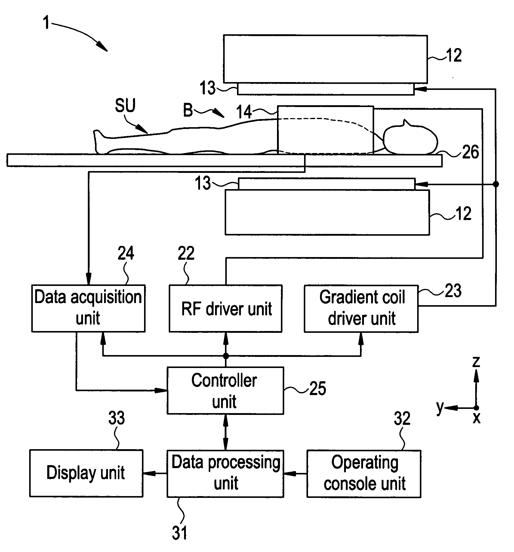
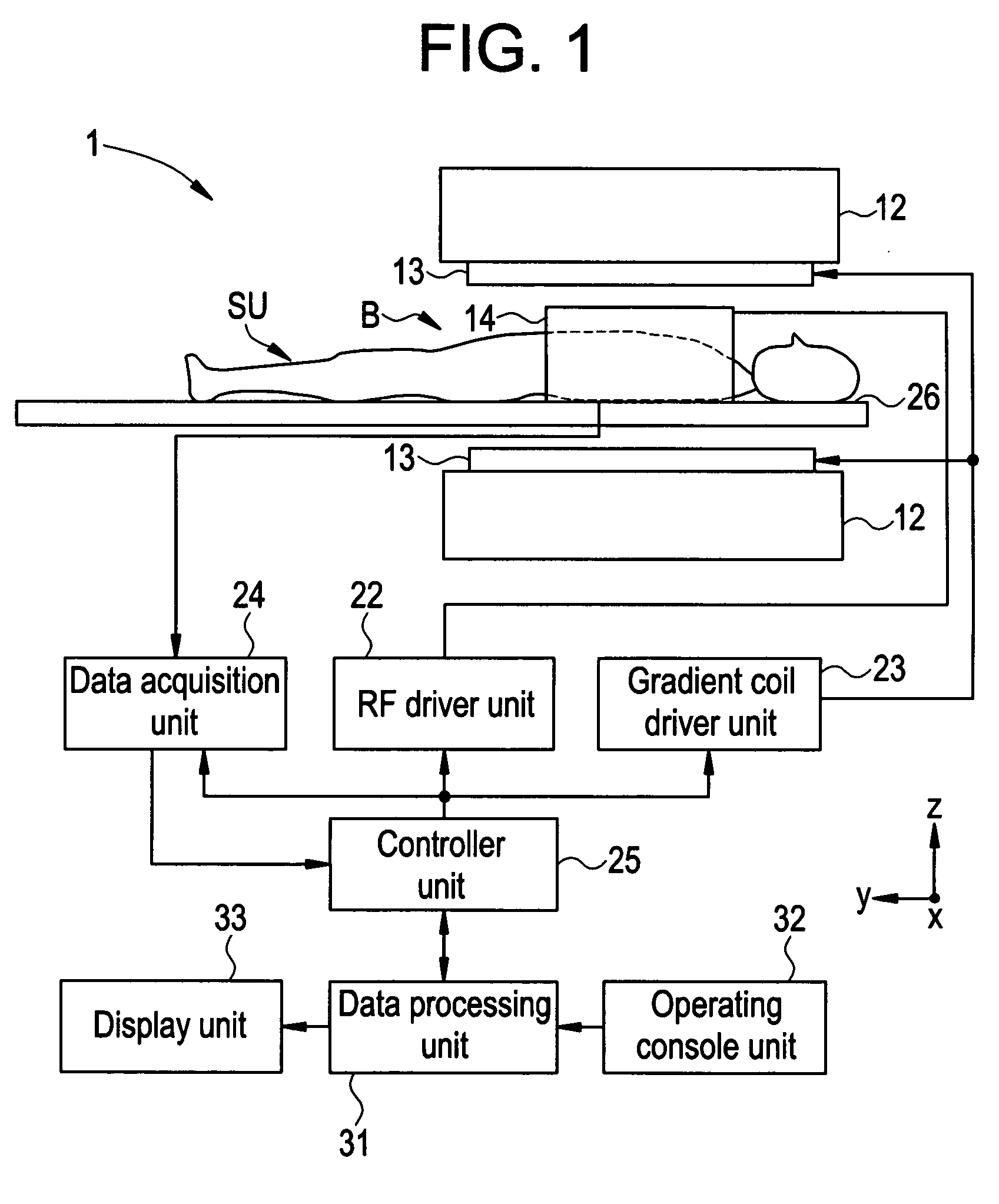
[0019] Now referring to FIG. 1, there is shown a schematic block diagram of a magnetic resonance imaging apparatus 1 in accordance with a preferred embodiment of the present invention.

[0020] As shown in FIG. 1, the magnetic resonance imaging apparatus 1 includes a magnetostatic field magnet unit 12, a gradient coil unit 13, an RF coil unit 14, an RF driver unit 22, a gradient coil driver unit 23, a data acquisition unit 24, a controller unit 25, a cradle 26, a data processing unit 31, an operating console unit 32, and a display unit 33. The magnetic resonance imaging apparatus 1 transmits electromagnetic waves to a subject SU placed in an imaging space B with magnetostatic field formed, to perform a scan for obtaining magnetic resonance signals from the subject SU, to reconstruct an image of the slice of the subject SU based on...


PUM


Abstract

The present invention provides a magnetic resonance imaging apparatus and a magnetic resonance imaging method which improve the imaging efficiency. A preview scan initiator unit is provided for initiating a preview scan for scanning the slice position set prior to performing the actual imaging scan. Based on the magnetic resonance signals from the subject obtained by the review scan initiated by the preview scan initiator unit, the preview image for the slice position set by the slice position setting unit is generated to display on the display screen by the display unit.

Description

CROSS REFERENCE TO RELATED APPLICATIONS [0001] This application claims the benefit of Japan Application No. 2005-136815 filed May 10, 2005. BACKGROUND OF THE INVENTION [0002] The present invention is related to a magnetic resonance imaging apparatus and magnetic resonance imaging method. [0003] A magnetic resonance imaging (MRI) apparatus is known which uses the nuclear magnetic resonance phenomenon (NMR) to take tomographic images of a subject. The MRI apparatus is widely used in such fields as medical and industrial. [0004] When taking a tomographic image of a subject by using an MRI apparatus, the subject is placed in the imaging field where a static magnetic field is formed, to align the spin direction of protons within the subject in the direction of magnetostatic field to obtain magnetic vector. Then, electromagnetic waves at a resonance frequency are emitted from an RF coil to generate the NMR phenomenon to modify the magnetic vector of the protons. The MRI apparatus then rec...

Claims


Application Information

Patent Timeline
Patent Type & Authority Applications(United States)
IPC IPC(8): G01V3/00
CPCG01R33/4835G01R33/546G01R33/543G01R33/54
Inventor IKEDA, HITOSHI
Owner GE MEDICAL SYST GLOBAL TECH CO LLC
Why Eureka
Try Eureka