Ghrelin binding nucleic acids

a nucleic acid and ghrelin technology, applied in the field of nucleic acids binding to ghrelin, can solve the problems of inducing overeating and hunger in test subjects

Inactive Publication Date: 2006-11-16
NOXXON PHARM AG
View PDF5 Cites 96 Cited by
  • Summary
  • Abstract
  • Description
  • Claims
  • Application Information

AI Technical Summary

Benefits of technology

[0094] The present invention is further illustrated by the figures, examples and the sequence listing from which further features, embodiments and advantages may be taken, wherein
[0095]FIG. 1 shows an alignment of sequences of RNA ligands binding to human ghrelin. In FIG. 1 the sequences are shown in the Sequence Listing with the following sequence identifiers: SEQ ID NO.Sequence5MS-P2-E36MS-P2-G27MS-P2-D28MS-P2-A39MS-P2-E110MS-P2-B111MS-P2-F112MS-P2-C313MS-P2-C214MS-P2-H215MS-P2-A416MS-P2-B217MS-P2-A218MS-P3-H319MS-P2-D1
[0096]FIG. 2 shows a competition assay of binding to ghrelin with truncated aptamers versus the known sequence SOT-C (B11trc).
[0097]FIG. 3 shows an alignment of chosen sequences of RNA ligands, which were published in patent application WO 2004 / 013274 A2. In FIG. 3 the sequences are shown in the Sequence Listing with the following sequence identifiers: SEQ ID NO:Sequence20SOT-C (B11trc)23SOT-D-00022SOT-D (C12)21F12
[0098]FIG. 4 shows calculated secondary structure of ghrelin binding RNA spiegelmer clone SOT-C, the secondary structure was calculated with the program “RNAfold” (Hofacker et al., 1994, Monatsh. Chem 124:167-188).
[0099]FIG. 5 shows calculated secondary structure of ghrelin binding RNA spiegelmer clone SOT-D-000 (SEQ ID NO: 23), the secondary structure was calculated with the program “RNAfold” (Hofacker et al., 1994, Monatsh. Chem 125:167-188).
[0100]FIG. 6 shows calculated secondary structure of ghrelin binding RNA spiegelmer clone SOT-D (SEQ ID NO: 22), the secondary structure was calculated with the program “RNAfold” (Hofacker et al., 1994, Monatsh. Chem 125:167-188).
[0101]FIG. 7 shows calculated secondary structure of ghrelin binding RNA spiegelmer clone F-12 (SEQ ID NO: 21), the secondary structure was calculated with the program “RNAfold” (Hofacker et al., 1994, Monatsh. Chem 125:167-188).
[0102]FIG. 8 shows an alignment of SOT-D-000 derivates which are the result of truncation and relational design experiments. In FIG. 8 the sequences are shown in the Sequence Listing with the following sequence identifiers: SEQ ID NO:Sequence23SOT-D-00024SOT-D-10025SOT-D-10126SOT-D-10227SOT-D-10428SOT-D-10629SOT-D-10830SOT-D-10931SOT-D-11032SOT-D-111
[0103]FIG. 9 shows a one-point measurement for the inhibition of ghrelin-induced Ca++-release by Spiegelmers SOT-C, SOT-D-000 and variants thereof at room temperature; cells were stimulated with 5 nM ghrelin preincubated at room temperature with various amounts of Spiegelmer SOT-C, SOT-D000 or a variant of D-000 the results show the percentage of fluorescence signal normalized to the signal obtained with no Spiegelmer.
[0104]FIG. 10 shows a presumptive secondary structure of ghrelin binding RNA spiegelmer clone SOT-D-109 (SEQ ID NO: 30).
[0105]FIG. 11 shows a dose-response curve for the inhibition of ghrelin-induced Ca++-release by Spiegelmers D-109 and 5′-PEGylated D-109 at room temperature; cells were stimulated with 5 nM ghrelin preincubated at room temperature with various amounts of Spiegelmer D-109 and 5′-PEGylated D-109; the results show the percentage of fluorescence signal normalized to the signal obtained with no Spiegelmer; Spiegelmer D-109 and its modified versions were found to inhibit ghrelin-induced Ca++-release with same IC50.
[0106]FIG. 12 shows calculated secondary structure of ghrelin binding RNA Spiegelmer clone SOT-E (SEQ ID NO: 33), the secondary structure was calculated with the program “RNAfold” (Hofacker et al., 1994, Monatsh. Chem 125:167-188).
[0107]FIG. 13 shows binding analysis of truncated variants of Spiegelmers SOT-E tested by cell culture experiments.
[0108]FIG. 14 shows a presumptive secondary structure of ghrelin binding RNA Spiegelmer SOT-E-19 (SEQ ID NO: 39).
[0109]FIG. 15 shows a presumptive secondary structure of ghrelin binding RNA spiegelmer clone SOT-E-21 (SEQ ID NO: 40).
[0110]FIG. 16 shows a presumptive secondary structure of ghrelin binding RNA spiegelmer clone SOT-E-33 (SEQ ID NO: 42).
[0111]FIG. 17 shows a presumptive secondary structure of ghrelin binding RNA spiegelmer clone SOT-E-25 (SEQ ID NO: 41).
[0112]FIG. 18 shows the Biacore 2000 sensorgrams indicating the KD values of D-ghrelin binding RNA clones SOT-C and SOT-E;
[0113]FIG. 19 shows a dose-response curve for the inhibition of ghrelin-induced Ca++-release by Spiegelmer SOT-E, SOT-E-19-5 ′-Amino, SOT-E-19-5′-PEG or SOT-D-109 at 37° C.; cells were stimulated with 2 nM human ghrelin preincubated at 37° C. with various amounts of Spiegelmer SOT-E, SOT-E-19-5′-Amino, SOT-E-19-5 ′-PEG or SOT-D-109; the results show the percentage of fluorescence signal normalized to the signal obtained with no Spiegelmer; Spiegelmer SOT-E-19 and its modified versions were found to inhibit ghrelin-induced Ca++-release with an IC50 of about 4 nM;
[0114]FIG. 20A shows the definition of the sequence boxes which are characteristic for ghrelin binding Spiegelmers;
[0117]FIG. 20D one variant of FIG. 20A;
[0117]FIG. 20D one variant of FIG. 20A;
[0117]FIG. 20D one variant of FIG. 20A;
[0118]FIG. 21 shows the sequences of SOT-D-109 (SEQ ID NO: 30), SOT-E (SEQ ID NO: 33), SOT-E-19 (SEQ ID NO: 39), SOT-E-21 (SEQ ID NO: 40), SOT-E-33 (SEQ ID NO: 42) and SOT-E-25 (SEQ ID NO: 41).
[0119]FIG. 22 shows the inhibition of growth hormone release after exogenous ghrelin administration by anti-ghrelin Spiegelmer SOT-E19-5′-PEG.
[0120]FIG. 23 shows an overview of different derivatives of SOT-E-19 which comprise additional nucleotides instead of the internal linker, and the observed IC50 value expressed in nM. In FIG. 23 the sequences are shown in the Sequence Listing with the following sequence identifiers: SEQ ID NO:Sequence33SOT-E39SOT-E-1943SOT-E19-L44SOT-E19-L145SOT-E19-L246SOT-E19-L347SOT-E19-L448SOT-E19-L549SOT-E19-L650SOT-E19-L7
[0121]FIG. 24 shows the result of a cellular competition assay with octanoyl-ghrelin, des-octanoyl-ghrelin, and Spiegelmer SOT-E-19, with combinations and concentrations of the components summarized below the bars.
[0122]FIG. 25 shows a standard curve of absorbance versus human ghrelin (octanoylated) concentration recorded by an EIA type detection assay whereas the ghrelin-binding Spiegelmer NOX—B11 was used to immobilize human ghrelin (octanoylated) thus allowing the quantification of human ghrelin (octanoylated).
[0123] FIGS. 26A-D shows various steps of method for quantification of octanoyl-ghrelin using a ghrelin binding nucleic acid according to the present invention.

Problems solved by technology

However, in biological samples a mixture of both the octanoyl ghrelin which is a form of a bioactive ghrelin, and the unmodified or des-octanoyl ghrelin is present.
Moreover, systemic ghrelin administration in humans cause sensations of hunger in the test subjects and induce overeating.

Method used


Image

  • Ghrelin binding nucleic acids
  • Ghrelin binding nucleic acids
  • Ghrelin binding nucleic acids

Examples


example 1

Ghrelin Binding Sequences

[0220] If not explicitly indicated to the contrary, the ghrelin used throughout these examples was ghrelin in its octanoylated form.

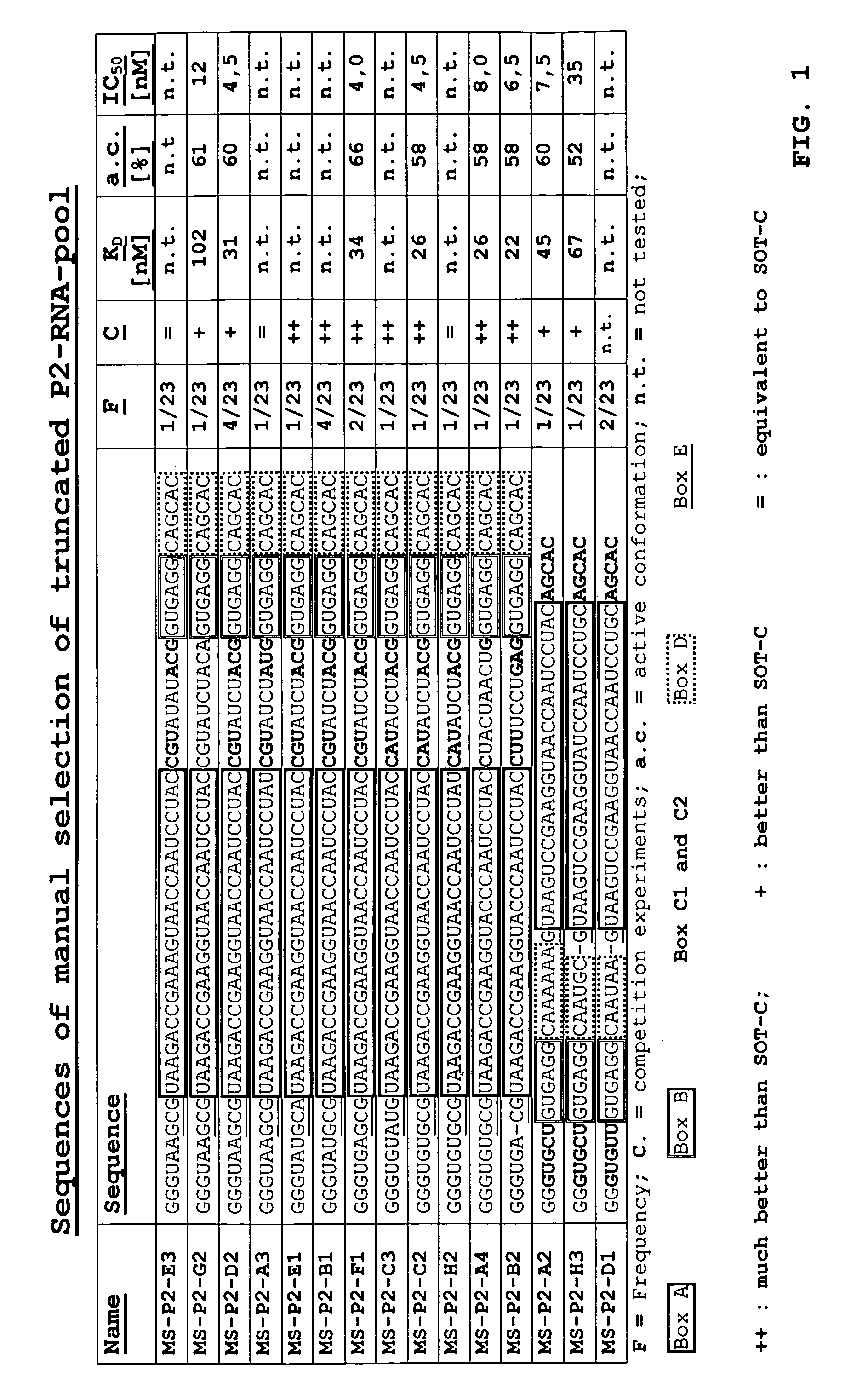
[0221] Using a technology derived from the one described in DE 10349441.3 an RNA in vitro selection directed to biotinylated human D-ghrelin was carried out. An enriched population of dsDNA molecules was cloned and sequenced. The result of the sequence analysis can be seen in FIG. 1.

[0222] All sequences comprise the Ghrelin-binding motif A (25 nucleotides) of the known Ghrelin-binding sequence SOT-C (B11trc; see patent application WO2004 / 013274 A2). Only in four out of these 23 clones motiv A is rather located at the 3′-end of the sequence. Within 19 sequences motif A is located at the 5′-end of the clones. Additionally motif called motiv D is located at the 3′-end of motif A.

[0223] Binding Characteristics of the Sequences

[0224] The 15 clones (FIG. 1) showing variations at different positions were chosen for ranking experim...

example 2

Binding Characterization of the Sequences

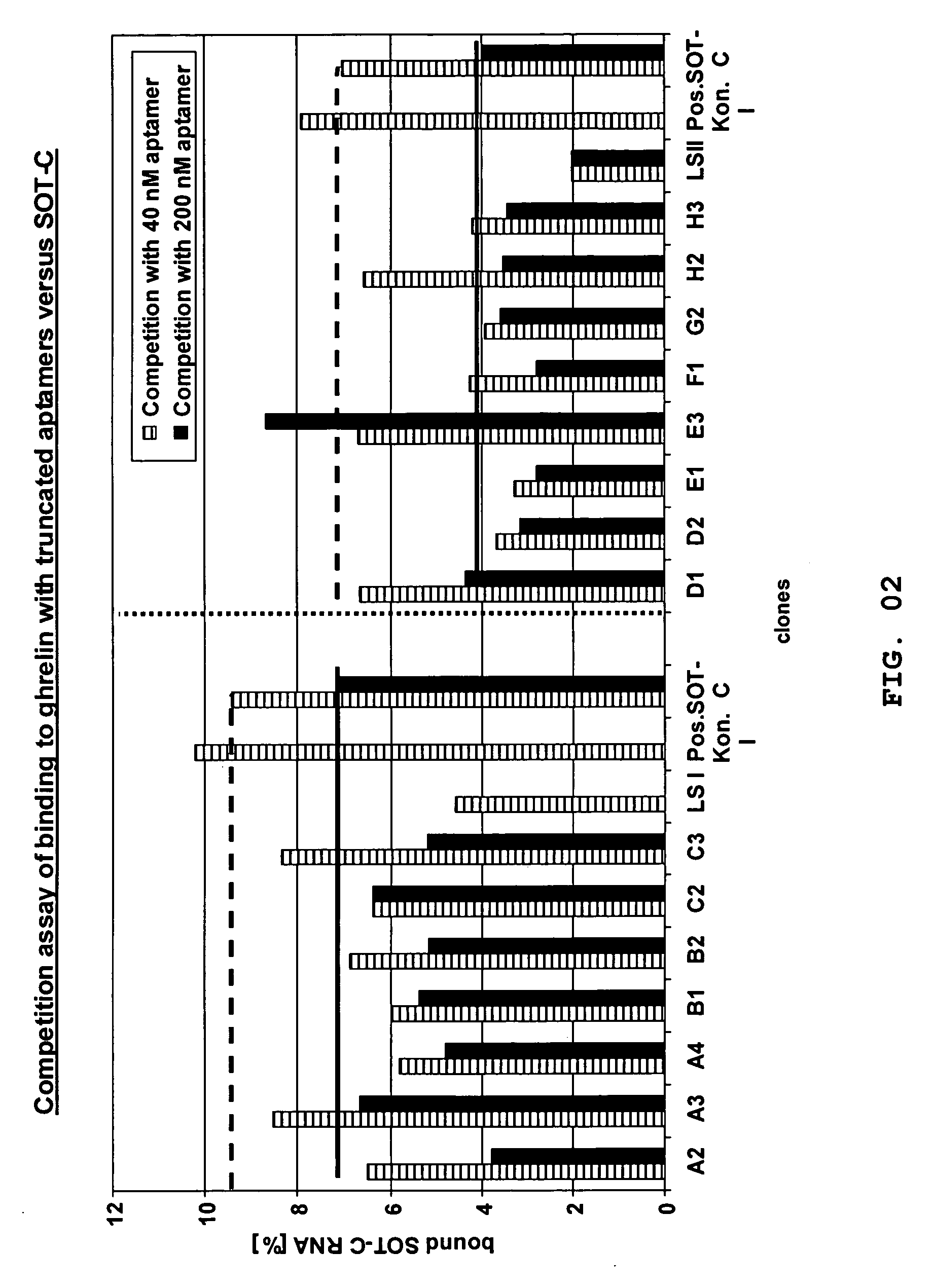
[0226] 2.1 Ranking of Radiolabeled Aptamers by Using the “Equilibrium Binding Assay”

[0227] Most of the clones that are disclosed herein and that are more particularly listed in FIG. 1 were ranked as radiolabeled aptamers (D-RNA) in respect of their binding behaviour towards biotinylated human D-ghrelin by using the “equilibrium binding assay”.

[0228] A ranking of the binding behaviour of the molecules towards human D-ghrelin was done. For this purpose, using standard protocols as described herein the identified aptamers were synthesized as truncated aptamers (without primer binding sites) as depicted in FIG. 1.

[0229] In the following the aptamer sequences were radiolabeled at the 5′-end with γ-P32-ATP using the following protocol.

Component[final]Oligonucleotide5 μMT4 Forward Reaction Buffer1x(Invitrogen)T4 Polynucleotide Kinase10 U / 10 μlreaction volume(Invitrogen)[γ-32P]-ATP1 μl / 10 μlreaction volume

[0230] The reaction was incubated for 1 ...

example 3

Definition of Ghrelin Binding Spiegelmer Motifs

[0244] 3.1 Truncation of SOT-D-000

[0245] 3.1.2 Terminal Truncation of Previously Selected Spiegelmer SOT-D-000

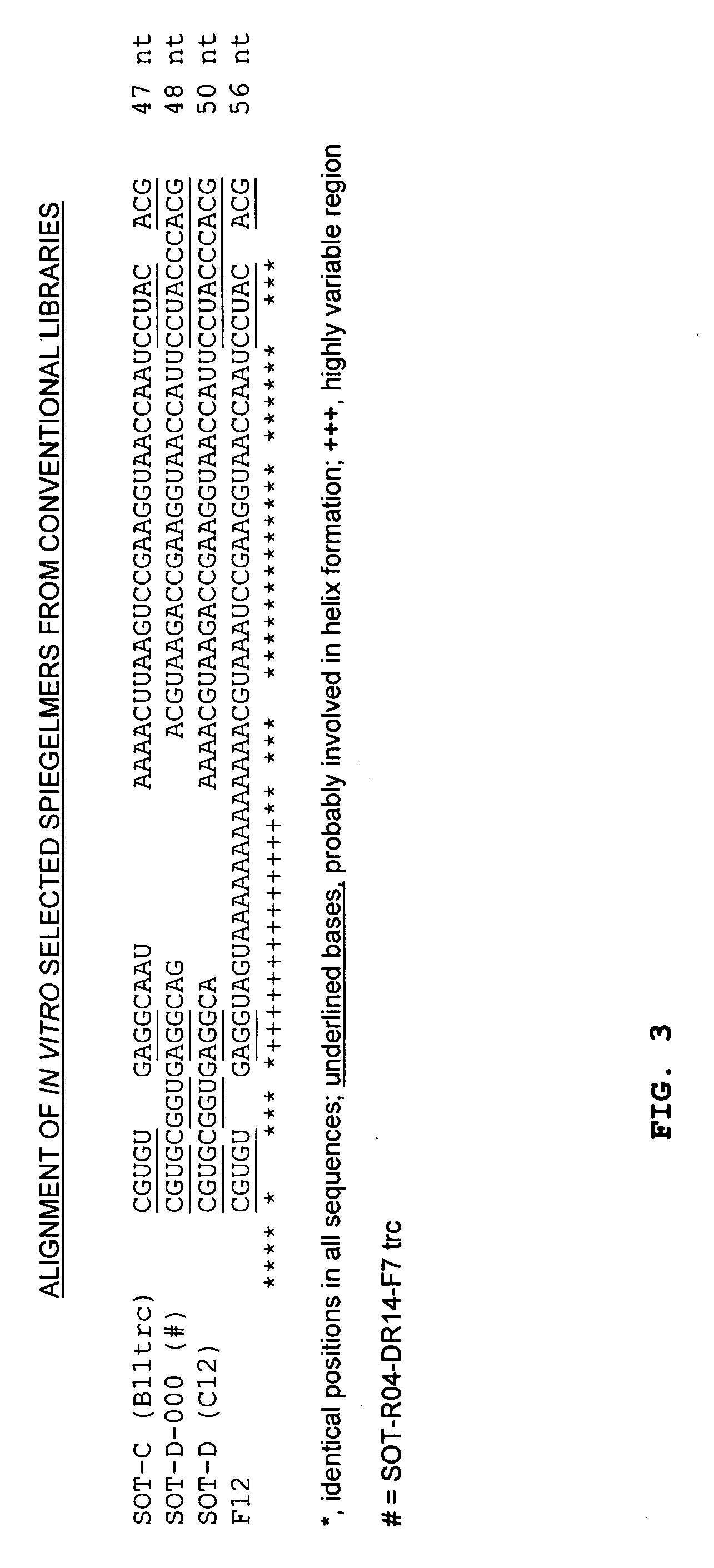
[0246] The ghrelin-binding Spiegelmers which are shown in FIG. 3 have been obtained previously, as already presented in patent application WO2004 / 013274 A2. In secondary structure predictions (minimum free energy conformations [Hofacker et al., 1994, Monatsh. Chem 125:167-188]), the possibility for canonical intramolecular base pairing between the 5′- and the 3′-terminus becomes obvious for all Spiegelmers (FIGS. 4-7; underlined bases in FIG. 3). The presence of such structural elements in a Spiegelmer may be vital for correct folding of the active three-dimensional structure, but for economical Spiegelmer synthesis, the molecules should be as short as possible. Spiegelmer SOT-D-000 (alias a truncated version of SOT-R04-DR14-F7 without primer binding sites) was chosen as basis for terminal truncation as it is the shortest sel...


PUM

PropertyMeasurementUnit
molecular weightaaaaaaaaaa

Abstract

The present invention is related to a nucleic acid, preferably binding to ghrelin, whereby the nucleic acid comprises a first stretch Box A, and a second stretch Box B, whereby the first stretch Box A comprises about 25 consecutive nucleotides, the second stretch Box B comprises about six to eight consecutive nucleotides, whereby a 3′-terminal stretch of nucleotides of the first stretch Box A hybridises with the second stretch Box B, whereby upon hybridisation a first double-stranded structure is formed, whereby such first double-stranded structure comprises a bulge.

Description

INCORPORATION OF SEQUENCE LISTING [0001] The contents of the following submission on compact discs are incorporated herein by reference in its entirety: two copies of the Sequence Listing (COPY 1 and COPY 2) and a computer-readable form of the Sequence Listing (CRF COPY), all on CD-ROMs, each containing: file name: 14167-1 Sequence Listing, date recorded: Apr. 7, 2006, size: 15 KB. FIELD OF THE INVENTION [0002] The present invention is related to nucleic acids binding to ghrelin, and their use in the manufacture of a medicament, and their use in the manufacture of a diagnostic agent. BACKGROUND OF THE INVENTION [0003] Ghrelin was identified as the natural ligand of the growth hormone secretagogue receptor 1a (GHSR1a). The receptor is most abundant in the pituitary gland and in hypothalamic parts of the brain, but can also be detected in other tissues at low concentrations. Since the late 70ies synthetic peptides and other compounds, named secretagogues had been shown to stimulate th...

Claims


Application Information

Patent Timeline
Patent Type & Authority Applications(United States)
IPC IPC(8): A61K48/00C12Q1/68C07H21/04C12N15/115
CPCC12N15/115C12N2310/16C12N2310/53C12N2310/351C12N2310/3183A61P1/00A61P3/00A61P3/10A61P35/00A61P3/04A61P43/00A61P5/02A61P5/06A61P5/08A61P9/00A61P9/12A61K31/713C12Q1/6811A61K31/715C07K14/61
Inventor JAROSCH, FLORIANEULBERG, DIRKMAASCH, CHRISTIANHELMLING, STEFFENKLUSSMANN, SVEN
Owner NOXXON PHARM AG
Why Eureka
Try Eureka