Epitope protection assay and method for detecting protein conformations

a technology of protein conformation and proteassay, which is applied in the field of proteassays for and achieves the effect of reducing reactions and preventing or reducing reactions

Inactive Publication Date: 2007-01-04
AMORFIX LIFE SCI
View PDF9 Cites 22 Cited by
  • Summary
  • Abstract
  • Description
  • Claims
  • Application Information

AI Technical Summary

Benefits of technology

[0043] The method of the invention has many advantages over existing technology. As noted above, the invention is optionally referred to as “EPA”, which in the case of prion protein disease detection is a simple, efficient method for detecting aggregated disease proteins such as PrPSc, the pathogenic molecule which is thought to constitute the infectious particle in prion diseases and polypeptide, associated with AD.

Problems solved by technology

Inaccessibility is often due to aggregation making the target epitope inaccessible.

Method used


Image

  • Epitope protection assay and method for detecting protein conformations
  • Epitope protection assay and method for detecting protein conformations
  • Epitope protection assay and method for detecting protein conformations

Examples


example 1

Peroxynitrite Reacts Differently with PrP in Normal and Acid Treated or Scrapie Brain Homogenate

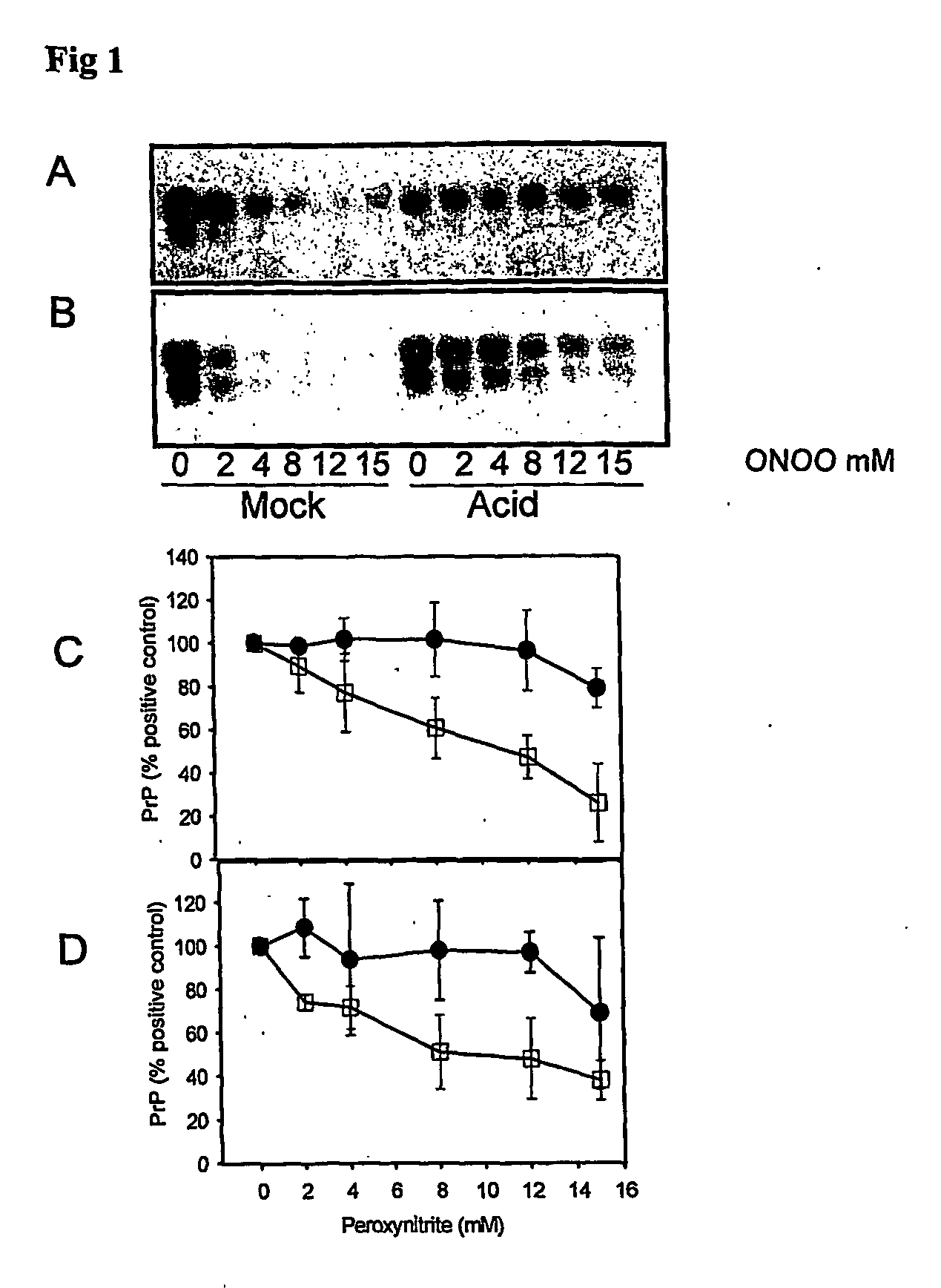
[0240] When brain homogenate is incubated at pH 3.5 in the presence of guanidine, PrP becomes detergent insoluble and is more susceptible to misfolding to a PK-resistant isoform in the presence of PrPSc (29). This acid treated PrP is a ‘model prion’ which is partially misfolded and / or aggregated resembling characteristics of PrPSc. When mock (□) and acid treated (●) brain homogenate is incubated with increasing concentrations of peroxynitrite and then subjected to immunoblotting, there is less PrP recognized by both 3F4 (FIGS. 1A and C) and 6H4 (FIGS. 1B and D) in mock treated brain homogenate than in acid treated brain homogenate. The PrP in the acid treated brain homogenate is protected from modification by peroxynitrite.

example 2

PrP in Scrapie Infected Hamster Brain is Protected from Modification by Peroxynitrite

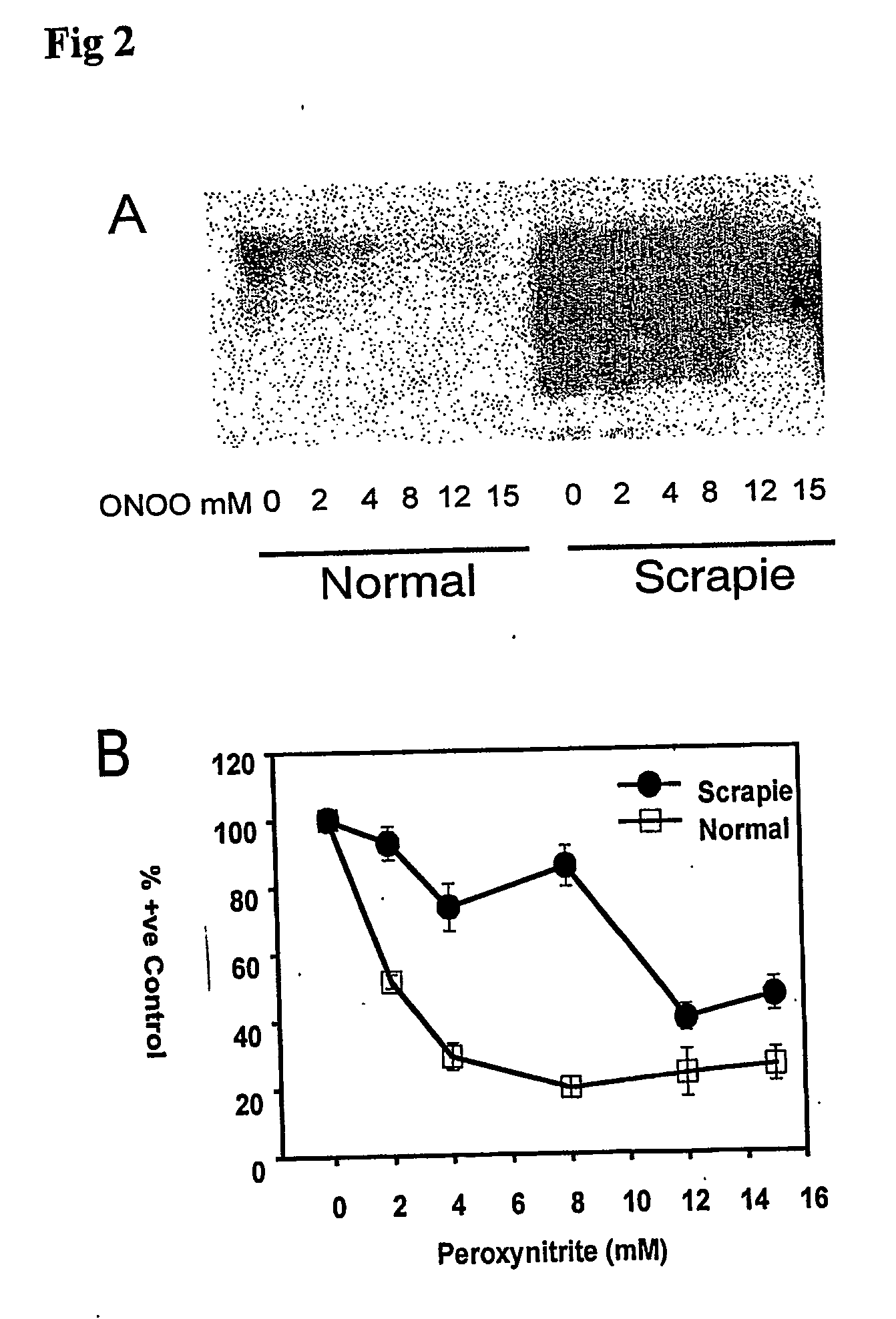
[0241] The epitope protection phenomenon for ‘model prions’ as observed in example 1 was also observed for authentic disease-misfolded prion protein in scrapie infected hamster (Ha) brain (FIGS. 2A and B). As with model prions, the 3F4 and 6H4 epitopes of PrP in HaSc brain homogenate are protected from modification by peroxynitrite. It is clear that ‘model prions’ and HaPrpsc share characteristics that provide protection from chemical modification by peroxynitrite, such as differential misfolding or aggregation.

example 3

Aggregation is Responsible for the Reduction in Peroxynitrite-Induced Epitope Modification of Misfolded PrP

[0242] To show that epitope protection of acid treated and scrapie brain was due to aggregation, samples were treated with peroxynitrite and then incubated with or without guanidine before immunoprecipitation. Treatment of the samples with guanidine dissociates aggregates of PrP (43-45) that protect the polypeptide from modification by peroxynitrite. Incubation of mock treated brain with 2.5 M guanidine after peroxynitrite treatment did not show an increase in 3F4 and 6H4 epitopes as revealed by immunoprecipitation (FIG. 3A lanes 1-4). However, when peroxynitrite-treated acid brain homogenate was incubated with guanidine, there was an increase in PrP that could be detected by immunoprecipitation with 3F4 and 6H4 immunobeads (FIG. 3A lanes 5-8). This shows that guanidine is able to dissociate aggregates of acid treated brain homogenate and release PrP that is protected from mo...


PUM

PropertyMeasurementUnit
pHaaaaaaaaaa
pHaaaaaaaaaa
volumesaaaaaaaaaa

Abstract

The invention relates to an epitope protection assay for use in diagnosis, prognosis and therapeutic intervention in diseases, for example, involving polypeptide aggregation, such as prion infections. The methods of the invention first block accessible polypeptide target epitope with a blocking agent. After denaturation of the polypeptide, a detecting agent is used to detect protein with target epitope that was inaccessible during contact with the blocking agent.

Description

FIELD OF THE INVENTION [0001] The invention relates to an epitope protection assay for use in diagnosis, prognosis and therapeutic intervention in diseases, for example, diseases involving polypeptide aggregation such as prion infections. BACKGROUND OF THE INVENTION Protein Misfolding and Aggregation [0002] Proteins can fold into complex and close-packed structures. Folding is not only crucial for biological activity but failure of proteins to fold properly or remain folded can give rise to disease (Dobson C M, Methods (2004) 34:4-14). Misfolding can in some cases cause protein aggregation which can further give rise to discrete deposits extracellularly (e.g., plaques) or intracellularly (e.g., inclusions in the cytosol or nucleus). [0003] Neurogenerative diseases such as Alzheimer's disease (AD), Parkinson's disease (PD), Huntington's disease (HD), amyotrophic lateral sclerosis (ALS) and prion diseases are characterized by neural deposits of misfolded aggregated protein. Type II d...

Claims


Application Information

Patent Timeline
Patent Type & Authority Applications(United States)
IPC IPC(8): G01N33/53G01N33/574G01N33/569G01N33/68
CPCA61K38/00C07K16/18G01N33/68G01N33/6845G01N2333/90283G01N2800/2828C07K16/40G01N33/581G01N33/6896A61P25/00C07K2317/31C07K2317/34C07K2317/54C07K2317/55C07K2317/622C07K2317/624C07K2317/626G01N33/58G01N2800/28
Inventor CASHMAN, NEILLEHTO, MARTY
Owner AMORFIX LIFE SCI
Why Eureka
Try Eureka