
Patsnap Eureka
For R&D, Patsnap Eureka makes reading and utilizing patents & technical documents easy.
Patsnap Eureka AIR
Designed for self-driven R&D workflows. Generate viable solutions, solve complex R&D challenges, empower your innovation with AI.

Patsnap Eureka Materials
Designed for material experts only. Revolutionize your material R&D, from search, analyze, to developing new materials.

TechResearch
Generate reliable direction feasibility study reports for your R&D in just a few steps.

TechSeek
Discover and master advanced knowledge NOW. Basics, ideas, possibilities, all at once.

TechMind
As an expert in R&D Theories, TechMind can generates customized viable solutions instantly.

TechRisk
Analyze your overall solution with one click, know your potential R&D risks in advance.

TechMonitor
Get weekly tech updates, stay abreast of the latest tech innovations and key insights.
Neural stem cells and use thereof for brain tumor therapy
- Summary
- Abstract
- Description
- Claims
- Application Information
AI Technical Summary
Benefits of technology
Problems solved by technology
Method used
Image

Examples
example 1
Migratory Capacity of NSCs in Culture
[0084] To determine properties of the NSCs in association with glioma cells, studies were initially performed in culture comparing the relative migratory capacity of NS-, (clone C17.2) to fibroblasts (the lacZ-expressing TR-10 fibroblast cell line) when cocultured with glioma cells. C17.2 and TR-10 cells were maintained in Dulbecco's modified Eagle's medium (DMEM; Mediatech, Washington, D.C.) supplemented with I 0% fetal calf serum (FCS; Sigma, St. Louise, Mo.), 5% horse serum (HS;Gibco), 1% Glutamine (2 mM; Gibco), 1% penicillin / streptomycin (Sigma). CNS-1 cells were stably transduced with the PGK-GFP-IRES-NeoR retroviral vector construct to express green fluorescent protein (GFP) as previously described [Aboody-Guterman et. al, 1997], and maintained in RPNH-1640 (Bio Whittaker) supplemented with 10% FCS and 1% penicillin / streptomycin (Sigrna). Cell structure studies were performed in 100 mm petri dishes under standard conditions: humidified, 3...
example 2
Transgene-Expressing NSCs Migrate Throughout and Beyond Invading Tumor Mass in Vivo
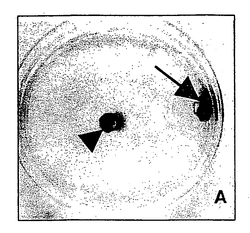
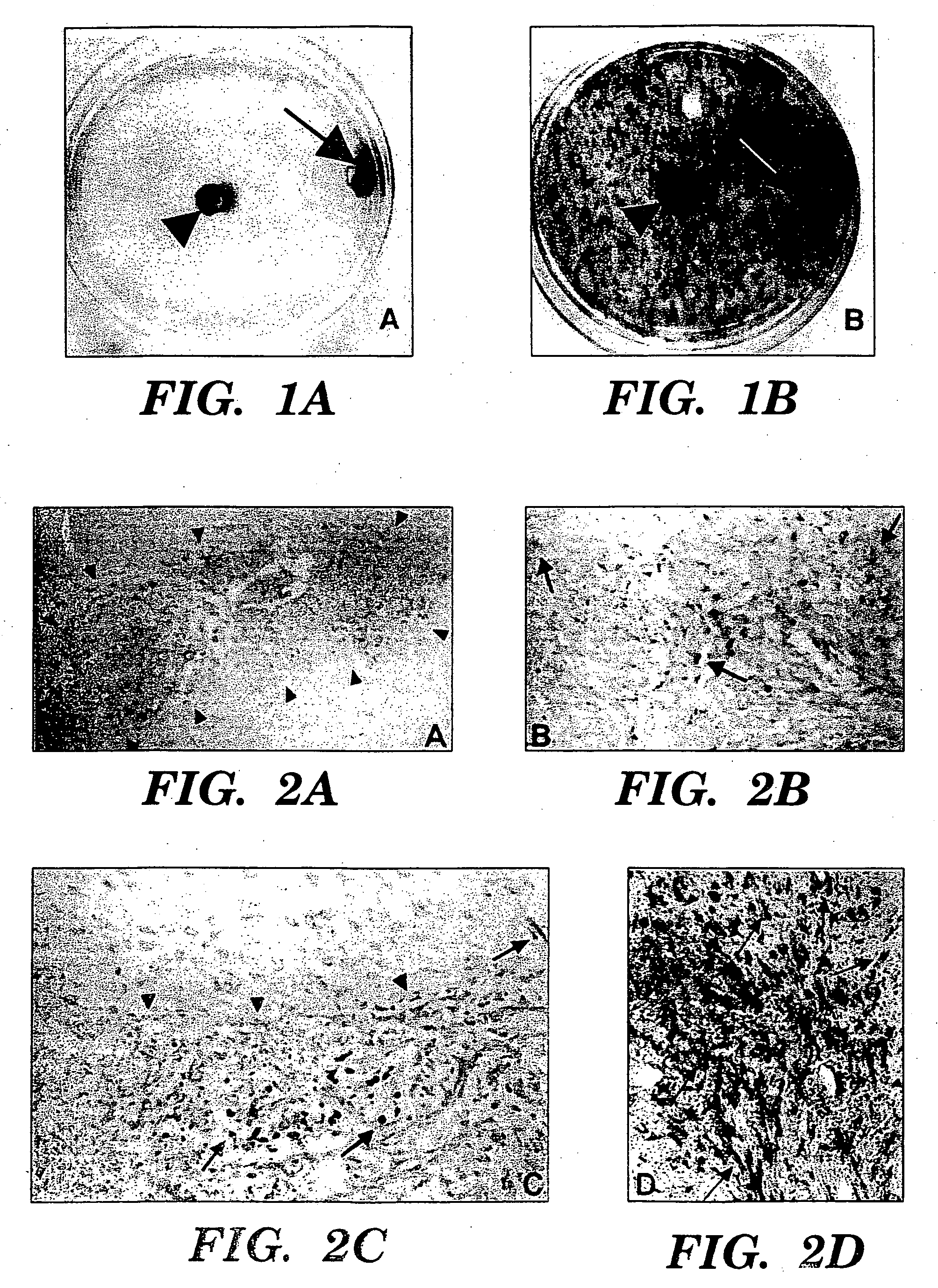
[0086] To determine the behavior of clone C17.2 NSCs introduced into brain tumors, experimental animals (syngeneic adult rats) first received an implant of 4×10⁴ D74 rat glioma cells in 1 μl injected into the right frontal lobe. Four days later, 1×10⁵ C17.2 NSCs in 1.5 μl PBS were injected at same coordinates directly into the D74 tumor bed. Animals were then sacrificed at days 2, 6, and 10 days post-intratumoral injection and cryostat sections of the brains were processed with X-gal histochemistry for P-galactosidase (β-gal) activity to detect donor-derived cells and counterstained with neutral red to detect tumor cells. Donor C 1 7.2 NSCs were found extensively dispersed throughout the tumor within a few days, spanning an 8 mm width of tumor as rapidly as 2 days after injection (FIGS. 2A, 2B). This is a much more extensive and rapid dispersion compared to previous reports of 3T3 fibroblasts grafted...
example 3
NSCs “Track” Infiltrating Tumor Cells
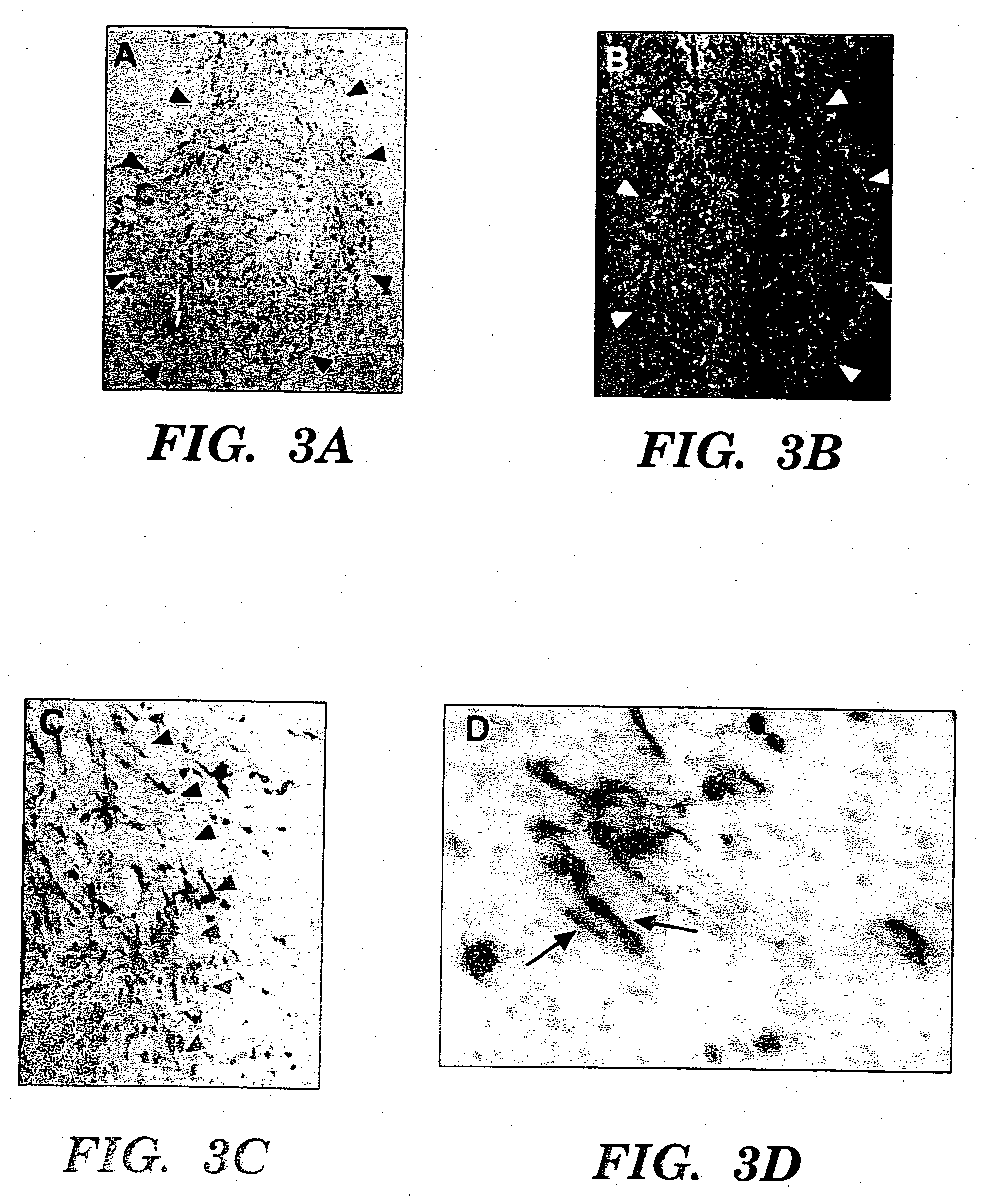
[0087] CNS-1 tumor cells were labeled by retroviral transduction with green fluorescent protein (GFP), prior to implantation, to better distinguish single cells away from the main tumor bed [Aboody-Guterman et al., NeuroReport 8: 3801-08, 1997]. GFP-expressing CNS-1 glioma cells (3×10⁴) in 1 μl PBS injected into right frontal lobe at stereotaxic coordinates 2 mm lateral to bregma, on coronal suture, 3 mm depth from dura. 4×10⁴ C17.2 or TR-10 cells in 1 μl PBS injected at same coordinates directly into tumor bed on day 6. 3-4 C17.2 animals (2 BUdR labeled, 1 BUdR pulsed) and 1-2 TR-10 control animals (1 BUdR labeled). Animals were sacrificed on days 9, 12, 16 and 21 post-tumor implantation. Cryostat sectioned, fixed brain tissue was stained either with 0-galactosidase (C17.2 cells blue) and neutral red (tumor cells dark red) or double immunofluorescence with Texas Red anti-p-galactosidase (C17.2 cells red) and FITC anti-GFP (tumor cells green). F...
PUM
| Property | Measurement | Unit |
|---|---|---|
| Molar density | aaaaa | aaaaa |
| Molar density | aaaaa | aaaaa |
| Fraction | aaaaa | aaaaa |
Abstract
Description
Claims
Application Information
 - R&D Engineer
- R&D Manager
- IP Professional
- Industry Leading Data Capabilities
- Powerful AI technology
- Patent DNA Extraction
Browse by: Latest US Patents, China's latest patents, Technical Efficacy Thesaurus, Application Domain, Technology Topic, Popular Technical Reports.
© 2024 PatSnap. All rights reserved.Legal|Privacy policy|Modern Slavery Act Transparency Statement|Sitemap|About US| Contact US: help@patsnap.com