Method and detector for identifying subtypes of human papilloma viruses

a detection method and human papilloma virus technology, applied in the field of human papilloma virus detection methods and detectors, can solve the problems of serious affecting diagnosis accuracy, inability to identify clinically important subtypes of human papilloma viruses contained in specimens, and inability to detect the conventional hpv detection kits without system control for checking house-keeping genes. , the effect of rapid and reliable detection and identification

Inactive Publication Date: 2007-02-08
LIN CHING YU +13
View PDF6 Cites 15 Cited by
  • Summary
  • Abstract
  • Description
  • Claims
  • Application Information

AI Technical Summary

Benefits of technology

[0015] The main purpose of the present invention is to provide a HPV detecting kit, which is able to diagnose multiple HPV subtypes (up to 39 different subtypes) at the same time, allowing the rapid and reliable detection and identification of HPV possibly present in a biological sample.
[0016] It is another object of the present invention to provide a rapid and reliable method to detect and identify the HPV present in a biological sample.
[0017] It is another object of the present invention to provide a HPV detecting kit with high specificity and accuracy, which includes an internal control to show whether the detecting process is well handled so that the detecting result is dependable.

Problems solved by technology

However, according to the comparison of the epidemiology research and the conventional HPV detecting kits, several clinically-important subtypes of human papilloma viruses contained in a specimen could not be identified by the conventional HPV detecting kits.
In addition, the conventional HPV detecting kits only tell the information of HPVs contained in a specimen by two categories, high risk HPVs or low HPVs, rather than tell the definite subtypes as which they are classified.
Therefore, except the high risk HPVs and the low risk HPVs, if other HPV subtypes are contained in the specimen, the conventional HPV detecting kits can not identify immediately, which would seriously affects the diagnosis accuracy.
Furthermore, the conventional HPV detecting kits lack the system control for checking the house-keeping genes contained in a specimen.
Without the system control, it will be hard to confirm whether the detecting protocols are precisely followed.
That is, the user can not tell the positive / negative result comes from the HPV subtypes presence / absence or comes from the incorrect protocols execution.
Therefore, the conventional detecting kit without the system control would not be able to provide a convincing result.
Therefore, how to simultaneously detect many HPV subtypes contained in a biological simple and design an accurate internal control in the detecting kits have become a major problem waited to be solved.

Method used


Image

  • Method and detector for identifying subtypes of human papilloma viruses
  • Method and detector for identifying subtypes of human papilloma viruses
  • Method and detector for identifying subtypes of human papilloma viruses

Examples


example i

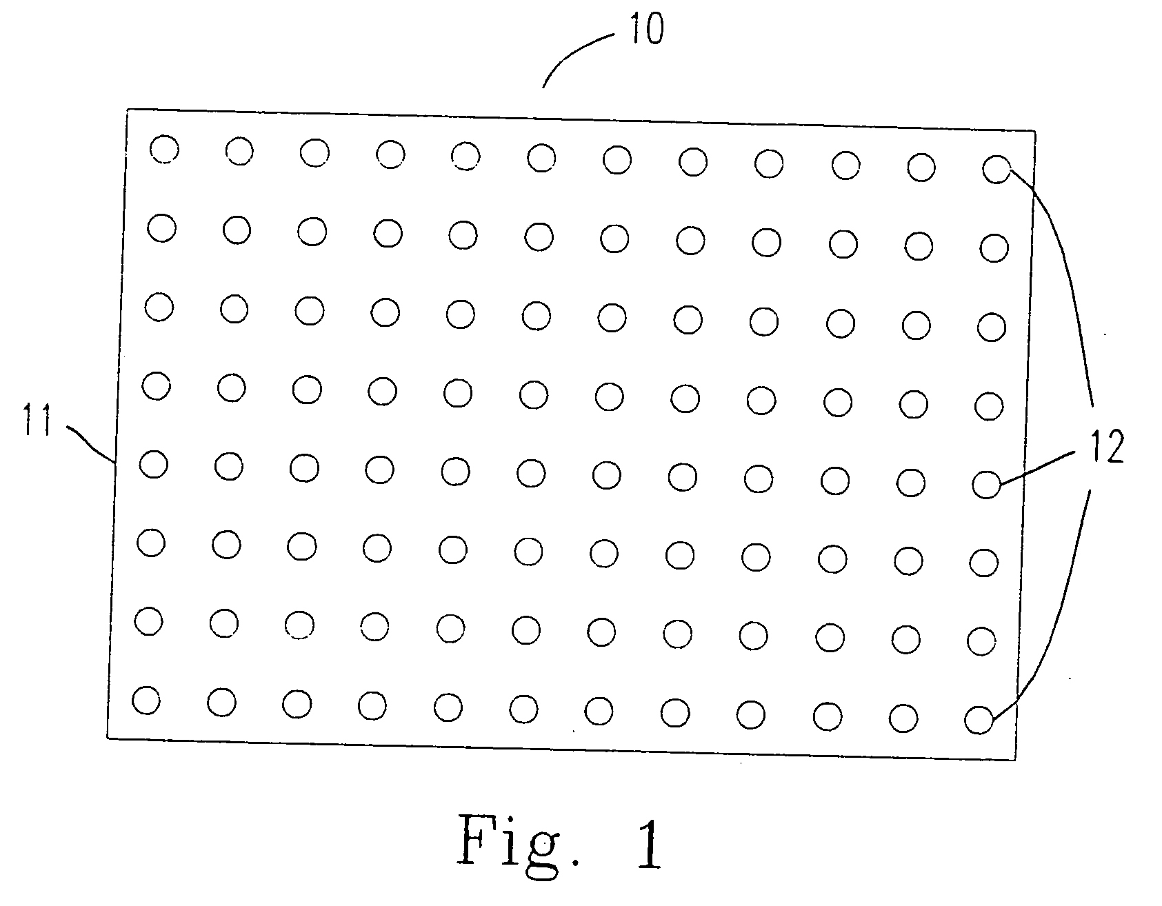
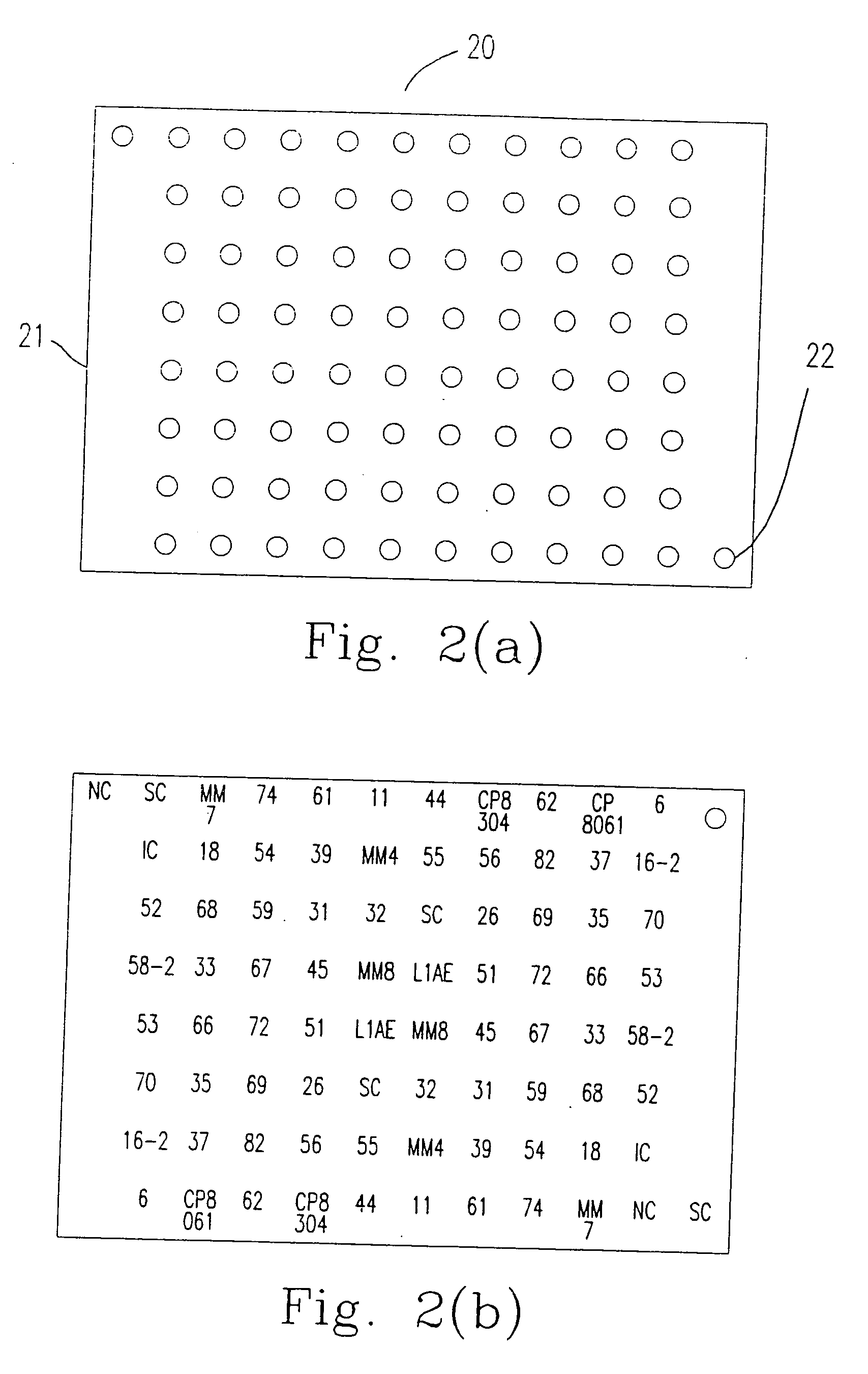
[0058] The method for immobilizing or mounting the above mentioned probes (oligonucleotides) on the carrier 11 (the nylon membrane) is described as follows.

[0059] 1.-TTTTTTTTTTTTTTT (SEQ ID NO 469) is added to the 3′ end of the oligonucleotide provided by the present invention by terminal transferase according to the following steps 1.1 to 1.3.

[0060] 1.1 Mixing the following components:

10X NEBuffer 45μl2.5 mM CoCl25μloligonucleotide5˜300pmol10˜300 mM dATP, dCTP, dTTP or dGTP1μlTerminal Transferase (20 U / μl)0.5˜5μl(NEW English BioLabs, M0252S)Add M.Q. H2O to final volume50μl

[0061] 1.2 The components are mixed at 37° C. for 15-60 minutes.

[0062] 1.3 10 μl of 0.2 M EDTA (pH 8.0) is added to the mixture to stop the reaction.

[0063] 2. The oligonucleotide having 3′ end labeling is mounted on the carrier 11 according to the following steps 2.1 to 2.3.

[0064] 2.1 The oligonucleotide having 3′ end labeling is mounted on the carrier 11 by a needle having a 400 μm wide head. The distance ...

example ii

[0067] According to another preferred embodiment of the present invention, the carrier 11 could be a glass plate. The method for immobilizing or mounting the above mentioned probes (oligonucleotides) on the carrier 11 (glass plate) is described as follows.

[0068] 1. The surface of the carrier 11 is treated according to the following steps 1.1 to 1.8.

[0069] 1.1 The carrier 11 is cleaned in non-fluorescent and soft cleaner.

[0070] 1.2 The clean carrier 11 is immersed in 10% NaOH.

[0071] 1.3 The carrier 11 is oscillated in double-distilled water, 1% HCl solution and methanol in sequence for 2 minutes, and dried in an oven.

[0072] 1.4 The carrier 11 is immersed in 1% 3-aminopropyltrimethoxysilane (APTMS) in 95% aqueous acetone at room temperature for about 2 minutes.

[0073] 1.5 The carrier 11 is washed in acetone, and the carrier 11 is dried in the oven at 110° C. for 45 minutes.

[0074] 1.6 The dried carrier 11 is immersed in 0.2% 1,4-phenylene diisothiocyanate, wherein the solvent is ...

example iii

[0085] 1. The biological sample obtained from the patient is treated according to the following steps 1.1 to 1.3.

[0086] 1.1 The cells are centrifuged at 1,500 rpm at 200□ for 5 minutes.

[0087] 1.2 The cell pellet is washed in 10 mM Tris (pH 8.5) and dissolved in 8 mM NaOH. Then, the solution is transfer to 1.5 mL micro-tube.

[0088] 1.3 A proper amount of TreTaq (1U / μl) solution is added to the micro-tube. The reaction is carried out at 95□ for 1 hour. The DNA contained in the sample is obtained after centrifugation at 13,500 rpm, 20□ for 5 minutes. The obtained DNA is preserved at −20□.


PUM

PropertyMeasurementUnit
distanceaaaaaaaaaa
widthaaaaaaaaaa
widthaaaaaaaaaa

Abstract

A detector for detecting and simultaneously diagnosing at least one subtype of human papilloma viruses (HPV) contained in a biological sample is provided. The detector comprises: a carrier, a plurality of micro-dots immobilized on the carrier, wherein each micro-dot is for identifying one particular HPV subtype, and the HPV subtype is one selected from a group consisting of 39 different HPV subtypes; and at least one oligonucleotide sequence contained in each the micro-dot that is specific to the one particular HPV subtype, wherein the at least one oligonucleotide sequence serves as a detection probe that hybridizes specifically with an L1 gene sequence of the one particular HPV subtype to form a hybridization complex as a detection indicator, so that each micro-dot identifies one particular HPV subtype via a corresponding oligonucleotide of the one particular HPV subtype, and thereby detecting and simultaneously identifying subtypes of human papilloma viruses.

Description

CROSS REFERENCE TO RELATED APPLICATION [0001] This application is a divisional of U.S. patent application Ser. No. 10 / 601,497, filed Jun. 23, 2003 which is a continuation in part of U.S. patent application Ser. No. 09 / 885,799 filed Jun. 20, 2001, now abandoned, the entirety of which is hereby incorporated by reference into this application.FIELD OF THE INVENTION [0002] The present invention relates to a method and a detector for detecting human papilloma viruses, and more particularly to a method and a detector for simultaneously detecting and identifying subtype of human papilloma viruses (HPV). BACKGROUND OF THE INVENTION [0003] In humans, more than 70 genetically distinct strains of human papilloma virus (HPV) have been identified based on DNA hybridization studies. According to some reports, different HPV types cause distinct diseases. For example, “Low-risk” HPVs, e.g., HPV 6 and HPV 11, cause benign hyperplasias such as genital warts, while “high-risk” HPVs, e.g., HPV-16, HPV-...

Claims


Application Information

Patent Timeline
Patent Type & Authority Applications(United States)
IPC IPC(8): C12Q1/70C12Q1/68C12P19/34
CPCC12Q1/708C12Q2565/501
Inventor LIN, CHING-YULIN, RUEY-WENYOU, CHIOU-MIENHUANG, HSING-HSUANLEE, BOR-HENGLEE, HSIEN-HSIUNGLIN, YU-JUFAN, CHIH-CHUNHSU, HAN-CHUANSHIH, CHIA-WENYEH, CHIH-HSINGKAO, YI-FENGPAN, CHIH-LONGCHAN, PETER
Owner LIN CHING YU
Why Eureka
Try Eureka