

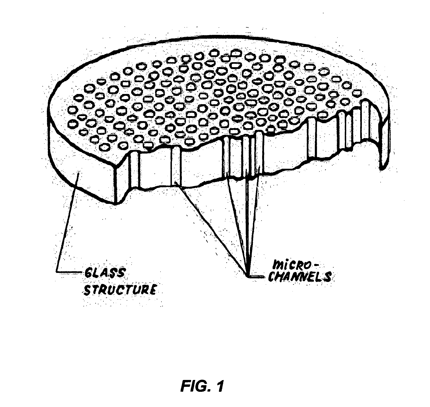
Device for rapid detection and identification of single microorganisms without preliminary growth
a technology of single microorganisms and detection devices, which is applied in the field of rapid detection and identification of single microorganisms without preliminary growth, can solve the problems of long (many hours or days) preliminary growth, the enumeration of initial contamination is not reliable with pcr usage, and the method of detection and/or identification of cells without preliminary growth usually requires expensive and sophisticated equipment and the work of high-level professionals. , to achieve the effect of rapid and reliable detection and/
- Summary
- Abstract
- Description
- Claims
- Application Information
AI Technical Summary
Benefits of technology
Problems solved by technology
Method used
Image

Examples
example 1
Detection of Microbial Contamination in Liquid Samples
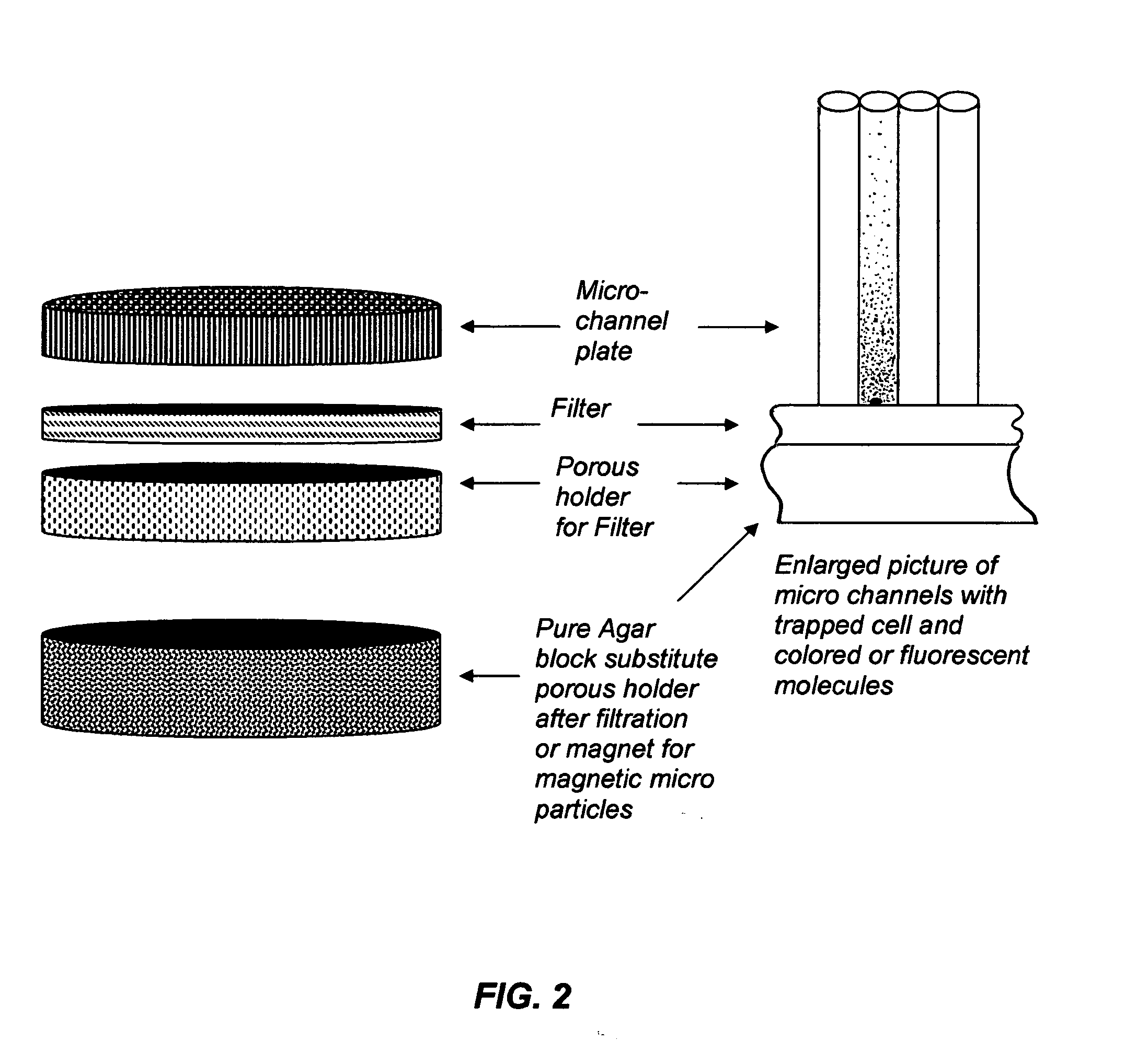
[0056] Many different liquid samples in food or pharmaceutical industry must be tested for the presence of bacteria or fungi. Approximately 100 ml of liquid sample, presumably containing microbes, is filtrated through the device shown in FIGS. 4, 5 and 6. The liquid is easily passed tough a black filter (size of pores is 0.2 microns, cellulosic or nitroellulosic) but cells are trapped at the bottom of the channels on the surface of the filter. After this device is untwisted and a porous disk is removed and substituted by an agar disk preliminarily filled with 4-Methylumbelliferyl phosphate and 4-Methylumbelliferyl acetate (0.1 ml of each with concentration 0.5 mg / ml). The mixture of these fluorogenic substrates guarantees that all live cells will be found because both substrates correspond to big groups of enzymes present in all live cells—Esterases and Phosphatases. Liquid from the agar block containing molecules of named fluo...
example 2
Identification of Escherichia coli O:157 in Samples
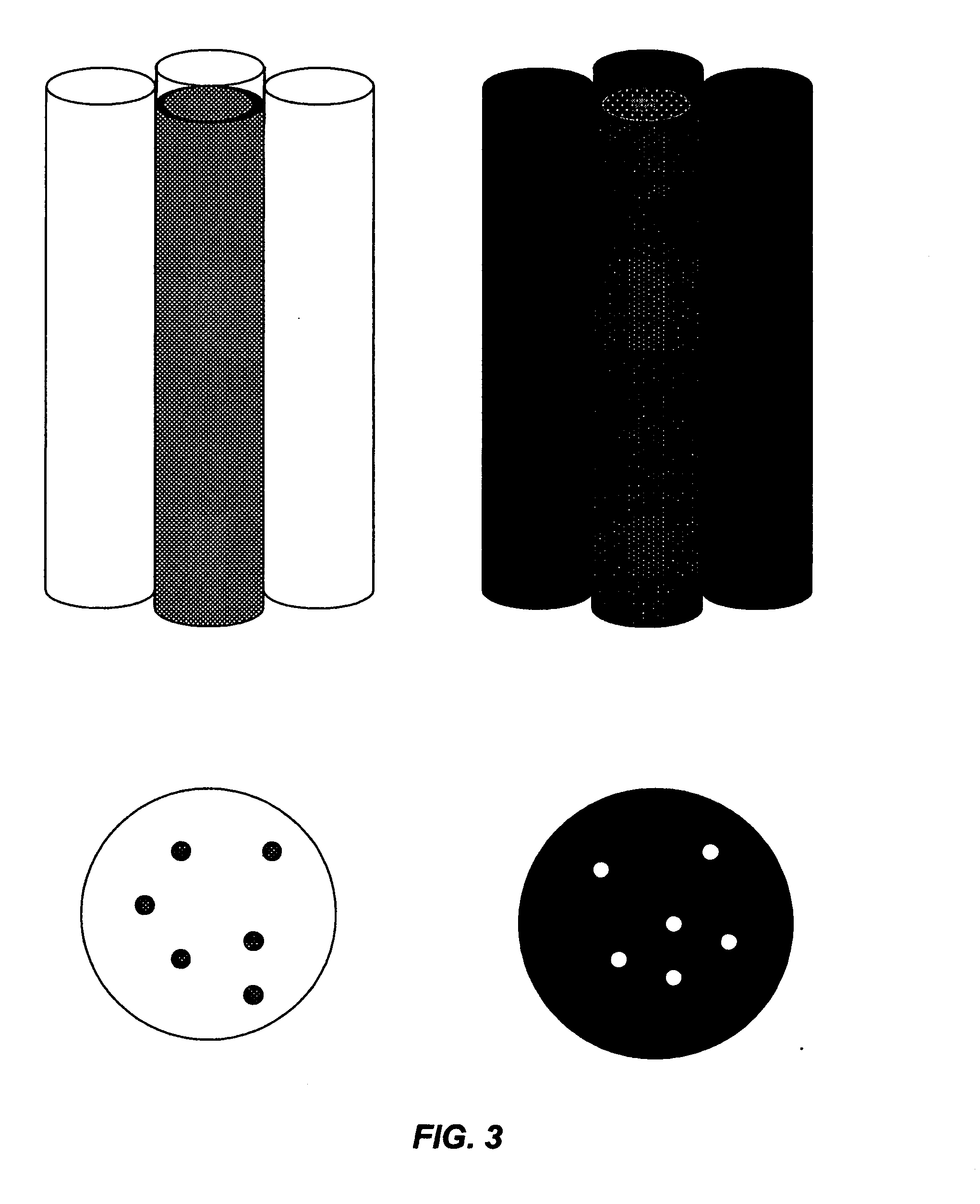
[0057] A 100 ml sample of liquid is filtrated through the device using a white nitrocellulose filter and colorless micro-channel plate. About 2 ml of standard conjugated antibody for E. coli O:157 antigens with Horseradish Peroxidase (HRP) are added to the device and slowly—part by part, in several minutes—filtrated through the device; the conjugate (Ab+HRP) is attached to the surface of E. coli O:157 if the cells are present in some of the channels; After that, 50 ml of distilled water is filtrated through the device in order to wash out the rest of the conjugate. An agar block containing: solution of 3,3′,5,5′-Tetramethylbenzidine is added to the device instead of porous holder for filter. Incubation is for 35-40 minutes at 40° C. After incubation, the device with filter is placed under a light microscope (multiptication=X100). Micro-channels containing E. coli O:157 appear as blue dots. Other micro-channels appear as white dots...
example 3
Detection and Identification by Coated Magnetic Particles
[0058] Detection and identification of bacteria, viruses, and biomolecules in a sample with the help of magnetic particles consists of several stages. First stage: addition of magnetic particles coated by antibody to the sample presumably containing detected organisms or biomolecules. In this stage, the object is attached to the magnetic particle by Ab-Ag interaction. Second stage: liquid containing magnetic particles together with mixture of other particles passes through the device (FIG. 10). Magnetic particles are separated in micro-channels by magnetic field, and other particles flow out through output channel. Third stage antibody-enzyme conjugate passes through array of micro-channels and is attached to antigens trapped on magnet particles: bacteria, viruses, or biomolecules. Fourth stage: substitute magnet with transparent porous holder for filter and wash out the surplus of conjugate by distilled water: Fifth stage: ...
PUM
| Property | Measurement | Unit |
|---|---|---|
| diameter | aaaaa | aaaaa |
| volumes | aaaaa | aaaaa |
| diameters | aaaaa | aaaaa |
Abstract
Description
Claims
Application Information