
Device for rapid detection and identification of single microorganisms without preliminary growth

a technology of single microorganisms and detection devices, which is applied in the field of rapid detection and identification of single microorganisms without preliminary growth, can solve the problems of long (many hours or days) preliminary growth, the enumeration of initial contamination is not reliable with pcr usage, and the method of detection and/or identification of cells without preliminary growth usually requires expensive and sophisticated equipment and the work of high-level professionals. , to achieve the effect of rapid and reliable detection and/

Inactive Publication Date: 2014-03-04
NANOLOGIX INC
View PDF25 Cites 5 Cited by
  • Summary
  • Abstract
  • Description
  • Claims
  • Application Information

AI Technical Summary

Benefits of technology

The patent describes a device that can quickly and reliably detect and identify cells in a sample using micro channels. The device filters microorganisms onto a special material and then fills the micro channels with a biochemical indicator that can change color or fluoresce when it comes into contact with enzymes in the cells. The device can detect as many as one single cell per sample. The invention provides a sensitive and efficient tool for identifying and counting cells in microbiological samples.

Problems solved by technology

These methods require hundreds to millions of pure cells for detection of a certain microorganism, and, therefore, long (many hours or days) preliminary growth.
In addition to other drawbacks, methods of detection and / or identification of cells without preliminary growth usually need very expensive and sophisticated equipment and the work of high-level professionals.
Enumeration of initial contamination is not reliable with PCR usage.
However, the amount of fluorescent substance present is restricted by volume of the cell body or cell surface.
The small size of the object (single microorganism) makes the detection of one cell very difficult, especially with large background fluorescence usually present in a majority of the samples.
Usage of flow cytometry in microbiology is not easy.
In microbiology these instruments are not often used because microorganisms can create clusters with other particles and can be confused with natural particles or dead cells.
Microbes are also much more diverse by size and shape than blood cells and, thus, mistakes occur very often.
Nevertheless, it is technologically complicated.
This method could be reproduced only in a laboratory by highly professional personnel with use of sophisticated and expensive equipment.

Method used


Image

  • Device for rapid detection and identification of single microorganisms without preliminary growth
  • Device for rapid detection and identification of single microorganisms without preliminary growth
  • Device for rapid detection and identification of single microorganisms without preliminary growth

Examples


example 1

Detection of Microbial Contamination in Liquid Samples

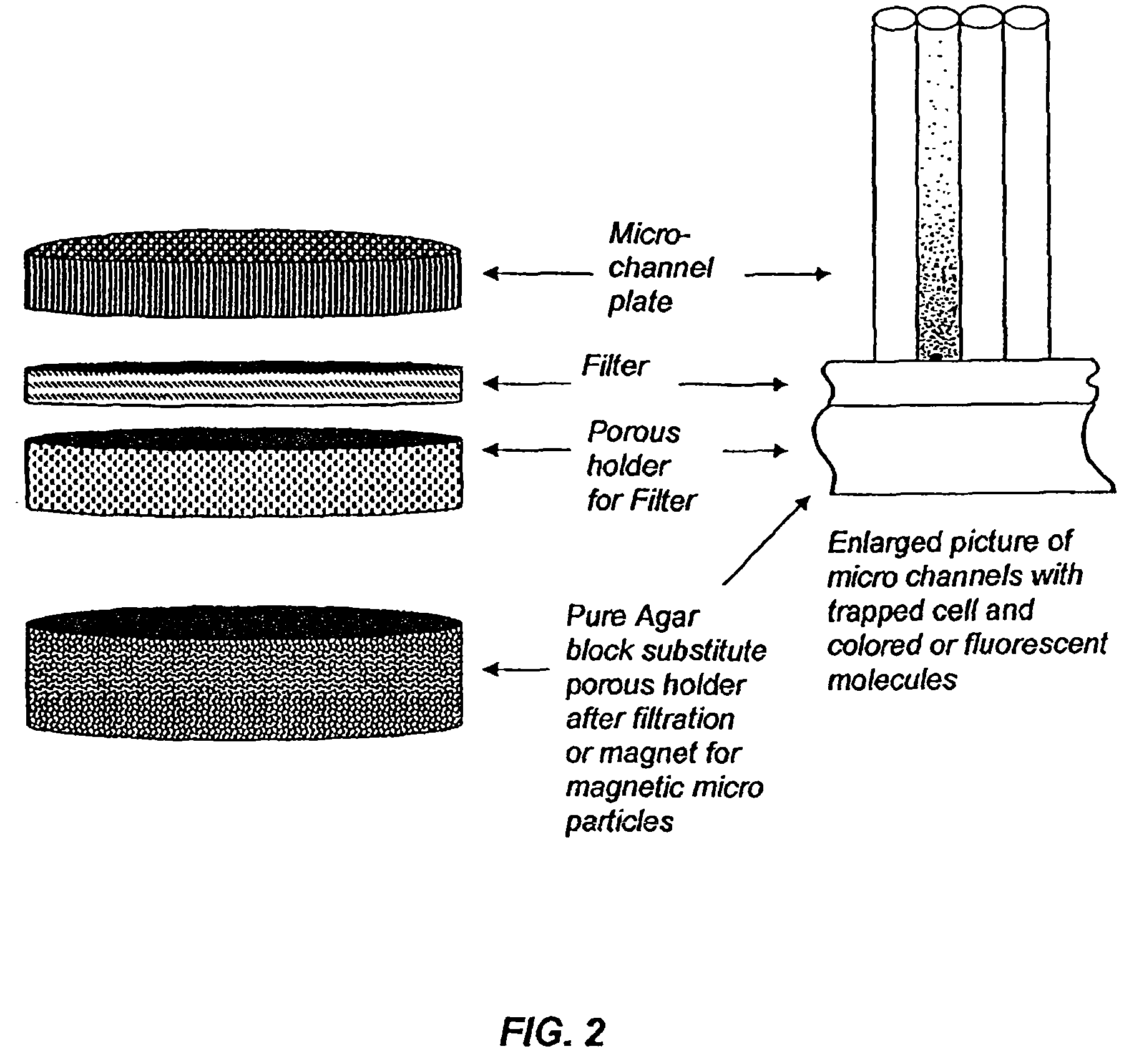
[0057]Many different liquid samples in food or pharmaceutical industry must be tested for the presence of bacteria or fungi. Approximately 100 ml of liquid sample, presumably containing microbes, is filtrated through the device shown in FIGS. 4, 5 and 6. The liquid is easily passed tough a black filter (size of pores is 0.2 microns, cellulosic or nitroellulosic) but cells are trapped at the bottom of the channels on the surface of the filter. After this device is untwisted and a porous disk is removed and substituted by an agar disk preliminarily filled with 4-Methylumbelliferyl phosphate and 4-Methylumbelliferyl acetate (0.1 ml of each with concentration 0.5 mg / ml). The mixture of these fluorogenic substrates guarantees that all live cells will be found because both substrates correspond to big groups of enzymes present in all live cells—Esterases and Phosphatases. Liquid from the agar block containing molecules of named fluorog...

example 2

Identification of Escherichia coli O:157 in Samples

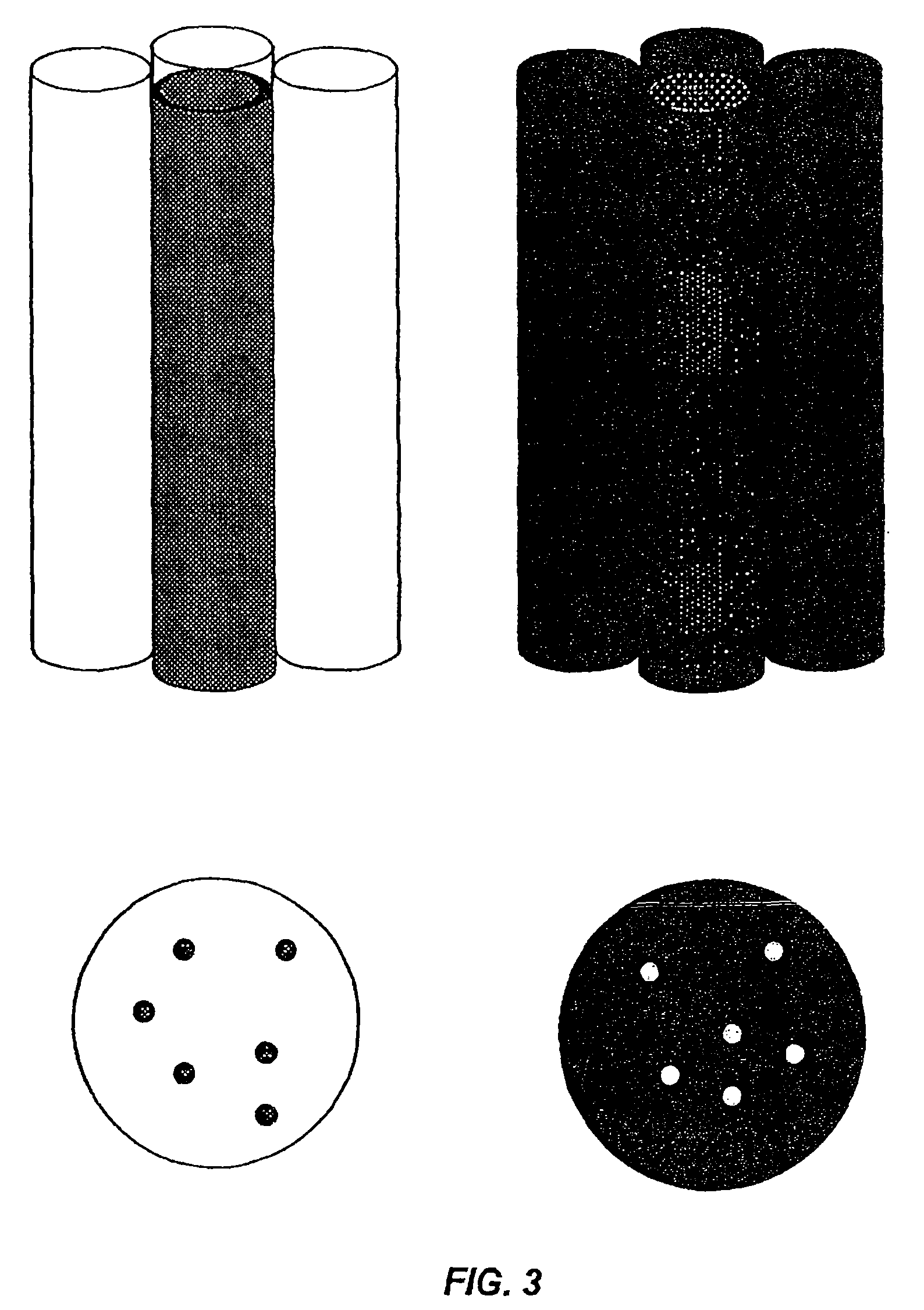
[0058]A 100 ml sample of liquid is filtrated through the device using a white nitrocellulose filter and colorless micro-channel plate. About 2 ml of standard conjugated antibody for E. coli O:157 antigens with Horseradish Peroxidase (HRP) are added to the device and slowly—part by part, in several minutes—filtrated through the device; the conjugate (Ab+HRP) is attached to the surface of E. coli O:157 if the cells are present in some of the channels; After that, 50 ml of distilled water is filtrated through the device in order to wash out the rest of the conjugate. An agar block containing: solution of 3,3′,5,5′-Tetramethylbenzidine is added to the device instead of porous holder for filter. Incubation is for 35-40 minutes at 40° C. After incubation, the device with filter is placed under a light microscope (multiptication=X100). Micro-channels containing E. coli O:157 appear as blue dots. Other micro-channels appear as white dots. E...

example 3

Detection and Identification by Coated Magnetic Particles

[0059]Detection and identification of bacteria, viruses, and biomolecules in a sample with the help of magnetic particles consists of several stages. First stage: addition of magnetic particles coated by antibody to the sample presumably containing detected organisms or biomolecules. In this stage, the object is attached to the magnetic particle by Ab-Ag interaction. Second stage: liquid containing magnetic particles together with mixture of other particles passes through the device (FIG. 10). Magnetic particles are separated in micro-channels by magnetic field, and other particles flow out through output channel. Third stage antibody-enzyme conjugate passes through array of micro-channels and is attached to antigens trapped on magnet particles: bacteria, viruses, or biomolecules. Fourth stage: substitute magnet with transparent porous holder for filter and wash out the surplus of conjugate by distilled water: Fifth stage: rem...


PUM

PropertyMeasurementUnit
diameteraaaaaaaaaa
angleaaaaaaaaaa
lengthaaaaaaaaaa

Abstract

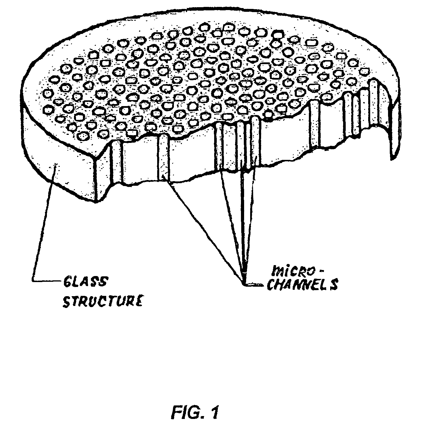
This invention describes a device consisting of a micro channel plate, filter, and porous holder for filter, which is substituted by a pure agar block during method performance, and supportive structural elements. The device is intended for rapid detection and / or identification of microorganisms. Microorganisms are trapped by filtration in long (diameter / length=1 / 10-1 / 100), cylindrical, parallel, micro-channels that are open from both sides and attached to a filter from one side. A micro channel plate houses a multiplicity of micro channels (possible diameter of each channel=1-30 μm, length 100-1000 μm, and number on centimeter2=100,000-1,000,000). The micro channel plate with cells trapped on the surface of the filter is attached to an agar block impregnated by artificial substrate(s) so that the molecules of the artificial substrates will fill all micro channels. Trapped cells produce colored or fluorescent molecules from artificial substrates. These molecules are collected in the very small volume of a micro channel. The extremely small volume of a micro channel (1 / 25 million part of milliliter) allows it to collect a detectable concentration of color or fluorescent substances in a very short time (several minutes). Even one cell from a filtrated sample can be detected by the enzyme—artificial substrate method and / or identified by enzyme immunoassay.

Description

[0001]The present application is a continuation application of U.S. patent application Ser. No. 10 / 141,677, filed May 9, 2002, now U.S. Pat. No. 7,781,159, which is incorporated by reference herein.BACKGROUND OF THE INVENTION[0002]1. Field of the Invention[0003]Modern microbiological diagnostics and analysis serves for detection, enumeration, and identification of microorganisms present in different samples. In the areas of medical diagnostics and veterinary medicine, these organisms are pathogenic or dangerous microbes in human or animal blood, internal organs, skin, tissues, respiratory organs, and so on. In the area of industrial microbiology, microbes commonly pollute technological processes, materials, equipment, and finished products. In environmental analysis, there is often microbial contamination of water, indoor and outdoor air, and various surfaces. In epidemiology and biodefense—highly contagious pathogenic microorganisms from human body or environment.[0004]Time, qualit...

Claims


Application Information

Patent Timeline
Patent Type & Authority Patents(United States)
IPC IPC(8): C12M1/36G01N33/53C12M1/34C12Q1/00C12M3/00C12Q1/02C12M1/00C12M1/38C12N11/16G01N33/543C12Q1/04G01N33/569B01L99/00C12N11/00G01N33/554
CPCC12Q1/04
Inventor GAZENKO, SERGEY
Owner NANOLOGIX INC
Learn More