System, method and apparatus for navigated therapy and diagnosis

a technology of navigation therapy and diagnostic equipment, applied in the field of system, method and apparatus for navigation therapy and diagnosis, can solve the problems of insufficient landmark identification, difficult directing the tip of the device into the location of interest, and iatrogenic damage to the tissue, etc., to achieve high torque transmission, high pushability, and high kink resistance

Active Publication Date: 2007-02-08
PHILIPS ELECTRONICS LTD
View PDF99 Cites 129 Cited by
  • Summary
  • Abstract
  • Description
  • Claims
  • Application Information

AI Technical Summary

Benefits of technology

[0012] In some embodiments, the properties of the transmission element may be tuned to provide high pushability, high torque transmission, high kink resistance, low ductility, and / or other characteristics necessary for transmitting the intended energy or material along the transmission element.

Problems solved by technology

Directing the tip of these devices into locations of interest, such as those identified preoperatively or intraoperatively through medical scans of the patient (such as CT, MR, PET, SPECT, x-ray etc.) can be difficult.
This is especially true if, among other things, the location of the instrument relative to a lesion or other area of interest cannot be visualized directly.
If the tip of an instrument is not properly positioned, multiple problems can arise such as, for example, iatrogenic damage to tissue can be imparted, samples of tissue can be obtained that are not the intended target of the procedure, or other problems.
Sometimes, the location of a target lesion or other area of interest is known on a preoperative scan but insufficient landmarks are identifiable during the actual surgical intervention to easily locate the lesion or area of interest.
When used, the lesion, area of interest, and / or instrument may be invisible or inaccessible to the “live” (i.e., intraoperative) imaging modality, may appear in insufficient resolution, or the image of the lesion might be degraded through the presence of intervening anatomy.
This may render it difficult or impossible to determine the location of the instrument relative to the lesion or area of interest using traditional intraoperative imaging.
Furthermore, some imaging modalities, such as those that are based on x-rays, may impart ionizing radiation or be otherwise harmful to patient and surgeon.
Even when the intraoperative imaging modalities are able to render a lesion or area of interest and instrument, the images produced can sometimes be difficult to interpret or can take excessive time to obtain.
Furthermore, the imaging modalities used for the initial preoperative scans may be inappropriate or otherwise unavailable for use intraoperatively (i.e., during the actual interventional procedure).

Method used


Image

  • System, method and apparatus for navigated therapy and diagnosis
  • System, method and apparatus for navigated therapy and diagnosis
  • System, method and apparatus for navigated therapy and diagnosis

Examples


Embodiment Construction

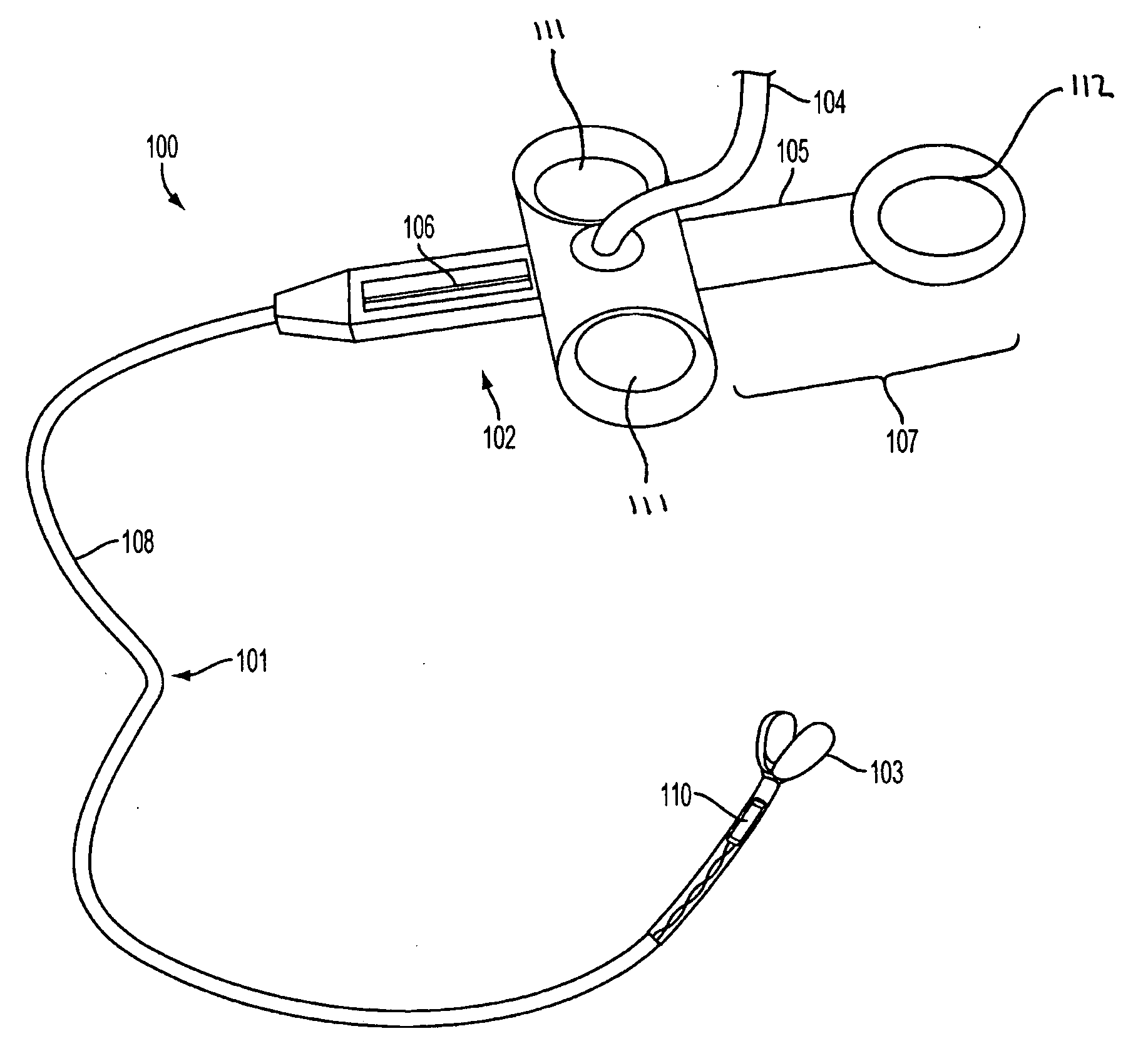
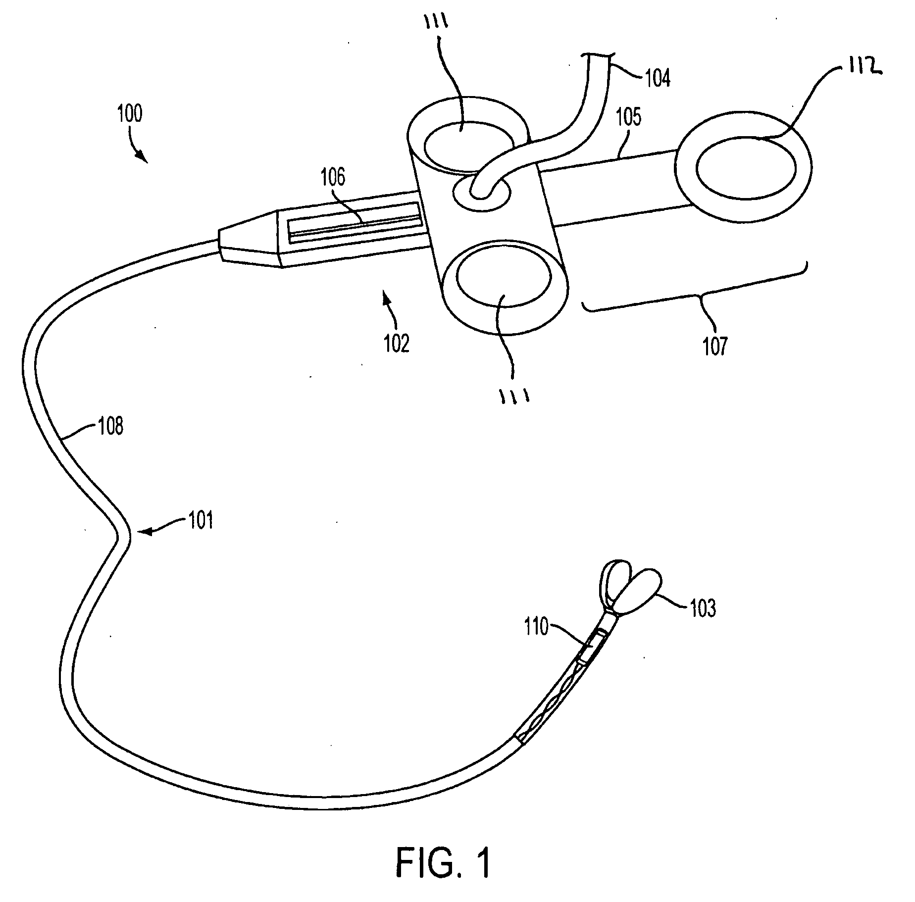
[0032] In one embodiment, the invention provides an image-guided medical instrument, which can be used in minimally invasive surgery. FIG. 1 illustrates an image-guided medical instrument 100, according to an embodiment of the invention. In some embodiments, image-guided medical instrument 100 may include a body member 101, a handle 102, a treatment apparatus 103, an operating element 107, and / or other elements.

[0033] In one embodiment, body member 101 may include one or more elongated flexible elements or materials such as, for example, elongated tubing, wires, and / or other elements. For example, in one embodiment, body member 101 may include tubing 108. Body member 101 may also include coiled springs, insulating and / or protective jackets, braided elements, and / or other elements.

[0034] In one embodiment, body member 101 may include first and second ends and may connect handle 102 to treatment apparatus 103 over or through the flexible elements comprising body member 101.

[0035] B...


PUM


Abstract

The invention provides an image-guided medical instrument that utilizes a tracking device to track the location of at least a portion of the instrument on at least one image of a patient's anatomy. The instrument may include a handle having an operating element, an elongated flexible body member connected to the handle, wherein the body member includes a lumen, a transmission element housed within the lumen of the body member, and a treatment apparatus connected to end of the transmission element, wherein actuation of the operating element actuates the treatment apparatus via the transmission element. The body member also includes at least one sensor element that is utilized by the tracking device to determine position information regarding the treatment apparatus that is used to display the location of the treatment apparatus on the at least one image of the patient's anatomy.

Description

RELATED APPLICATIONS [0001] This application claims priority to U.S. Provisional Application No. 60 / 692,272, filed Jun. 21, 2005, which is hereby incorporated by reference herein in its entirety.FIELD OF THE INVENTION [0002] The invention relates to a system, method and apparatus for navigated therapy and diagnosis. BACKGROUND OF THE INVENTION [0003] Access to natural and artificial passages in the body for treatment or investigation for possible treatment traditionally make use of elongated instruments that are inserted into the body. These instruments can be inserted into the body alone (e.g., as in a biopsy needle) or may make use of other implements such as an endoscope (e.g., a bronchoscope or colonoscope) through which the elongated instruments are inserted. Typical devices for access to natural or artificial passages in the body may include, for example, spring loaded biopsy “guns,” endoscopic biopsy forceps, biopsy needles, polypectomy snares, cytology brushes, biliary guide...

Claims


Application Information

Patent Timeline
Patent Type & Authority Applications(United States)
IPC IPC(8): A61B5/05
CPCA61B1/018A61B5/064A61B5/065A61B10/0275A61B10/06A61B17/32056A61B2019/5483A61B18/22A61B19/52A61B19/5244A61B2019/5251A61B2019/5276A61B2019/5475A61B18/18A61B34/20A61B90/36A61B2034/2051A61B2090/378A61B2090/3975A61B2090/3983
Inventor GLOSSOP, NEIL DAVID
Owner PHILIPS ELECTRONICS LTD
Why Eureka
Try Eureka