
Low profile intravascular ultrasound catheter

a low-profile, catheter technology, applied in the field of catheters, can solve the problem that catheters cannot be used to access desired sites within the patient's body through narrow blood vessels, and achieve the effects of enhancing catheter's ability to navigate, facilitating access to sites, and increasing flexibility

Active Publication Date: 2009-06-25
BOSTON SCI SCIMED INC
View PDF5 Cites 38 Cited by
  • Summary
  • Abstract
  • Description
  • Claims
  • Application Information

AI Technical Summary

Benefits of technology

[0006]In a preferred embodiment, a low profile intravascular catheter is adapted to navigate through a guide catheter that is 5 French (approximately 0.066 inch) or smaller in diameter. This allows the catheter to more easily access sites within the vascular system (e.g., coronary vessel) through the radial artery.
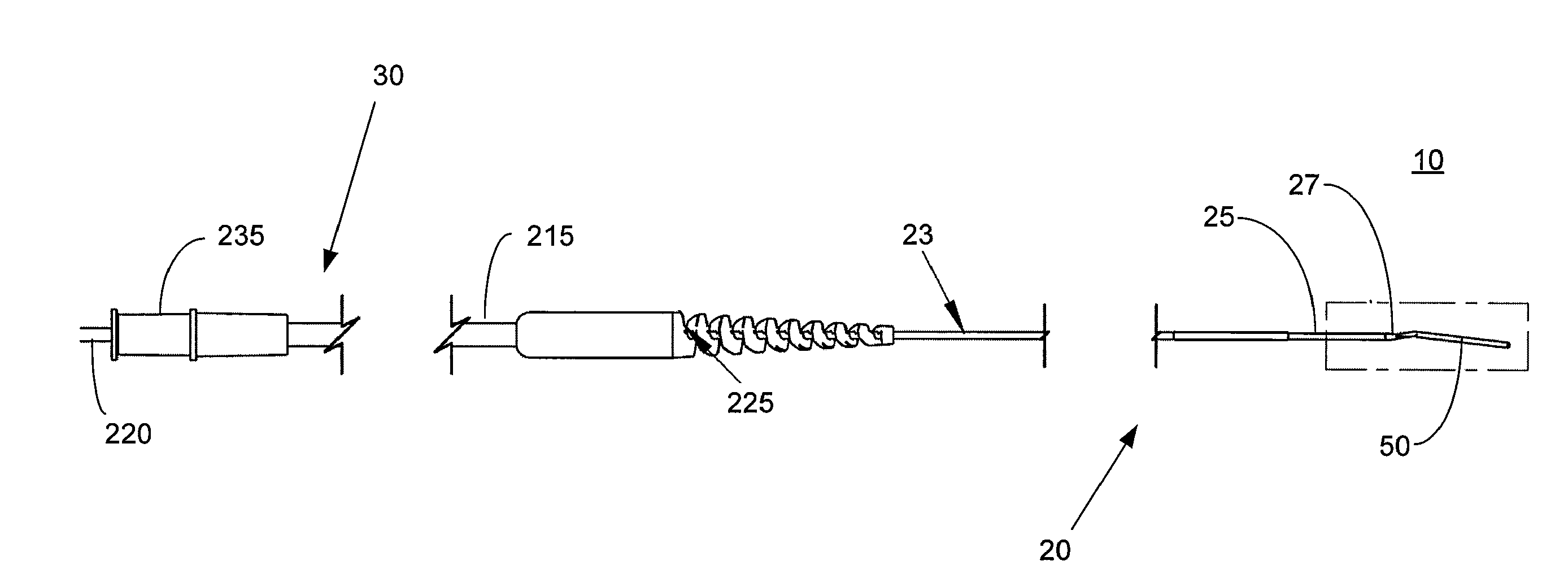
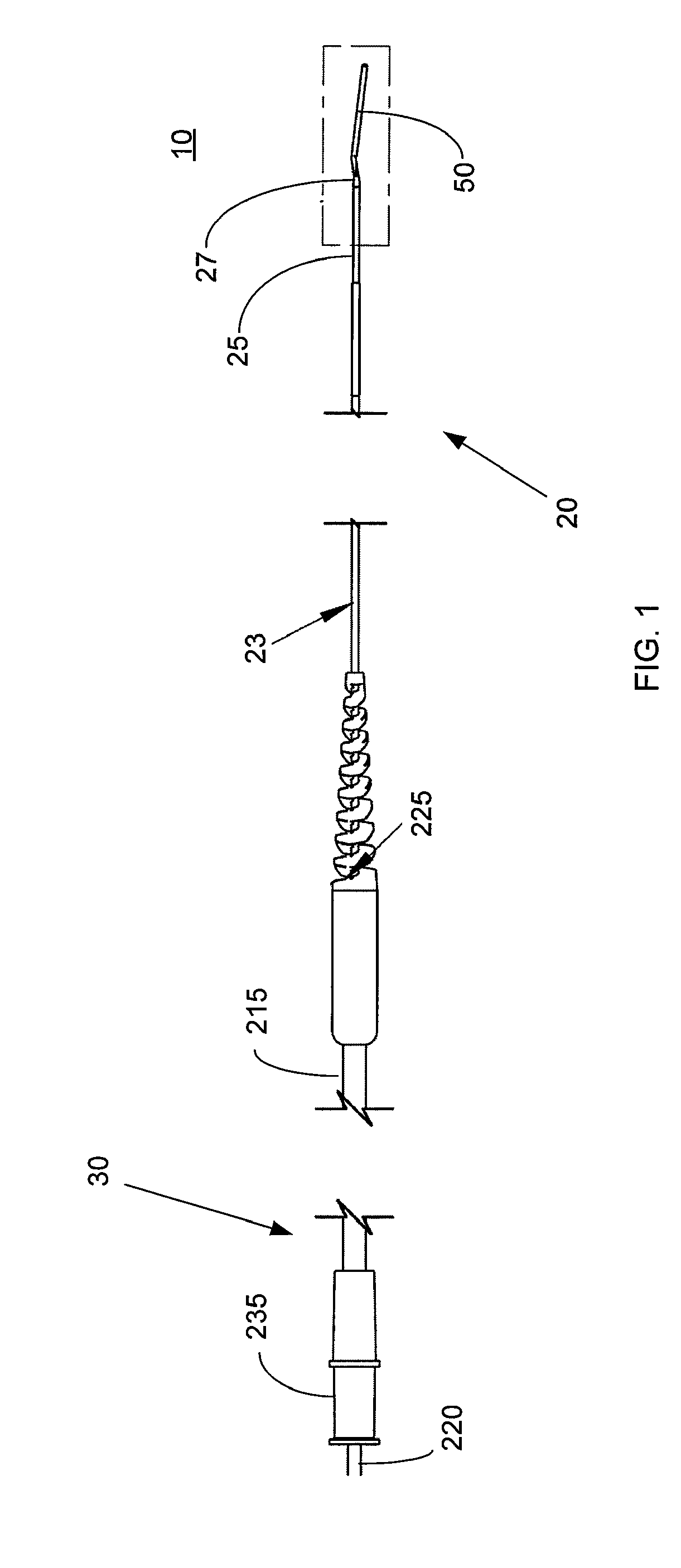
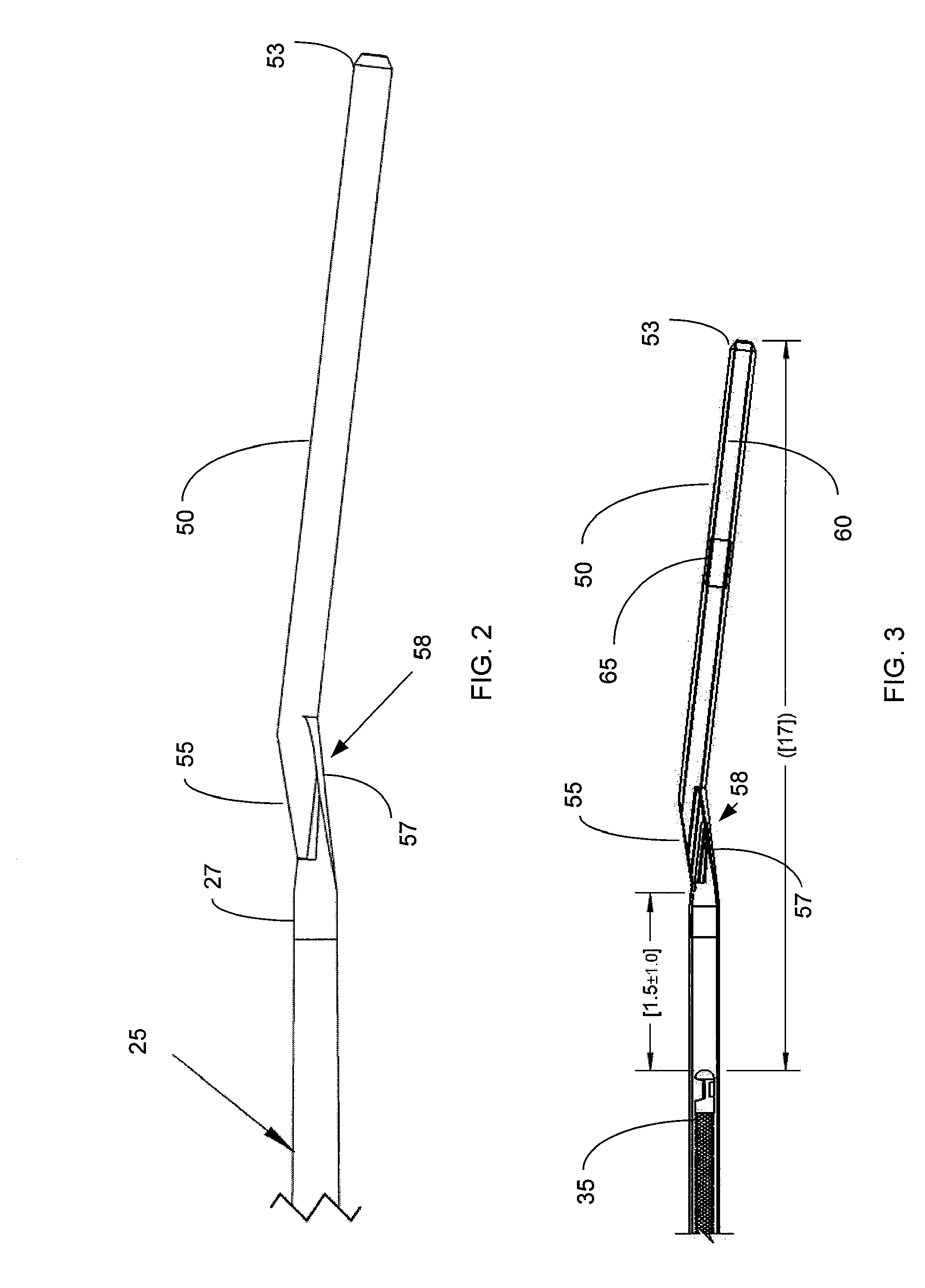
[0007]In an exemplary embodiment, a low profile catheter comprises an elongated catheter sheath, a short guidewire receiver attached to the distal end of the catheter sheath, and a telescope assembly at the proximal end of the catheter sheath. The catheter sheath comprises a main portion and a tapered portion located distally to the main portion. The tapered portion has an outer diameter and wall thickness that gradually decreases along the length of the tapered portion toward the distal end of the catheter. The tapered portion provides increased flexibility toward the distal end of the catheter enhancing the catheter's ability to navigate the tortuous path of a blood vessel and facilitating lesion crossibility. The tapered portion also provides a gradual increase in stiffness toward the main portion of the catheter sheath for increased pushability of the catheter through the blood vessel.
[0008]In one embodiment, a rotatable and translatable imaging core is received within a lumen of the catheter sheath for obtaining ultrasound images at different positions within the patient's body. In an embodiment, the imaging core comprises an ultrasound transducer with a rectangular face to maximize the ultrasound emitting surface area of the transducer with respect to the catheter size. In another embodiment, the length of the guidewire receiver attached to the distal end of the sheath is kept short to allow the imaging core to be advanced farther distally with respect to the distal end of the catheter.
[0009]In one embodiment, the catheter comprises a telescope assembly at the proximal end of the catheter sheath. The telescope comprises a female telescoping tube and a male telescoping tube adapted to slid within the female telescoping tube to retract and extend the telescope assembly. In an embodiment, the catheter sheath extends through the female telescoping tube to provide enhanced support for the imaging core within the female telescoping tube.

Problems solved by technology

A problem with existing catheters is that they are adapted for use with guide catheters that are 6 French or greater in diameter.
As a result, these catheters can not be used to access desired sites within the patient's body through narrower blood vessels, e.g., the radial artery, without risk of vessel spasm.

Method used


Image

  • Low profile intravascular ultrasound catheter
  • Low profile intravascular ultrasound catheter
  • Low profile intravascular ultrasound catheter

Examples


Embodiment Construction

[0021]FIG. 1 shows a low profile intravascular ultrasound catheter 10 according to an embodiment of the present invention. The low profile ultrasound catheter 10 is adapted access sites within the patient's body through narrow blood vessels, e.g., the radial artery. In the preferred embodiment, the sheath 20 of the catheter 10 is adapted to navigate through a guide catheter that is 5 French (approximately 0.066 inch) or smaller in diameter. Thus, the low profile catheter 10 is preferably 5 French compliant. This allows the catheter 10 to more easily access sites within the vascular system (e.g., coronary vessel) through the radial artery. Access through the radial artery provides for greater patient comfort during a follow up procedure, in which an area of the vascular system that has undergone an operation is examined post operation.

[0022]The catheter 10 comprises an elongated catheter sheath 20 having a distal portion 27, a tapered portion 25, and a main portion 23. The catheter s...


PUM


Abstract

The present invention provides low profile intravascular ultrasound catheters adapted to access sites within the patient's body through narrow blood vessels, e.g., the radial artery. In an embodiment, a low profile catheter comprises an catheter sheath, a short guidewire receiver attached to the distal end of the catheter sheath, and a telescope assembly at the proximal end. The catheter sheath comprises a main portion and a tapered portion for increased flexibility toward the distal end of the catheter. In one embodiment, a rotatable and translatable imaging core is received within the catheter sheath for ultrasound imaging. A short guidewire receiver is used to allow the imaging core to be advanced farther distally with respect to the distal end of the catheter. In an embodiment, the catheter sheath extends through a portion of the telescope assembly to provide enhanced support of the imaging core within the telescope assembly.

Description

FIELD OF THE INVENTION[0001]The present invention relates to catheters, and more particularly to low profile intravascular catheters.BACKGROUND INFORMATION[0002]Intravascular ultrasound imaging systems (IVUS) are used to obtain ultrasound images inside a patient's body. An IVUS system typically includes an ultrasound catheter having a flexible catheter sheath adapted for insertion into the vascular system of the patient. To obtain ultrasound images, the catheter comprises an imaging core received within a lumen of the catheter sheath. The imaging core comprises an ultrasound transducer connected to the distal end of a flexible drive cable that extends to the proximal end of the catheter through the catheter lumen. The drive cable is used to rotate and longitudinally translate the transducer within the catheter lumen to obtain images at different positions within the patient's body.[0003]An IVUS catheter is commonly advanced through a guide catheter to a desired site within the patie...

Claims


Application Information

Patent Timeline
Patent Type & Authority Applications(United States)
IPC IPC(8): A61B8/12
CPCA61B8/12A61B8/445A61B8/4461A61B8/4483A61M25/0054A61M2025/0183A61M2025/0059A61M2025/0098A61M2025/0175A61M2025/018A61M2025/0042
Inventor ZELENKA, ROBERTOKEEFFE, DANIELCARS, MIKAELMCEVERS, BRUCE
Owner BOSTON SCI SCIMED INC
Learn More