
Method for determining co-receptor selectivity of human immunodeficiency virus-1
- Summary
- Abstract
- Description
- Claims
- Application Information
AI Technical Summary
Benefits of technology
Problems solved by technology
Method used
Image

Examples
example i
Experimental Procedures and Materials
1. Pairwise Local and Global Superimposition
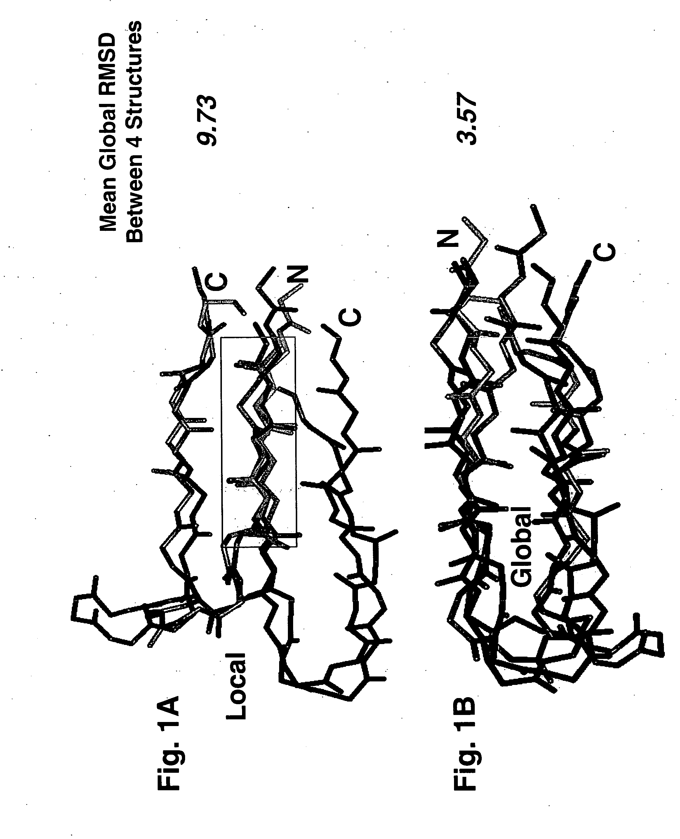
[0060] Global superimpositions were performed by minimization of root-mean-square-deviation (RMSD) of each rigid body pair of β-hairpins [63-66]. Local superimpositions were performed by a modified method of differences between interatomic distances as implemented in the ICM software suite [67, 68]. Each method results in an RMSD score and, as shown in Table 2 The former corresponds to superimposing the whole structure in every pairwise comparison, while the latter corresponds to superimposing only the N-terminal strands between pairs of structures in some of the comparisons. The sequence alignment from each valid pairwise global alignment was then extracted and concatenated into the equivalent of a single multiple sequence alignment, more accurately termed a “structure-based residue equivalency table,” shown in Table 1A / 1B.
2. Protein Structural Modeling
[0061] All modeling manipulations and graphi...
example ii
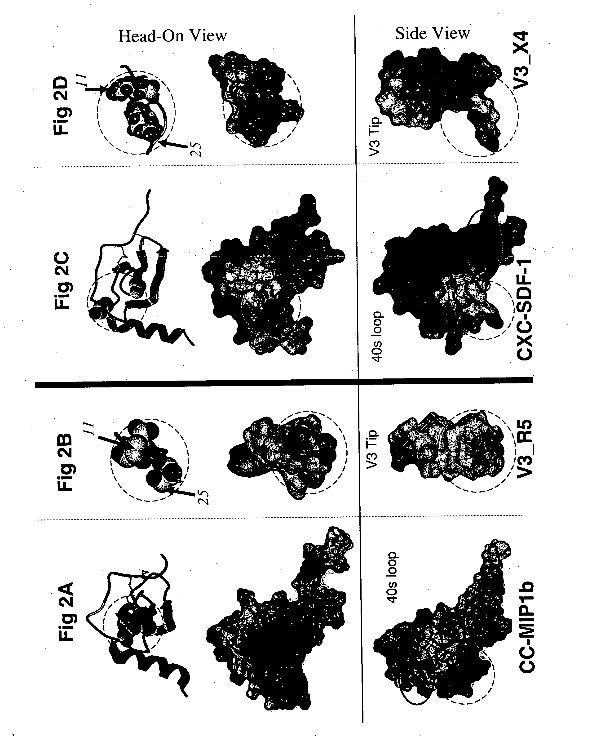
Structural Basis of the 11 / 25 Rule
[0066] To investigate the structural basis of the 11 / 25 rule, the 3D organization of the following structures was compared atom-by-atom. All of these bind to the receptors for either CC or CXC chemokines: the β2-β3 hairpins of the CC chemokines RANTES, MIP-1α, and MIP-1β, the β2-β3 hairpin of the CXC chemokine SDF-1, and the β-hairpin structures assumed by the V3 peptides from HIV-1 strains IIIB and MN complexed with neutralizing mAbs 0.5β and 447-52D, respectively [27-29, 48, 49]. Optimal superimposition of two protein structures is frequently ambiguous [50]. Usually the choice is between the best local and the best global superimposition (see Methods), and this was the case in this study. The optimal local superimposition which superimposes the 3 or 4 residue segment of the structures with the best RMSD, a quantitative measure of the aggregate distances between atoms of two 3D structures, was previously shown in [28] and was found between the N-t...
example iii
Determination of Location and Orientation of Surface: Residues 11, 24 and 25
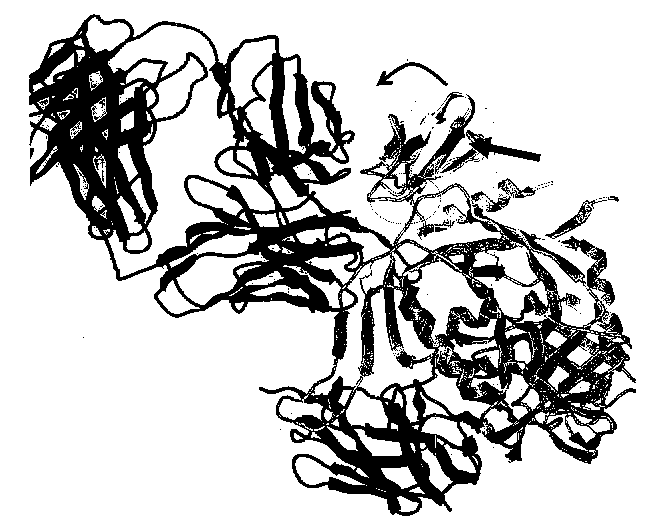
[0070] To determine the location and orientation of this surface in its full biological context, an energy minimized model was built of SDF-1 fused to the stem of V3 on the full crystallographic structure of gp120 [37, 38], a construct that is known to be infective [20]. The model shows that SDF-1 can only fit without clashing in one orientation (FIG. 3). This orientation finds the 11 / 25 patch identified here in contact with the gp120 bridging sheet and facing the space presumably occupied by the chemokine receptor (which, in the crystal structure, is occupied by mAb 17b). mAb 17b recognizes an epitope in the bridging sheet of gp120 involved in binding to the chemokine receptor. Interestingly, this model places the β-turn at the tip of the β2-β3 hairpin (the 40s loop) on the opposite side of the molecule at a distance that is 25 Å away from the presumed chemokine receptor location on gp120 [51]. These data ...
PUM
| Property | Measurement | Unit |
|---|---|---|
| Ratio | aaaaa | aaaaa |
| Selectivity | aaaaa | aaaaa |
Abstract
Description
Claims
Application Information