Sterile shell for an ultrasonic probe and method of using same

a technology of ultrasonic probes and sterile shells, applied in the field of ultrasonic guidance systems, can solve the problems of user-dependent procedures, difficult to accurately track the path and position of medical devices, and incur possible additional liability for hospitals, etc., to facilitate the placement of ultrasound monitors and avoid unnecessary surgery

Inactive Publication Date: 2007-04-05
ULTRASOUND VENTURES
View PDF38 Cites 63 Cited by
  • Summary
  • Abstract
  • Description
  • Claims
  • Application Information

AI Technical Summary

Benefits of technology

[0017] The present invention is directed to an ultrasound needle guidance system that facilitates placement of an ultrasound monitor over a patient and ensures accurate and simple needle placement in a target of interest within a patient's body. According to an embodiment of the invention, a hand-held ultrasonic probe includes a needle guidance position that holds a needle. The needle is orientated transverse to the scanning plane of the transducer. The needle can be rotated through a continuous range of angles and these angular changes can be tracked and displayed as a cross-hair (or other type of visual indicia) on a nearby monitor screen with the ultrasonic image. In this way, a health professional can accurately track and locate a needle to ensure precise placement at a target within a patient's body.
[0025] Preferably a removable sterile clip is used to mount the needle to the probe. In this aspect of the invention, the clip is configured to minimize mechanical noise associated with removal of the probe from the needle. As such, it is preferred to use a clip that does not rely on a mechanical engagement to retain the needle in the needle clip.
[0026] In another embodiment of the invention, an ultrasonic probe includes a needle clip that has a cradle for a needle and an arm having a first end coupled to the cradle and a second end forming a cover. In this embodiment, the cover is manually movable between a first position opening the cradle and a second position closing the cradle. Also, the cover is detached from the cradle in the second position and when the cover is in the second position, the cover and cradle together form a passageway for a needle shaft disposable between the cover and cradle such that the passageway allows movement of the needle in a first direction and substantially prohibits movement of the needle in a second direction that is perpendicular to the first direction.
[0027] In another embodiment of the invention, a method of releasably fastening a needle to an ultrasonic probe includes the steps of providing a needle clip on the probe, the needle clip including a displaceable arm and a cradle adapted to receive a needle shaft, placing the needle shaft within the cradle, applying pressure to the arm such that the arm moves from a first position distal of the cradle to a second position proximal to and mechanically decoupled from the cradle, whereupon the needle is held between the cradle and arm, and relieving the pressure on the arm, whereupon the arm moves from the second position to the first position. Alternatively, finger pressure may move the arm away from the cradle so that when the finger pressure is relieved, the needle is retained within the cradle.

Problems solved by technology

It is sometimes difficult to accurately track the path and position of the medical device after it has entered the patient's body on the monitor.
Such an error-prone, user-dependent procedure is painful for the patient, time consuming for the health professional, and incurs possible additional liability for the hospital with each use.
There are multi-planar ultrasound imaging devices capable of producing a three-dimensional image of the body, which may be capable of more accurately locating the position of an invasive medical device, but these types of devices are typically expensive to operate, and require a relatively high degree of skill and training to operate.
In the “transverse” type, the medical device, e.g., needle, is orientated out of plane and is sometimes disfavored because visualization of the needle is not reliable as it passes through the patient's body.
The “longitudinal” type has the added advantage of seeing the entire length of the needle because it is inserted in plane with the ultrasound beam; however, it can be difficult to keep the needle in the plane of the transducer image due to operator skill and inherent needle-bending when passing through tissue.
While the longitudinal type device is preferred because there is greater chance of tracking the needle, it is also more difficult to position the needle at the target when the needle is planar with the image.
This procedure can cause complications as it is often necessary to maintain precise positioning of the needle within the body.
When the door is being unlatched, there can be unacceptable motion of the transducer (and therefore of the needle) as a result of overcoming mechanical resistance in the latch.
Additionally, after inserting the needle into the target, the probe must be rocked to pry the needle from the needle guide, potentially disrupting the needle-target interaction.
Also, these devices have cords connecting the ultrasound probe to the monitor which are typically much longer than needed for most procedures because it must be sufficiently lengthy for extreme cases.
As a result, the cord can often obstruct the probe's user.
Additionally, the probe cannot be maintained in a sterile condition when it is placed on a holder provided with the system.

Method used


Image

  • Sterile shell for an ultrasonic probe and method of using same
  • Sterile shell for an ultrasonic probe and method of using same
  • Sterile shell for an ultrasonic probe and method of using same

Examples


first embodiment

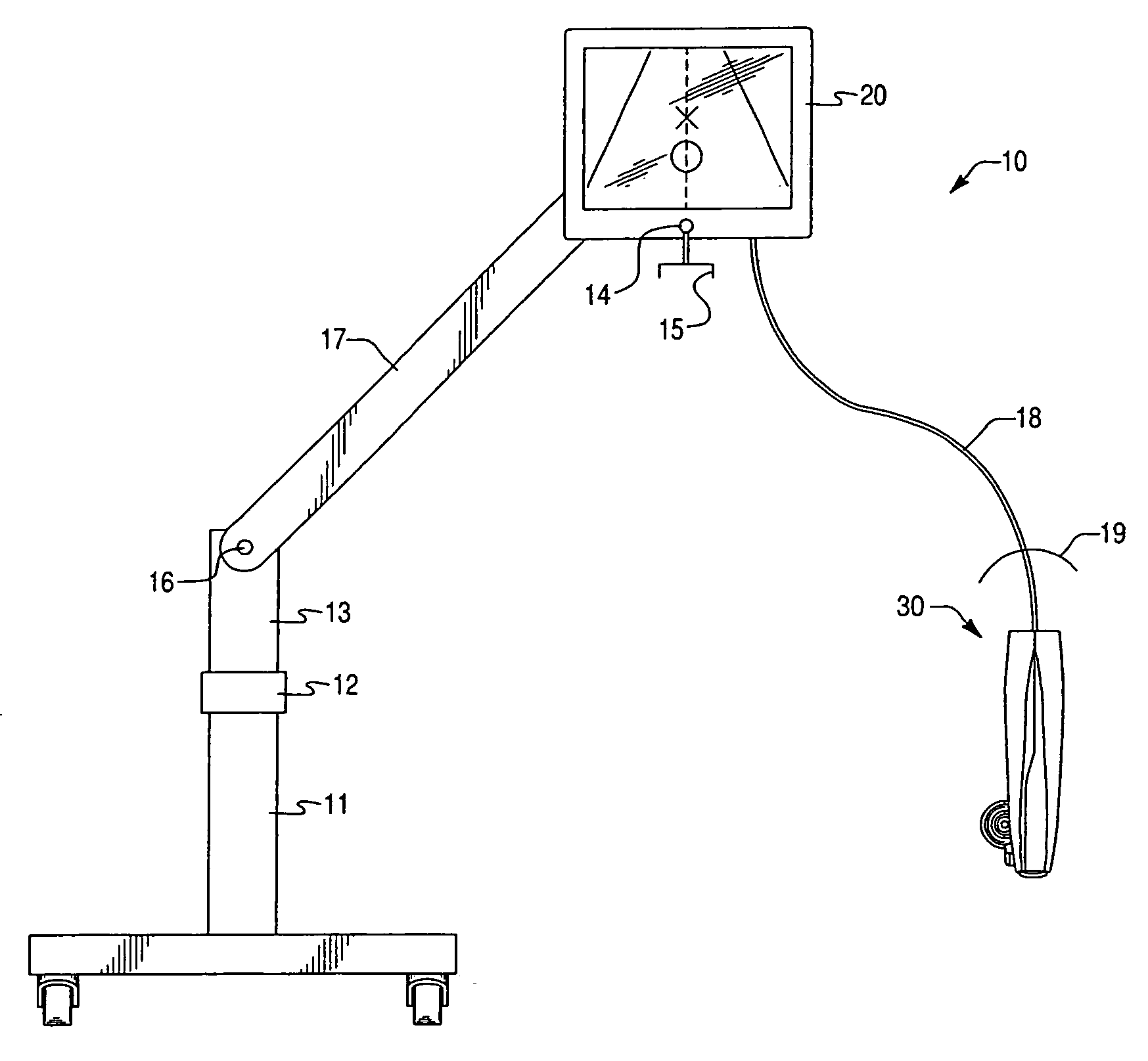
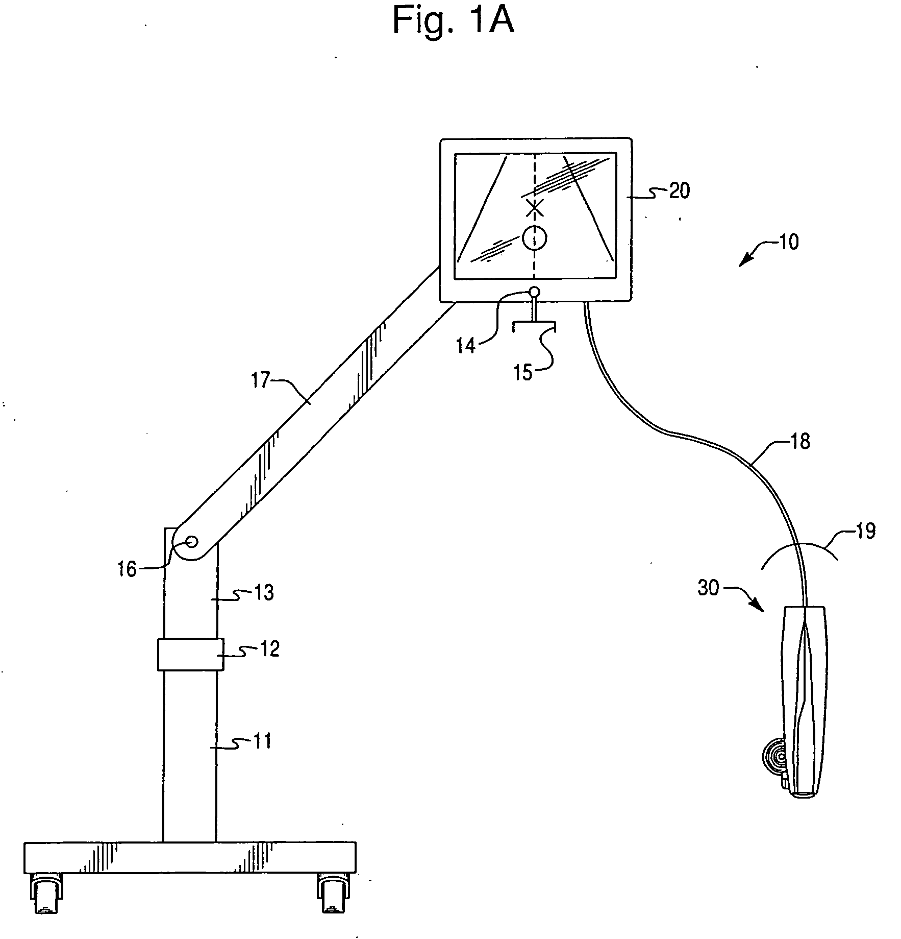
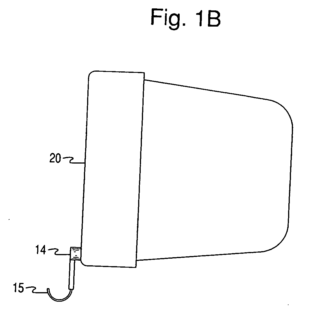
[0060] A needle clip 200 is attached to clip connector 44 by placing clip connector 44 within a hollow post 224 formed on needle clip 200 and engaging a snap-fit provided by depressions 44a formed on clip connector 44 and mating ledges formed on inner surfaces of post 224. Other means may be used for disengagably mounting clip 200 to clip connector 44. Needle clip 200 may also include a skirt formed near a lower end of post 224. The skirt is intended to cover opening 35 when needle clip 200 is mounted to clip connector 44, without obstructing rotation of needle clip 200 about probe 30, so as to further reduce the chance of contamination during a procedure. A shaft of the needle is received in a cradle portion 204 of needle clip 200 and releasably held therein by a fastening arm 216 during the procedure. The snap-fit engagement between post 224 and clip connector 44 is preferably easily releasable so as to enable a health professional to remove needle clip 200 from clip connector 44 ...

third embodiment

[0082] a needle clip 500 will now be described with reference to FIG. 10. In this embodiment, a fastening arm 516 is slidingly received in a grooved section 522 of first part 201 that is disposed adjacent to cradle 204. A flex member 524 is attached to fastening arm 516 at a lower surface 516a and is adapted to abut a surface 524a of probe 30, which causes flex member 524 to flex towards finger rest 520 when finger pressure is applied at finger rest 520. Before finger pressure is applied, cover 516b does not cover cradle 204. When finger pressure is applied, cover 516b extends over cradle 204, thereby trapping needle shaft between cradle 204 and cover 516b. While finger pressure is applied, flex member 524 is maintained in a flexed state. When finger pressure is removed, flex member 524 will move cover 516b back to its original position (i.e., not covering cradle 204) as it returns to its undeformed state. Needle shaft may then be separated from probe 30. This embodiment, like the o...


PUM


Abstract

A compact ultrasound needle guidance system and method of use is described. The needle guidance system has components to adjustably target a needle's destination in the plane of a two-dimensional ultrasound image before insertion of a needle into a patient. Needle movement is tracked using a position detector that provides a visual display of the needle path on the ultrasonic image.

Description

CROSS REFERENCE & RELATED APPLICATIONS [0001] This application is a continuation co-pending U.S. application Ser. No. 11 / 508,300, filed on Aug. 23, 2006 by Robert Park et al., entitled ULTRASOUND GUIDANCE SYSTEM, the entire contents of which is incorporated by reference, and for which priority is claimed under 35 U.S.C. §120. This application claims priority under 35 U.S.C. § 119(e) to U.S. Provisional Application No. 60 / 808,552 filed May 26, 2006, and U.S. Provisional Application No. 60 / 714,192 filed Sep. 2, 2005.FIELD OF INVENTION [0002] The present invention relates generally to ultrasound systems and, more particularly, to ultrasound guidance systems. BACKGROUND OF INVENTION [0003] As an inexpensive and noninvasive technique, ultrasound is useful as a medical imaging modality able to provide real time feedback in a two-dimensional fashion at a patient's bedside. Ultrasound facilitates dozens of procedures performed in hospitals and clinics every day, with these procedures rangin...

Claims


Application Information

Patent Timeline
Patent Type & Authority Applications(United States)
IPC IPC(8): A61B8/14
CPCA61B8/0833A61B8/0841A61B8/4218A61B2017/3413A61B8/4472A61B17/3403A61B19/081A61B8/4455A61B46/10
Inventor PARK, ROBERTMOSLER, THEODORE J.JARNAGIN, SCOTT P.KORGOI, TODD M.
Owner ULTRASOUND VENTURES
Why Eureka
Try Eureka