
Ultra-high-specificity device and methods for the screening of in-vivo tumors

a high-specificity, tumor-based technology, applied in the field of ultra-high-specificity in-vivo tumor screening, can solve the problems of false-positive tests having significant negative consequences, poor specificity, and inability to be used as widely as desired, and achieve non-electrical transmission of measuring photons, safe deployment, and simple

Inactive Publication Date: 2007-04-26
J FITNESS LLC
View PDF8 Cites 25 Cited by
  • Summary
  • Abstract
  • Description
  • Claims
  • Application Information

AI Technical Summary

Benefits of technology

[0016] Used as an adjunct to conventional in-vivo tumor screening tools, the device and methods of the present invention enable an earlier detection of cancer in many patients, without substantially increasing the burden of false-positive referrals. In the setting of an established screening program, traditional sensitivity-weighted detection can be sacrificed in order to add specificity-weighted detection to a screening tool without reducing the overall sensitivity of the program, with the new screening tool added as an adjunct to the established screening programs. By losing sensitivity as a guiding feature, changes can be made to the device of the present invention, such as lower spatial resolution, that facilitate manufacture, cost-effectiveness, ease of use, speed of use, and other beneficial changes.
[0018] One advantage is that a patient, physician, or surgeon can obtain real-time feedback regarding the discovery of local tumors in high-risk patients and respond early.
[0019] Another advantage is that the device and methods of the present invention may be safely deployed to patients at home or hospitals as a screening tool, to give long-term tumor-specific feedback as needed.
[0020] A further advantage is that the device and methods of the present invention can be actively coupled to a therapeutic device, such as a tumor ablation device, to provide feedback to the removal or ablation function, based upon the detection and degree of the local tumor.
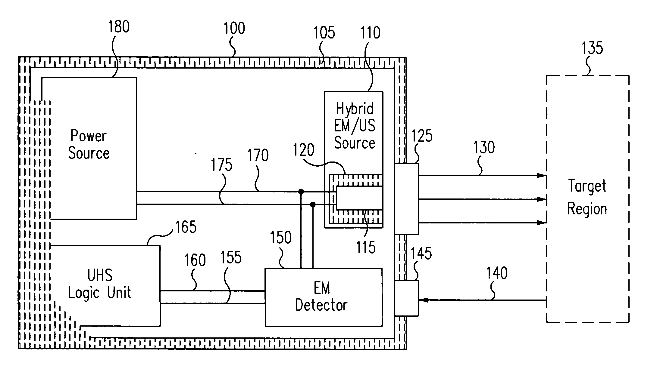
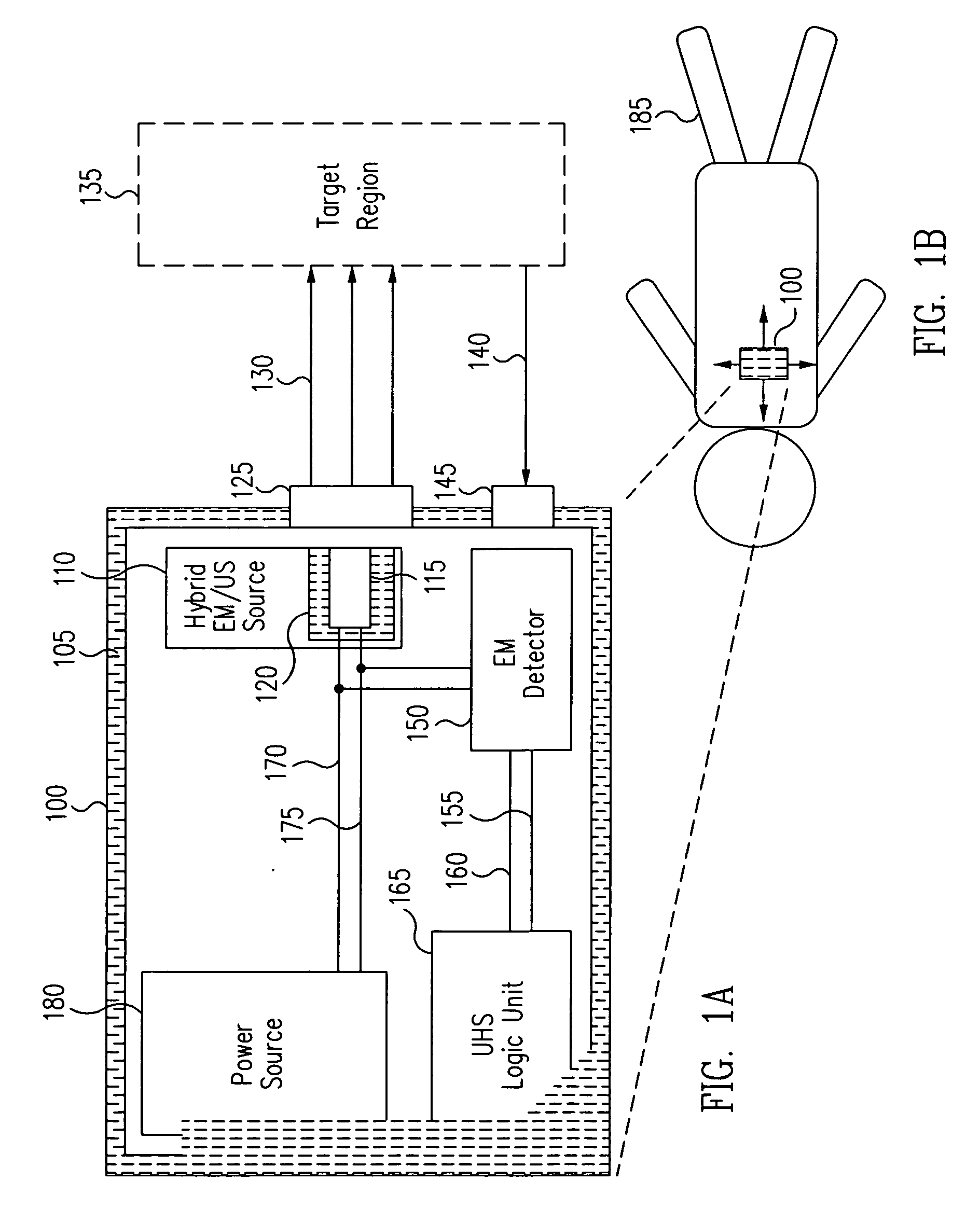
[0021] Yet another advantage is that the device of the present invention may be constructed to detect tumors using EM radiation, which allows for the simple, safe, and non-electrical transmission of measuring photons.

Problems solved by technology

However, their use is not as widespread as desired as their performance suffers from poor specificity.
False-positive tests have significant negative consequences, including unnecessary invasive tests, patient and family anxiety and pain, disruption of work, rising medical costs, and a fundamental loss of confidence in the medical testing itself when the workup reveals there wasn't any cancer there in the first place.
That means that for the vast majority of women, a positive screening test led to an unnecessary work-up.
These women who then later find more lumps, their doctors who have referred patients to find only benign lumps, and the radiologists who have been burned by falsely seeing too many early lesions, are each more hesitant to declare a lesion cancerous in the future, with obvious results.
This, the traditional goal of maximum sensitivity comes, in our view, at major personal and societal costs in terms of poor specificity and high false-positive rates.
It has also been shown that tumors are often hypoxic and / or hyperemic.
However, such published methods do not constitute clinically approved (e.g., FDA- or CE-approved), enabling instruments.
For example, United States Patent Publication No. 2005 / 0197583 discloses the use of optics to create two optical data sets, with a processor arranged to calculate congruence of the two optical data sets to detect abnormal tissue (such as tumors in an examined tissue), but does not teach or suggest maximization of specificity as a method to perform large-scale screening with an acceptably low false-positive rate.
EP 1008326, all teach optical methods for monitoring cancerous tissues, but do not teach maximization of specificity, nor are adaptations of the device needed for inducing acceptance as a screening tool taught or suggested.
All of the above known devices are limited in being designed to have a high sensitivity.
Because of the trade-off between sensitivity and specificity mentioned herein above, none of these prior art references disclose a means or arrangement designed to achieve high specificity, nor do they allow for a specific tumor detection in the setting of a large-population screening tool.
In short, the prior art lacks a unit arranged and optimized for the processing of different optical information for the purpose of achieving high specificity.

Method used


Image

  • Ultra-high-specificity device and methods for the screening of in-vivo tumors
  • Ultra-high-specificity device and methods for the screening of in-vivo tumors
  • Ultra-high-specificity device and methods for the screening of in-vivo tumors

Examples


Embodiment Construction

[0030] Generally, in accordance with exemplary embodiments of the present invention, a device and methods for the ultra-high-specificity (“UHS”) screening of in-vivo tumors in a target tissue site are provided. Tumors, as used herein, generally refer to a malignant tissue, or other type of cancer, to be diagnosed in a tissue, which may be any material from a living animal, plant, viral, or bacterial subject, with an emphasis on mammals, especially humans. A target tissue may be a tissue material to be detected, imaged, or studied. In the accompanying examples described herein below, one target tissue may be the breast.

[0031] In accordance with the present invention, the screening of in-vivo tumors can be applied in a large population. The device of the present invention can be used as an adjunct screening tool that is used in addition to other standard screening tools already in use. An adjunct screening tool has the disadvantage that it adds to the number of tests performed, but i...


PUM


Abstract

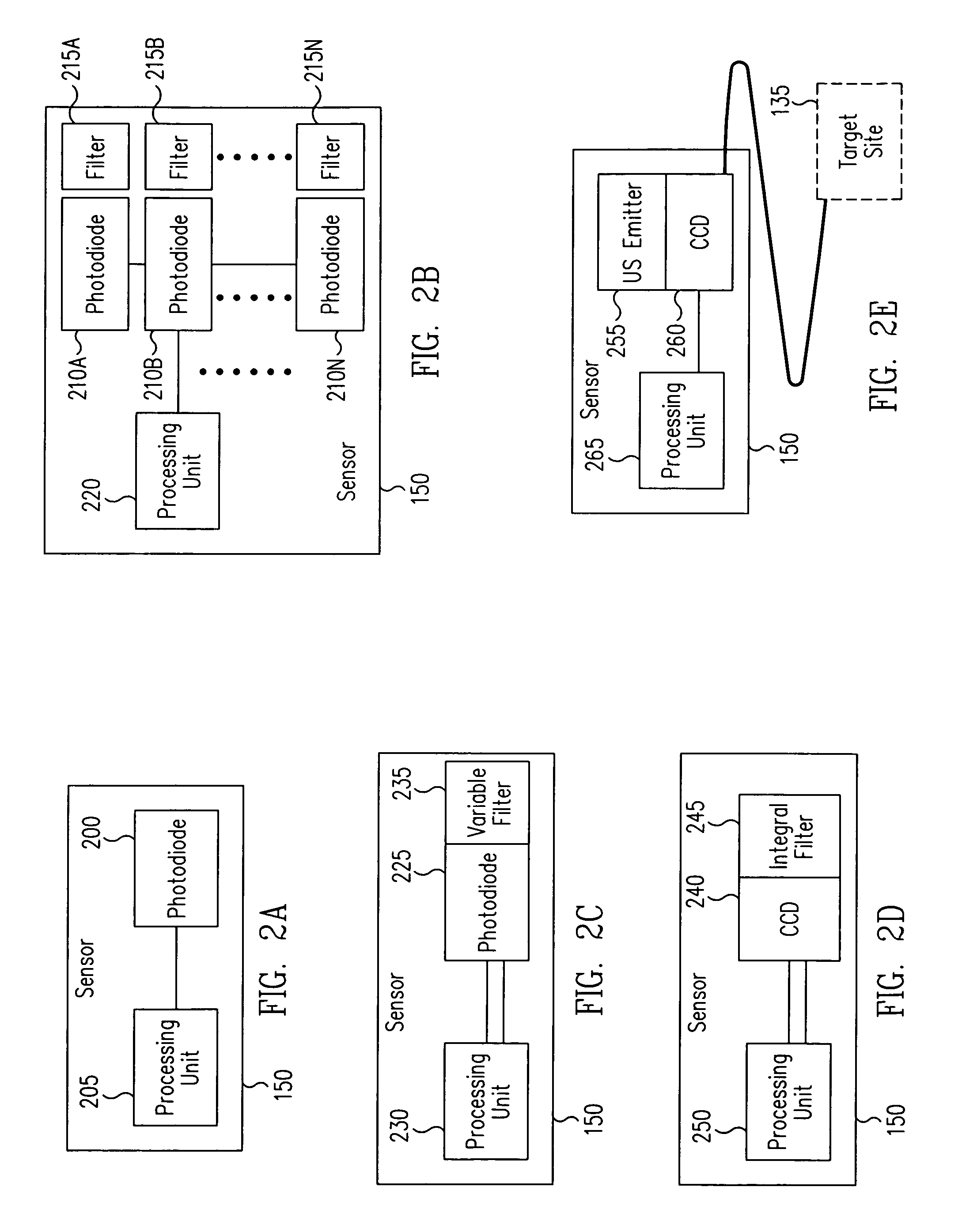
A device and a method for the screening of in-vivo tumors in a target tissue are provided. The device and method provide a local measure of a risk of tumor presence in the target tissue with high specificity. The local measure may be based upon a non-linear combination of local hemoglobin and tissue oxygen saturation and other tissue characteristics.

Description

FIELD OF THE INVENTION [0001] The present invention relates generally to a device and methods for performing in-vivo tumor screening. In particular, the device and methods of the present invention provide ultra-high-specificity in-vivo tumor screening using a portable, non-invasive hybrid electromagnetic and ultrasound scanner with a ultra-high-specificity logic unit that screens for the presence of a tumor with a low false-positive error rate, thus permitting a wide use in large population screening while minimizing false referrals for invasive follow-up testing. BACKGROUND INFORMATION [0002] Most tumors are detected very late, typically only after a cancer is well-established. For example, the average breast tumor size in the U.S. at first discovery is 2.0 cm wide for women and 2.5 cm for men, a surprisingly large lump. In contrast, early tumors often go undetected. The continued existence of frequent late cancer diagnosis is critical, as early diagnosis resulting from widespread ...

Claims


Application Information

Patent Timeline
Patent Type & Authority Applications(United States)
IPC IPC(8): A61B5/05A61B6/00
CPCA61B5/0091A61B5/0095A61B5/4312A61B8/0825
Inventor BENARON, DAVID A.PARACHIKOV, ILIAN H.FIERRO, MICHAEL R.FRIEDLAND, SHAI
Owner J FITNESS LLC
Learn More