Server-client architecture in medical imaging

a server-client and medical imaging technology, applied in the field of server-client architectures in medical imaging, can solve the problems of limiting the flexibility of user's workflow, relying on transferring patient data ahead of time to each client workstation, and consuming a large amount of time, so as to reduce the time before the client can start to operate autonomously, improve performance, and improve the overall transfer rate

Inactive Publication Date: 2007-05-24
TOSHIBA MEDICAL VISUALIZATION SYST EURO
View PDF38 Cites 69 Cited by
  • Summary
  • Abstract
  • Description
  • Claims
  • Application Information

AI Technical Summary

Benefits of technology

[0026] Because the data set is initially processed by the server computer, the server-generated rendered images quickly become available for display to a user. This is because it will generally be faster for the server computer to process the data set and transmit the resulting images to the client, than for the data set to be transferred to the client computer for local processing and generation of images for display. However, because the medical imaging data set itself is also transferred to the client computer, the client is able to operate independently of the server once the data set has been transferred. However, unlike in the traditional thick-client approach, this kind of autonomous operation by the client computer is achieved with the user still able to view the results of processing of the data set during the time in which it is being transferred to his location.
[0036] The method may further comprise waiting for a period of time before initiating transfer of the volume data set to a client computer over the network. This can ensure data are not transferred if a user is only viewing a patient's data for a short period.

Problems solved by technology

This can take a significant amount of time due to the large size of typical medical imaging data sets.
The main drawback of the thick-client approach is the reliance on transferring patient data ahead of time to each client workstation.
This limits the flexibility of the user's workflow (e.g. by requiring them to indicate the day before what work they will be doing the following day), and cannot cope with emergencies, such as an emergency medical case.
However, this is undesirable because the patient data for any given study is large (from hundreds of megabytes up to a few gigabytes) and thus transmitting patient data on demand requires the user to wait up to several minutes for the data to arrive over the network.
In the thick-client architecture, if the rules for distributing data ahead of time are not adequately effective, the user has to wait an objectionable amount of time for patient data to be sent to the client when demanded.
In the thin-client architecture, the system is able to access patient data quickly, but as the number of clients increases, the server begins to encounter scalability limitations (i.e. difficulty adding more client workstations to an existing network or implementing a new network with a large number of client) in respect of both processing power and network bandwidth (because the server is doing the work for all clients and the network is continually transferring results to the clients).
Thus it is not possible to keep adding client workstations as demand increases without decreasing performance for each user.
Furthermore, because in a thin-client network the data processing that happens in response to user's input occurs remotely from the client workstation, there can be significant latency in responding to user inputs.
This makes the system appear to be slow and unresponsive and can be frustrating for users.
Thus when implementing a network, deciding between a thick-or a thin-client architecture involves a trade off between potentially long waiting times to open a study (thick-client) and problems with scalability and latency during use (thin-client).
The approach is not practically applicable to rendering medical image data because medical imaging data typically comprise a continuous array of voxels representing a patient's body, or part thereof, that does not present any obviously advantageous decompositions between parts that can be rendered on the server and parts that can be rendered on the client.

Method used


Image

  • Server-client architecture in medical imaging
  • Server-client architecture in medical imaging
  • Server-client architecture in medical imaging

Examples


Embodiment Construction

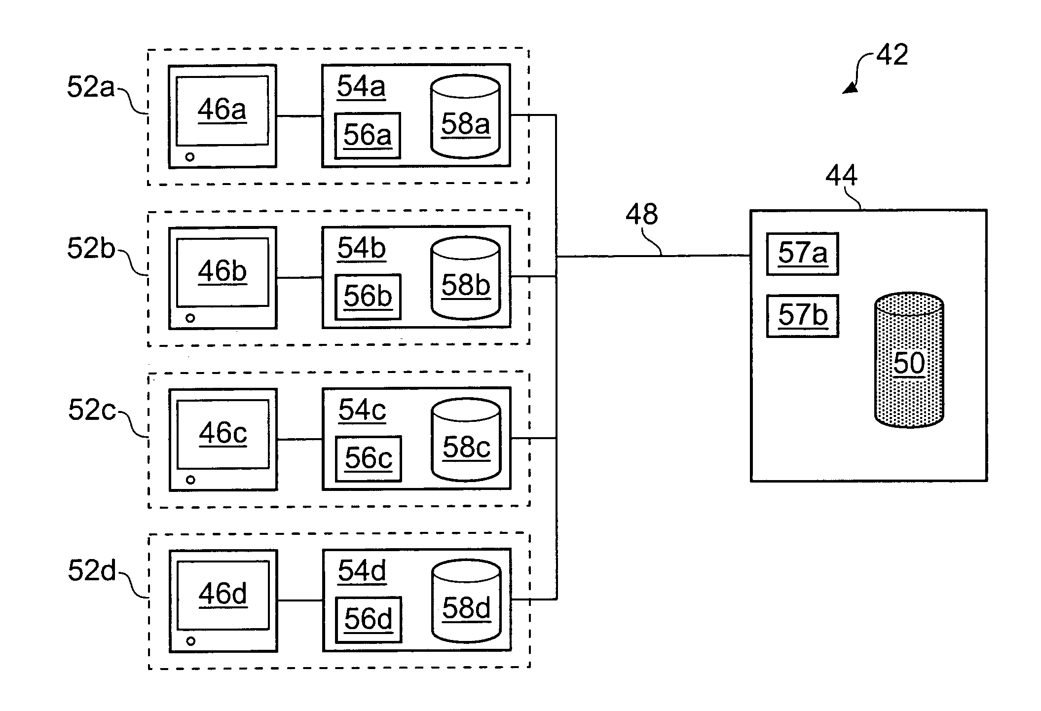
[0049]FIG. 5 schematically shows a computer network 42 for analyzing (e.g. processing and viewing) medical image volume data according to an embodiment of the invention. The data are analyzed using a software application. A typical software application supported by the network will comprise a suite of programs which allow a user to view and manipulate patient data as required for a given clinical study. An example of this type of application is Voxar3D™ provided by Barco N.V. A common task will be to render 2D images from 3D medical imaging data. The network 42 is deployed in a hospital, or similar environment. However, it could also be a distributed network (e.g. using the internet) that is not linked to any particular physical location. The network 42 comprises a server 44 and a number of (in this case four) client computer workstations (“clients”) 52a-d. The server 44 and clients 52a-d are in data communication through a conventional LAN network interconnection 48.

[0050] The ser...


PUM


Abstract

A method of processing medical imaging volume data in a computer network is described. The method comprises loading a medical imaging data set to be processed to a server computer, processing the data set on the server computer, e.g. by executing a software application, and generating corresponding server-generated results. The server-generated results, e.g. rendered images, may then be transmitted to a client computer for display to a user. This allows users to quickly view the results of the processing because they have not had to wait for the data set to be transferred to their local machine before locally processing the data. However, while this is happening, the data set itself is also transmitted, e.g. as a background operation, to the client computer. Thus eventually the client computer has access to a local copy of the data set and may start processing the data set itself, thus freeing up server resources.

Description

BACKGROUND OF THE INVENTION [0001] The invention relates to server-client architectures in medical imaging, and in particular to processing of medical image data in a network environment. [0002] Computer networks allow peripheral devices, data and computing applications to be shared amongst a number of users. This allows centralized management of a computing environment, helps reduce unnecessary duplication of computer resources, and can aid collaboration between users. For example, in a hospital or similar environment, it is common for patient data, e.g. medical image data, to be stored in a central data store from where it may be retrieved and subsequently processed, analyzed, viewed etc. by users working at computer workstations at different locations in the hospital (or working remotely over an appropriate link). This approach avoids the need for data to be stored locally to each of the users who might require it. Accordingly, the overall storage requirement of the computing sys...

Claims


Application Information

Patent Timeline
Patent Type & Authority Applications(United States)
IPC IPC(8): G06T17/00G06T15/08
CPCG06F19/321G06T15/08G16H10/60G16H30/40
Inventor TURNER, DAVIDPAPAGEORGIOU, PAVLOSCHIN, CONRADYATES, CARTERKIMPE, TOM
Owner TOSHIBA MEDICAL VISUALIZATION SYST EURO
Why Eureka
Try Eureka